
Reproductive Pathology — MCQs

On this page
Blood chimerism is most likely to occur in which of the following types of twin pregnancies?
Choriocarcinoma is differentiated from invasive mole (chorioadenoma destruens) by which of the following?
A female patient reports smoking 20 cigarettes a day. To which of the following conditions is she maximally predisposed?
Which of the following features is NOT evaluated for the histological grading of breast carcinoma?
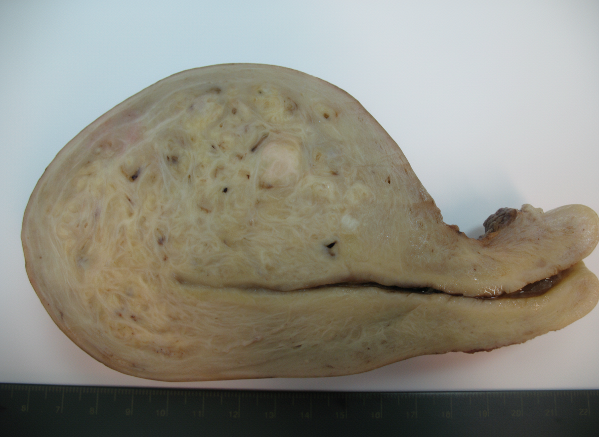
A hysterectomy specimen from a 38-year-old woman shows the following gross appearance. What is the most likely diagnosis?
The type of ductal carcinoma in situ (DCIS) most likely to result in a palpable abnormality in the breast is:
Which of the following is not a sex cord-stromal tumor?
Biopsy from a testicular mass following orchidectomy showed sheets of uniform cells containing abundant clear cytoplasm with prominent nucleus. Which is the histopathological diagnosis?
Which of the following is a germ cell tumor?
Which testicular tumor is rare in childhood?
Practice by Chapter
Diseases of Male Genital Tract
Practice Questions
Testicular Tumors
Practice Questions
Prostate Pathology
Practice Questions
Diseases of Female Genital Tract
Practice Questions
Cervical Pathology and Neoplasia
Practice Questions
Endometrial Pathology
Practice Questions
Ovarian Diseases and Tumors
Practice Questions
Gestational Trophoblastic Disease
Practice Questions
Placental Pathology
Practice Questions
Sexually Transmitted Infections
Practice Questions
Want unlimited practice?
Get full access to all questions, explanations, and performance tracking.
Scan to download app