
Reproductive Pathology — MCQs

On this page
A 35-year-old woman presents with a firm, painless, round lump in her right breast following a traumatic injury. Which statement is false regarding the condition she is experiencing?
Which of the following is NOT associated with arrhenoblastoma?
Most commonly, this tumor is due to carcinoma of which of the following organs?
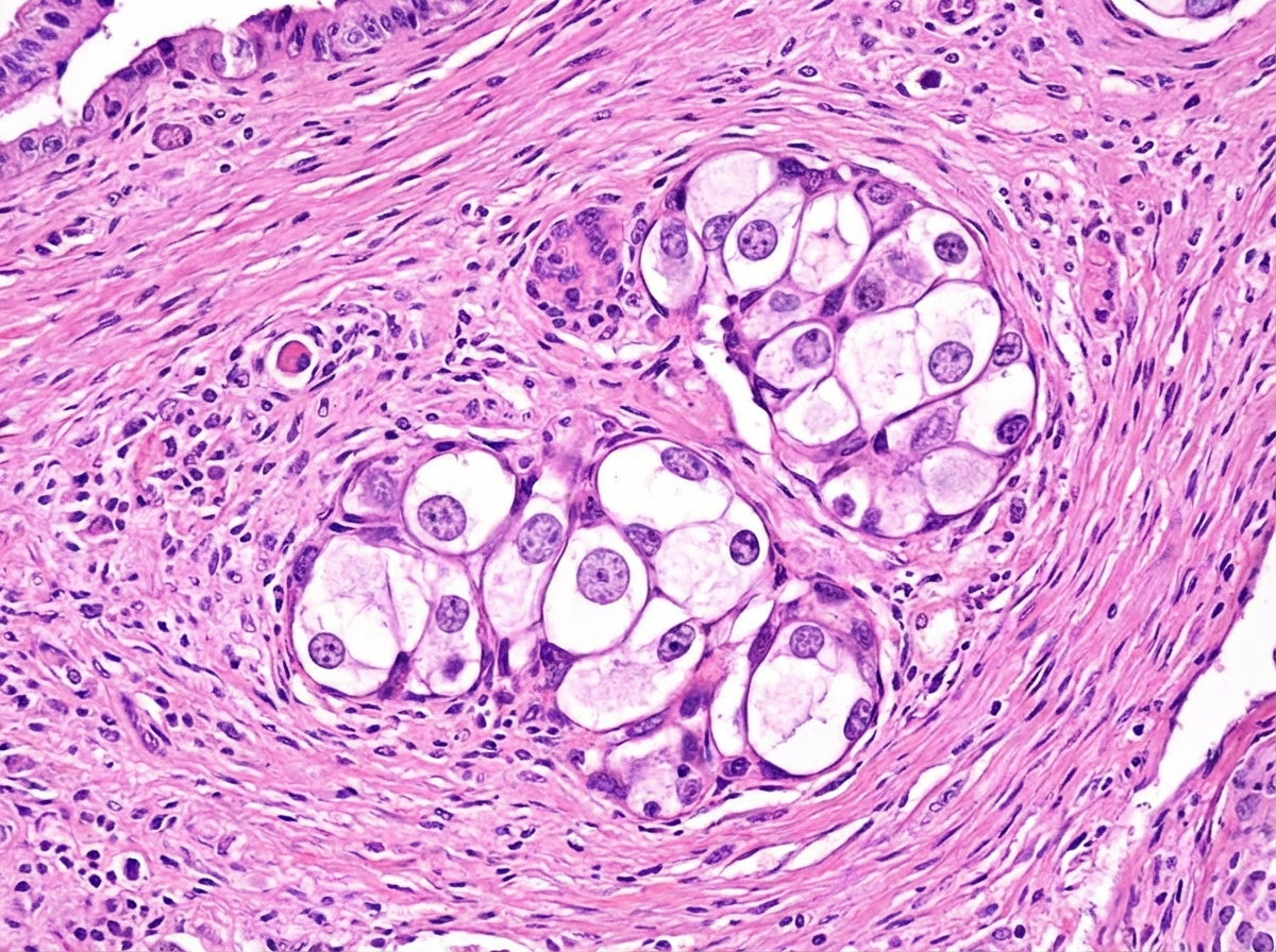
A 32-year-old male presents with a few weeks' history of progressive breast tissue enlargement. On examination, a painless testicular mass is noted. Hormonal investigations reveal low serum LH and testosterone levels. What is the likely cause in this case?
Which of the following is NOT associated with breast cancer?
Fallopian tube dysmotility is seen in which of the following conditions?
Koilocytes with perinuclear halo on Pap smear are pathognomic of which condition?
Adenoacanthoma is which type of uterine cancer?
Which of the following is NOT a sex cord-stromal tumor of the ovary?
Which tumor secretes placental alkaline phosphatase?
Practice by Chapter
Diseases of Male Genital Tract
Practice Questions
Testicular Tumors
Practice Questions
Prostate Pathology
Practice Questions
Diseases of Female Genital Tract
Practice Questions
Cervical Pathology and Neoplasia
Practice Questions
Endometrial Pathology
Practice Questions
Ovarian Diseases and Tumors
Practice Questions
Gestational Trophoblastic Disease
Practice Questions
Placental Pathology
Practice Questions
Sexually Transmitted Infections
Practice Questions
Want unlimited practice?
Get full access to all questions, explanations, and performance tracking.
Scan to download app