
Reproductive Pathology — MCQs

On this page
Which of the following structures is not affected by Gonococcus?
Paget's disease of the breast is a manifestation of which of the following?
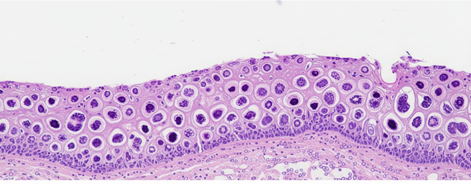
A cervical lesion is shown, which is similar to that obtained in a cone cervical biopsy from a 28-year-old sexually active woman who had had a "positive" Pap smear. What type of cervical change is often characterized by the lesion shown?
A patient presents with an abdominal mass. Investigations reveal bilateral ovarian masses with smooth surfaces. Microscopy shows cells with signet ring shapes. What is the most likely diagnosis?
Which of the following is NOT a type of primary uterine carcinoma?
P57KIP2 immunostaining is helpful in diagnosing which of the following conditions?
Gleason's classification is used for which of the following conditions?
A 33-year-old woman in her third trimester of pregnancy (gravida I, para 0) is rushed to the emergency room after suffering a seizure. The patient is hypertensive and laboratory studies show that the patient manifests nephritic syndrome. What is the appropriate diagnosis?
Which of the following is a marker for testicular tumor?
Which gene is involved in endometrial carcinoma?
Practice by Chapter
Diseases of Male Genital Tract
Practice Questions
Testicular Tumors
Practice Questions
Prostate Pathology
Practice Questions
Diseases of Female Genital Tract
Practice Questions
Cervical Pathology and Neoplasia
Practice Questions
Endometrial Pathology
Practice Questions
Ovarian Diseases and Tumors
Practice Questions
Gestational Trophoblastic Disease
Practice Questions
Placental Pathology
Practice Questions
Sexually Transmitted Infections
Practice Questions
Want unlimited practice?
Get full access to all questions, explanations, and performance tracking.
Scan to download app