
Reproductive Pathology — MCQs

On this page
Which of the following is NOT a prognostic factor of breast carcinoma?
What is the most common type of breast carcinoma?
Which of the following is true about a complete mole?
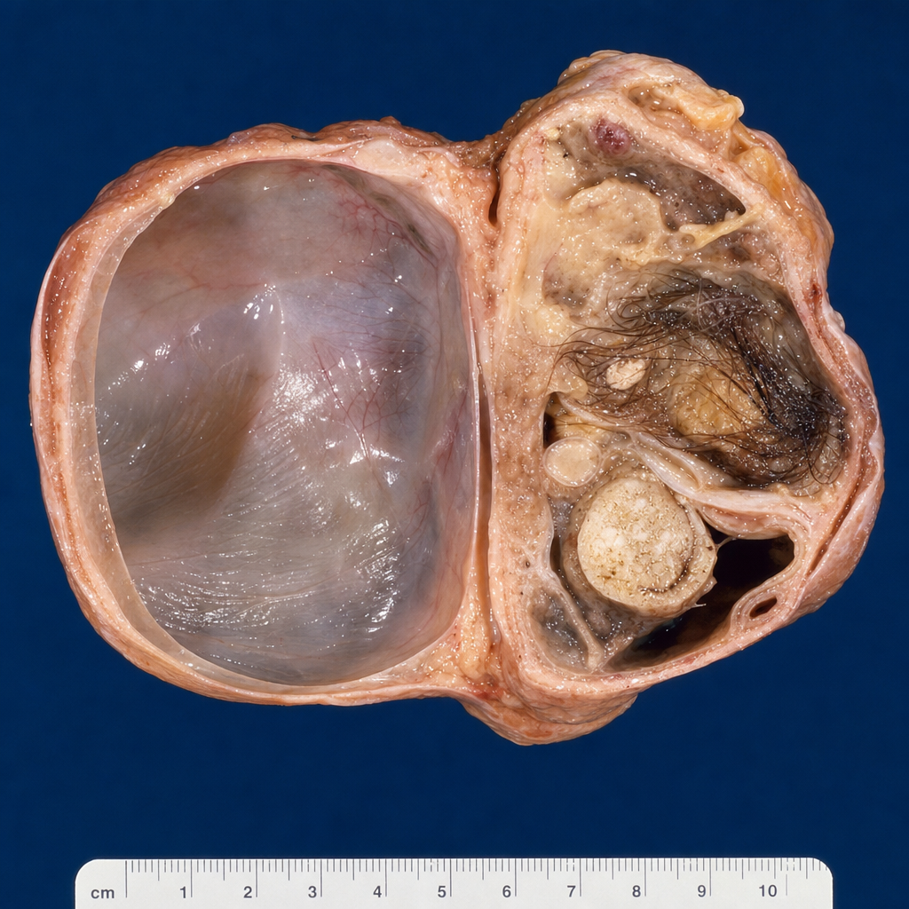
In the provided cut section of the ovary shown below, which of the following represents a common germ cell tumor?
A 32-year-old woman has been attempting to become pregnant for the past 2 years without success. She also has had extremely painful menstrual cramping of many years' duration. An exploratory laparoscopy demonstrated multiple red-blue nodules covering the surface of her ovaries and uterine ligaments. These findings are most likely indicative of which of the following conditions?
All of the following ovarian tumours arise from surface epithelium EXCEPT?
In which of the following infectious diseases is the testis involved but the epididymis spared?
A 24-year-old woman with a history of heavy and painful menstrual periods has been having difficulty conceiving. A bimanual pelvic examination and ultrasound demonstrate a mass in the uterus presumed to be a leiomyoma. This mass is a:
A 27-year-old woman in the third trimester of her third pregnancy discovers a lump in her left breast. On physical examination, a 2-cm, discrete, freely movable mass beneath the nipple is palpable. After the birth of a term infant, the mass appears to decrease in size. The infant is breastfed without difficulty. What is the most likely diagnosis?
Which of the following is an exception for secreting hormones?
Practice by Chapter
Diseases of Male Genital Tract
Practice Questions
Testicular Tumors
Practice Questions
Prostate Pathology
Practice Questions
Diseases of Female Genital Tract
Practice Questions
Cervical Pathology and Neoplasia
Practice Questions
Endometrial Pathology
Practice Questions
Ovarian Diseases and Tumors
Practice Questions
Gestational Trophoblastic Disease
Practice Questions
Placental Pathology
Practice Questions
Sexually Transmitted Infections
Practice Questions
Want unlimited practice?
Get full access to all questions, explanations, and performance tracking.
Scan to download app