Which type of amyloid is associated with long-term hemodialysis?
Kimmelstiel-Wilson lesion is characteristic of which condition?
Loss of foot processes seen on electron microscopy of renal biopsy is a classical feature in which of the following?
Class IV lupus nephritis is:
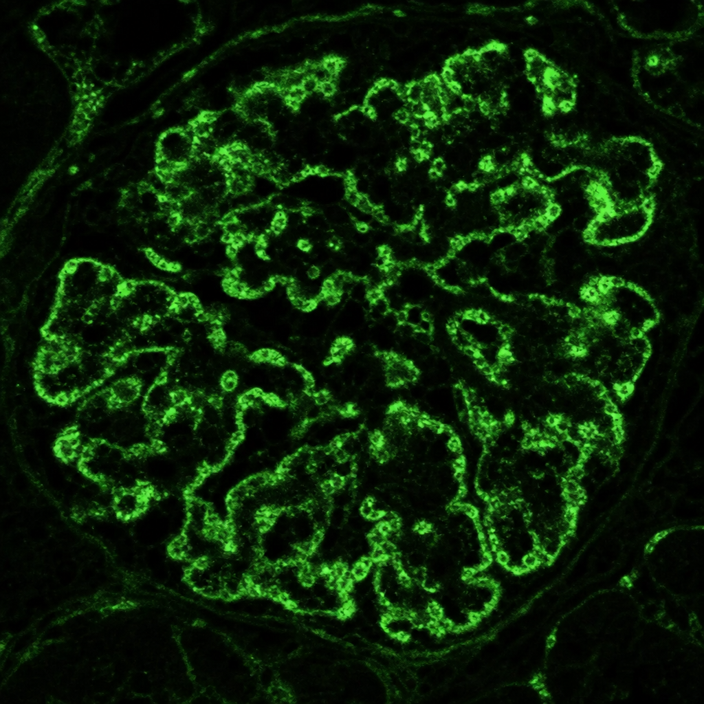
A patient presents with pulmonary hemorrhage and is P-ANCA positive. What is the most likely diagnosis?
All the following are features of Polycystic disease of kidneys EXCEPT:
In a case of glomerulonephritis (GN), C3 is normal in all the following except?
IgA nephropathy is not associated with which of the following?
A 7 year old boy presented with generalized edema. Urine examination revealed marked albuminuria. Serum biochemical examinations showed hypoalbuminemia with hyperlipidemia. Kidney biopsy was undertaken. On light microscopic examination, the kidney appeared normal. Electron microscopic examination is most likely to reveal
Which condition is characterized by wire loop lesions in the glomeruli?
Explanation: ***Beta 2 microglobulin*** - Accumulates in patients undergoing **long-term hemodialysis** and is primarily responsible for **dialysis-related amyloidosis** [1][4]. - It forms amyloid deposits, particularly affecting **joints and skin**, due to its impaired clearance during dialysis [1]. *ATTR* - Refers to **transthyretin amyloidosis**, which is primarily associated with familial or age-related amyloid deposits rather than hemodialysis [1]. - This type typically presents with **cardiac and neurological** symptoms, distinct from the findings in hemodialysis-associated conditions. *AL* - Stands for **light chain amyloidosis**, resulting from monoclonal immunoglobulin light chains, often seen in **multiple myeloma** [2]. - It is unrelated to hemodialysis; thus, patients do not typically develop AL amyloidosis due to kidney replacement therapy. *AA* - Associated with **acute phase reactants** like serum amyloid A, typically linked to chronic inflammatory diseases, not hemodialysis [3]. - AA amyloidosis is seen in conditions such as **rheumatoid arthritis**, making it distinct from beta 2 microglobulin's role in dialysis patients [3]. **References:** [1] Kumar V, Abbas AK, et al.. Robbins and Cotran Pathologic Basis of Disease. 9th ed. Diseases of the Immune System, p. 266. [2] Kumar V, Abbas AK, et al.. Robbins and Cotran Pathologic Basis of Disease. 9th ed. Diseases of the Immune System, pp. 266-267. [3] Kumar V, Abbas AK, et al.. Robbins and Cotran Pathologic Basis of Disease. 9th ed. Diseases of the Immune System, pp. 267-268. [4] Cross SS. Underwood's Pathology: A Clinical Approach. 6th ed. (Basic Pathology) introduces the student to key general principles of pathology, both as a medical science and as a clinical activity with a vital role in patient care. Part 2 (Disease Mechanisms) provides fundamental knowledge about the cellular and molecular processes involved in diseases, providing the rationale for their treatment. Part 3 (Systematic Pathology) deals in detail with specific diseases, with emphasis on the clinically important aspects., pp. 136-140.
Explanation: ***Diabetic nephropathy*** - Kimmelstiel-Wilson lesions are pathognomonic for **diabetic nephropathy**, appearing as nodular glomerulosclerosis [1]. - These lesions result from **hyperglycemia** leading to mesangial expansion and thickening of the glomerular basement membrane [2]. *HIV nephropathy* - Characterized by **focal segmental glomerulosclerosis** (FSGS) rather than Kimmelstiel-Wilson lesions. - Often presents with **proteinuria** and occurs in the context of **immune suppression**. *MPGN* - Membranoproliferative glomerulonephritis (MPGN) is associated with **proliferative glomerular changes** rather than Kimmelstiel-Wilson lesions. - Patients typically display **nephritic syndrome** with hematuria and decreased complement levels. *Hypertensive nephropathy* - Usually leads to **arteriosclerosis and hyaline changes** in the kidney, not Kimmelstiel-Wilson lesions. - Nephropathy manifests with **chronic kidney disease** and associated hypertension, without the specific lesion noted. **References:** [1] Kumar V, Abbas AK, et al.. Robbins and Cotran Pathologic Basis of Disease. 9th ed. The Endocrine System, pp. 1121-1122. [2] Kumar V, Abbas AK, et al.. Robbins and Cotran Pathologic Basis of Disease. 9th ed. The Endocrine System, p. 1121.
Explanation: ***Minimal change disease*** - **Loss of foot processes** (podocyte effacement) is the hallmark ultrastructural finding in **minimal change disease** on electron microscopy [1]. - This effacement of podocyte foot processes leads to increased permeability of the **glomerular filtration barrier** to albumin, causing **nephrotic syndrome** [1], [2]. *IgA nephropathy* - Characterized by **IgA immune complex deposition** in the **mesangium** on immunofluorescence. - Electron microscopy typically shows **mesangial immune deposits**, not primarily foot process effacement. *Membranous nephropathy* - Identified by the presence of **subepithelial immune deposits** and **thickening of the glomerular basement membrane** (GBM) [3]. - On electron microscopy, these deposits are visible, often with overlying **spikes** of GBM material separating them. *Rapidly progressive glomerulonephritis* - Defined by the rapid loss of renal function and the presence of **crescents** in more than 50% of glomeruli on light microscopy [2]. - While there may be secondary podocyte changes due to severe inflammation, **foot process effacement** is not its primary diagnostic feature. **References:** [1] Kumar V, Abbas AK, et al.. Robbins and Cotran Pathologic Basis of Disease. 9th ed. The Kidney, pp. 927-928. [2] Cross SS. Underwood's Pathology: A Clinical Approach. 6th ed. Common Clinical Problems From Diseases Of The Urinary And Male Genital Tracts, pp. 527-528. [3] Cross SS. Underwood's Pathology: A Clinical Approach. 6th ed. Common Clinical Problems From Diseases Of The Urinary And Male Genital Tracts, pp. 532-533.
Explanation: ***Diffuse lupus nephritis*** - This is the most **severe** and common form of lupus nephritis, characterized by **widespread inflammation** and proliferation affecting more than 50% of glomeruli [1]. - Patients typically present with **nephrotic-range proteinuria**, **hematuria**, and significant **renal impairment** [1]. *Proliferative lupus nephritis* - This is a general term describing **increased cellularity** within the glomeruli. - While Class IV (diffuse) lupus nephritis is a proliferative form, "proliferative lupus nephritis" alone is not a specific class in the ISN/RPS classification system without further qualification (e.g., focal or diffuse). *Mesangial lupus nephritis* - This refers to Classes I and II, characterized by **mesangial immune deposits** and/or mild mesangial hypercellularity. - It is typically a **milder form** with a better prognosis, unlike the severe presentation of Class IV [1]. *Membranous lupus nephritis* - This corresponds to Class V lupus nephritis, distinguished by **subepithelial immune deposits** and thickening of the glomerular basement membrane. - Patients often present with **nephrotic syndrome** but typically have less severe inflammation or cellular proliferation compared to Class IV. **References:** [1] Kumar V, Abbas AK, et al.. Robbins and Cotran Pathologic Basis of Disease. 9th ed. Diseases of the Immune System, pp. 230-232.
Explanation: ***Microscopic polyangiitis*** - This condition is characterized by **pulmonary hemorrhage** (often manifesting as diffuse alveolar hemorrhage) and **P-ANCA positivity**, which is typically associated with antibodies against **myeloperoxidase (MPO)**. [1] - It is a **small-vessel vasculitis** that frequently affects the kidneys (glomerulonephritis) and lungs without granuloma formation. *Churg-Strauss syndrome* - While Churg-Strauss syndrome (now known as **Eosinophilic Granulomatosis with Polyangiitis**, EGPA) can be P-ANCA positive, it is typically associated with a history of **asthma**, **allergic rhinitis**, and **eosinophilia**. [1] - Pulmonary involvement often includes **infiltrates** and nodules, but diffuse alveolar hemorrhage with severe pulmonary hemorrhage is less common as the primary presentation compared to MPA. *Wegener granulomatosis* - Wegener granulomatosis (now known as **Granulomatosis with Polyangiitis**, GPA) primarily presents with **upper and lower respiratory tract granulomatous inflammation** and **glomerulonephritis**. - It is typically associated with **C-ANCA positivity** (antibodies against proteinase 3, PR3), not P-ANCA. *Polyarteritis nodosa (PAN)* - Polyarteritis nodosa is a **medium-vessel vasculitis** that typically affects the **kidneys, gastrointestinal tract, skin, and nervous system**. [1] - It is classically **ANCA-negative** and does not typically cause pulmonary hemorrhage or diffuse alveolar hemorrhage.
Explanation: ***Erythrocytosis*** - While other renal conditions like **renal cell carcinoma** can cause erythrocytosis due to increased **erythropoietin** production, it is generally **not a typical feature** of Polycystic Kidney Disease (PKD). - Patients with PKD usually have **normal or even low erythropoietin levels** despite compromised kidney function, and anemia is more common, particularly as **renal failure progresses**. *Renal failure* - **Progressive cyst growth** leads to replacement of normal kidney parenchyma, inevitably culminating in **end-stage renal disease** [1] in the majority of patients. - This is a hallmark feature, often necessitating **dialysis or transplant** later in life for individuals with autosomal dominant polycystic kidney disease (ADPKD) [2]. *Haematuria* - **Gross or microscopic hematuria** is a common symptom in PKD, often resulting from **cyst rupture** [1], bleeding into a cyst, or the passage of a calculus due to urinary stasis. - It can be a presenting symptom and can cause significant pain and anxiety for patients. *Hypertension* - **Hypertension** is an early and frequent complication of PKD, often preceding any significant decline in glomerular filtration rate. - It is primarily caused by activation of the **renin-angiotensin-aldosterone system (RAAS)** [3] due to arterial compression and ischemia from expanding cysts. **References:** [1] Kumar V, Abbas AK, et al.. Robbins and Cotran Pathologic Basis of Disease. 9th ed. The Kidney, pp. 951-955. [2] Cross SS. Underwood's Pathology: A Clinical Approach. 6th ed. Common Clinical Problems From Diseases Of The Urinary And Male Genital Tracts, pp. 544-545. [3] Cross SS. Underwood's Pathology: A Clinical Approach. 6th ed. Common Clinical Problems From Diseases Of The Urinary And Male Genital Tracts, pp. 520-521.
Explanation: ***Diffuse lupus nephritis*** - This condition is characterized by **immune complex deposition** in the glomeruli [2], leading to activation of the **classical complement pathway** and consumption of C3, resulting in low C3 levels. [3] - Patients with diffuse lupus nephritis often present with **systemic lupus erythematosus (SLE)** symptoms, and **hypocomplementemia** is a hallmark. [3] *Goodpasture's syndrome* - This is an **autoimmune disease** targeting the **glomerular basement membrane (GBM)** and lung alveolar basement membranes. [1] - It involves **anti-GBM antibodies** directly, rather than widespread immune complex formation and complement consumption, so C3 levels are typically normal. [1] *IgA nephropathy* - Characterized by the deposition of **IgA immune complexes** in the glomeruli. [2] - While there is immune activity, C3 levels are generally **normal** because the alternative complement pathway, which primarily involves C3, is not extensively activated or consumed. *HSP* - **Henoch-Schönlein Purpura (HSP)** is a form of **IgA vasculitis** that can affect the kidneys, causing a glomerulonephritis similar to IgA nephropathy. - Similar to IgA nephropathy, C3 levels are usually **normal** in HSP-associated glomerulonephritis as the primary immune mechanism does not typically involve significant C3 consumption. **References:** [1] Kumar V, Abbas AK, et al.. Robbins and Cotran Pathologic Basis of Disease. 9th ed. The Kidney, p. 915. [2] Kumar V, Abbas AK, et al.. Robbins and Cotran Pathologic Basis of Disease. 9th ed. The Kidney, p. 911. [3] Kumar V, Abbas AK, et al.. Robbins and Cotran Pathologic Basis of Disease. 9th ed. The Kidney, pp. 910-911.
Explanation: ***Decreased complement level*** - IgA nephropathy is typically associated with **normal serum complement levels** (C3 and C4), which is an important distinguishing feature. - Unlike post-streptococcal glomerulonephritis or lupus nephritis where complement levels are **low/decreased**, IgA nephropathy does not cause systemic complement consumption. - While complement activation does occur locally in the glomerulus (via lectin and alternative pathways), it does not lead to a decrease in serum complement levels. *Focal mesangial proliferation* - This is a **common histological finding** in IgA nephropathy, reflecting the proliferative response to IgA deposition in the mesangium. - The mesangial cells proliferate in an attempt to clear the immune deposits. *Gross hematuria concurrent with upper respiratory infection* - This is a **classic clinical presentation** of IgA nephropathy, often referred to as **synpharyngitic hematuria**. - The episode of gross hematuria typically occurs **within 1-2 days** of the onset of an upper respiratory tract infection, distinguishing it from post-streptococcal glomerulonephritis where hematuria appears 1-3 weeks later. *Immunofluorescence deposits contain IgA and IgG* - The defining feature of IgA nephropathy on immunofluorescence is the **predominant deposition of IgA**, often accompanied by C3. - While IgA is the primary immunoglobulin, **IgG and IgM can also be present** in variable amounts, but IgA must be the dominant or co-dominant immunoglobulin for the diagnosis.
Explanation: ***Fusion of foot processes of the glomerular epithelial cells*** - The clinical presentation of generalized edema, marked albuminuria, hypoalbuminemia, and hyperlipidemia in a young child strongly indicates **minimal change disease** (MCD), which is the most common cause of **nephrotic syndrome** in children [1]. - While light microscopy is normal in MCD, electron microscopy characteristically reveals **effacement or fusion of the foot processes** of the podocytes, which leads to increased glomerular permeability to proteins [1]. *Rarefaction of glomerular basement membrane* - **Rarefaction** or thinning of the glomerular basement membrane is associated with **Alport syndrome** or diseases causing basement membrane injury, which typically present with progressive kidney disease and hematuria. - This finding is not characteristic of minimal change disease or the nephrotic syndrome described. *Deposition of electron dense material in the basement membrane* - The deposition of **electron-dense material** within the glomerular basement membrane is characteristic of immune complex-mediated diseases like **membranoproliferative glomerulonephritis** (MPGN) or **post-infectious glomerulonephritis**. - These conditions often present with hematuria and are not typically associated with normal light microscopy in the context of nephrotic syndrome. *Thin basement membrane* - A **thin basement membrane** is a defining feature of **thin basement membrane disease** (also known as benign familial hematuria). - This condition is primarily associated with **microscopic hematuria** and does not typically cause nephrotic range proteinuria or generalized edema. **References:** [1] Kumar V, Abbas AK, et al.. Robbins and Cotran Pathologic Basis of Disease. 9th ed. The Kidney, pp. 922-923.
Explanation: ***SLE*** - Wire loop lesions in glomeruli are a classic histological finding in **Systemic Lupus Erythematosus (SLE)**, indicating severe glomerulonephritis. - These lesions result from **subendothelial immune complex deposits**, leading to a distinctive appearance on renal biopsy [1]. *Amyloidosis* - Characterized by **polarized light appearance** of apple-green birefringence due to amyloid deposits, not wire loop lesions. - Renal involvement occurs with **nodular glomerulosclerosis**, shown by different histological patterns. *Nephrotic* - Refers to a syndrome characterized by **heavy proteinuria**, **hypoalbuminemia**, and **edema**, but not specifically wire loop lesions. - The histopathology typically reveals **minimal change disease** or **focal segmental glomerulosclerosis**, not wire loops. *IgA nephropathy* - Generally presents with **IgA deposits** in mesangial regions causing **hematuria** and mild proteinuria. - It does not exhibit wire loop lesions, which are characteristic of **lupus nephritis** [1]. **References:** [1] Kumar V, Abbas AK, et al.. Robbins and Cotran Pathologic Basis of Disease. 9th ed. Diseases of the Immune System, p. 232.
Get full access to all questions, explanations, and performance tracking.
Start For Free