A characteristic feature of nephritic syndrome in children is:
Which of the following is the LEAST common characteristic of nephrotic syndrome?
A kidney biopsy under electron microscopy shows subepithelial 'spike' formation. Which immunofluorescence pattern would confirm membranous nephropathy?
Child with proteinuria, generalized edema, hypoproteinemia, and hyperlipidemia - most common cause is?
Which of the following is NOT a feature of nephrotic syndrome?
In glomerulus subendothelial deposits are seen in?
Which of these conditions is classified as a nephritic syndrome?
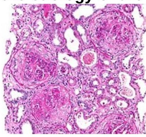
A 51-year-old person came with a complaint of hematuria. On examination, he was normotensive and had pedal edema. Investigations revealed the patient had no glucosuria and had a creatinine value of 9mg%. Renal biopsy is as shown below. Which of the following investigations should be done to identify the etiology of the disease?

Loss of foot processes seen on electron microscopy of renal biopsy is a classical feature in which of the following?
A 7-year-old boy presented with generalized edema. Urine examination revealed marked albuminuria. Serum biochemical examinations showed hypoalbuminemia with hyperlipidemia. Kidney biopsy was undertaken. On light microscopic examination, the kidney appeared normal. Electron microscopic examination is most likely to reveal:
Explanation: ***RBC casts in urine*** - The presence of **red blood cell (RBC) casts** in urine is a **pathognomonic sign** of **glomerulonephritis**, which is the underlying pathology in nephritic syndrome. - This indicates **glomerular inflammation** and bleeding, where RBCs leak through damaged glomeruli and are molded into casts within the renal tubules. *WBC casts in urine* - **White blood cell (WBC) casts** are characteristic of **pyelonephritis** (kidney infection) or other severe interstitial nephritis, indicating inflammation within the renal tubules. - While infection can sometimes accompany nephritic syndrome, WBC casts are not a primary diagnostic feature of the syndrome itself. *Lipid casts in urine* - **Lipid casts** and **fatty oval bodies** are typically associated with **nephrotic syndrome**, resulting from significant proteinuria and hyperlipidemia. - They signify severe disruption of glomerular filtration leading to lipid excretion, which is not the defining feature of nephritic syndrome. *Albumin in urine* - While **proteinuria (albumin in urine)** is present in nephritic syndrome, it is a non-specific finding and is also a hallmark of **nephrotic syndrome**, where it is much more severe. - The *quality* of the proteinuria (e.g., whether it contains RBCs) is more indicative for differentiating nephritic from nephrotic syndrome.
Explanation: ***Lipiduria*** - While lipiduria can occur in nephrotic syndrome, particularly with **severe proteinuria**, it is not a universally present clinical diagnostic criterion. - Its presence is a consequence of lipoprotein filtration rather than a direct defining feature of the syndrome itself. *Hypoalbuminemia* - This is a cardinal feature, defined as **serum albumin < 3.0 g/dL**, resulting from significant urinary protein loss. - It drives the generalized edema characteristic of nephrotic syndrome by reducing plasma oncotic pressure [1]. *Hyperlipidemia* - This is a very common characteristic due to increased hepatic synthesis of lipoproteins and decreased catabolism, often presenting as elevated **cholesterol and triglycerides**. - It is a risk factor for cardiovascular complications in nephrotic patients [1]. *Proteinuria > 3.5 gm per 1.73 m2 per 24 hours* - This is the **defining feature** of nephrotic syndrome, indicating significant damage to the glomerular filtration barrier [1]. - Massive proteinuria leads to the other key clinical manifestations of the syndrome.
Explanation: ***Granular IgG along GBM*** - **Membranous nephropathy** is characterized by the immune complexes forming **subepithelial deposits** along the glomerular basement membrane (GBM) [1], [2]. - These deposits appear as **granular IgG** (and often C3) on immunofluorescence, corresponding to the "spike" formation seen on electron microscopy [1]. *Linear IgG along GBM* - This pattern is characteristic of **Goodpasture syndrome** (anti-GBM disease), where antibodies directly bind to the GBM in a uniform, linear fashion [3], [4]. - The electron microscopy findings in Goodpasture syndrome do not show subepithelial "spike" formation but rather a normal or thickened GBM. *Mesangial IgA deposits* - This is the hallmark of **IgA nephropathy** (Berger's disease), where IgA immune complexes accumulate predominantly in the mesangium [2]. - IgA nephropathy would not typically present with subepithelial "spike" formation on electron microscopy. *C3 deposits only* - While C3 can be present in membranous nephropathy, finding "C3 deposits only" without significant IgG or other immunoglobulins suggests conditions like **C3 glomerulopathy** or dense deposit disease. - These conditions have distinct electron microscopy findings and a different pathogenesis compared to membranous nephropathy. **References:** [1] Kumar V, Abbas AK, et al.. Robbins and Cotran Pathologic Basis of Disease. 9th ed. The Kidney, p. 921. [2] Kumar V, Abbas AK, et al.. Robbins and Cotran Pathologic Basis of Disease. 9th ed. The Kidney, p. 911. [3] Kumar V, Abbas AK, et al.. Robbins and Cotran Pathologic Basis of Disease. 9th ed. The Kidney, p. 909. [4] Cross SS. Underwood's Pathology: A Clinical Approach. 6th ed. Common Clinical Problems From Diseases Of The Urinary And Male Genital Tracts, pp. 526-527.
Explanation: **Minimal change nephrotic syndrome** - This is the most common cause of **nephrotic syndrome** in children, characterized by the classic tetrad of **proteinuria**, **hypoalbuminemia**, **edema**, and **hyperlipidemia**. - The disease involves **effacement of podocyte foot processes**, visible on electron microscopy, but the glomeruli appear normal on light microscopy. *Mesangial glomerulonephritis* - This condition involves immune complex deposition in the **mesangium**, often presenting with **hematuria** and proteinuria, but not typically the full nephrotic picture in children. - It's a form of **glomerulonephritis**, distinct from the podocytopathy seen in minimal change disease. *FSGN* - **Focal segmental glomerulosclerosis (FSGS)** is a common cause of nephrotic syndrome, but it's less common than minimal change disease as the initial presentation in young children. - It is characterized by **segmental scarring of glomeruli**, which can be seen on light microscopy. *IgA nephropathy* - This is a common cause of **glomerular hematuria**, often presenting after an upper respiratory infection. - While proteinuria can occur, it's typically not severe enough to cause the full-blown **nephrotic syndrome** with generalized edema, hypoproteinemia, and hyperlipidemia in children.
Explanation: ### Original Explanation ***Hematuria*** - **Hematuria** is a characteristic feature of **nephritic syndrome**, which involves glomerular inflammation and damage leading to blood in the urine [1]. - In contrast, **nephrotic syndrome** is primarily characterized by increased glomerular permeability to protein, not red blood cells, resulting in significant **proteinuria** [1]. *Edema* - **Edema** is a hallmark of nephrotic syndrome, resulting from severe **hypoalbuminemia** that reduces plasma oncotic pressure [2]. - This leads to fluid extravasation into the interstitial spaces, causing generalized swelling. *Hypoalbuminemia* - **Hypoalbuminemia** is a defining feature of nephrotic syndrome, caused by excessive urinary loss of albumin due to widespread glomerular damage [2]. - Reduced serum albumin levels contribute to the characteristic edema and increased lipid synthesis by the liver. *Proteinuria* - **Proteinuria**, specifically *massive proteinuria* (>3.5 g/day in adults), is the cardinal feature of nephrotic syndrome [1], [2]. - It signifies significant damage to the glomerular filtration barrier, allowing large amounts of protein to leak into the urine.
Explanation: ***MPGN type I*** - **Subendothelial deposits** are a hallmark of MPGN type I, often associated with **immune complex deposition** [1]. - This condition can present with **hematuria**, **proteinuria**, and can be triggered by infections or autoimmune diseases [1]. *Good pasture syndrome* - Primarily involves **anti-GBM antibodies** leading to **glomerulonephritis** and pulmonary hemorrhage, not subendothelial deposits. - Typically, it presents with **crescent formation** in the glomeruli rather than deposits. *MPGN type II* - Characterized by **dense deposit disease**, it features **intramembranous** rather than subendothelial deposits [1]. - It is often associated with **C3 nephritic factor** and does not show classic subendothelial pathology. *IgA nephropathy* - Characterized by **IgA deposits** primarily in the **mesangium**, not subendothelially. - It presents with **hematuria** and recurrent episodes of **macrohematuria**, especially after infections. **References:** [1] Kumar V, Abbas AK, et al.. Robbins and Cotran Pathologic Basis of Disease. 9th ed. The Kidney, pp. 925-927.
Explanation: ***Post infectious Glomerulonephritis*** - Characterized by **hematuria, hypertension, and edema**, typically following an infection, such as streptococcal pharyngitis [2]. - Immune-mediated response leads to **decreased GFR** and signs of nephritic syndrome [1][2]. *Focal segmental glomerulosclerosis* - Primarily causes **nephrotic syndrome**, characterized by proteinuria and edema rather than hematuria [2]. - Often associated with **secondary causes** like obesity or HIV, not typically post-infectious. *Membranous Glomerulopathy* - Results in significant **proteinuria** and is classified as a **nephrotic syndrome** rather than a nephritic one [2][3]. - It presents with **edema and hypoalbuminemia**, lacking the hallmark features of hematuria. *Minimal change disease* - Predominantly causes **nephrotic syndrome** with heavy proteinuria and little to no hematuria [2]. - Young children are commonly affected, and it responds well to **corticosteroid therapy** [1].
Explanation: ***ANTI GBM antibodies*** - The presence of hematuria and renal failure suggests a possible **glomerulonephritis**, where **anti-GBM (Glomerular Basement Membrane) antibodies** would help confirm conditions like Goodpasture syndrome [2]. - This test specifically identifies **anti-GBM disease** [1,2], which is crucial in guiding management for this patient with suspected renal pathology [3]. *ANA* - **Antinuclear antibody (ANA)** testing is typically used for autoimmune diseases like **Systemic Lupus Erythematosus** but is less specific for glomerular diseases. - In this context, ANA would not specifically help in identifying the **etiology of renal failure** associated with hematuria. *Urine immunoelectrophoresis* - This test is primarily useful for detecting **light chains** in conditions like **multiple myeloma** and may not be relevant to general hematuria or renal failure. - It is not a direct test for **glomerular disease etiology** related to hematuria and edema. *HIV RNA* - While **HIV** can lead to renal complications, including **HIV-associated nephropathy**, this test is not the first line for etiological determination in this specific presentation. - Negative **HIV serology** doesn't rule out renal disease caused by other factors, making this test less relevant here. **References:** [1] Cross SS. Underwood's Pathology: A Clinical Approach. 6th ed. Common Clinical Problems From Diseases Of The Urinary And Male Genital Tracts, pp. 526-527. [2] Cross SS. Underwood's Pathology: A Clinical Approach. 6th ed. Common Clinical Problems From Diseases Of The Urinary And Male Genital Tracts, pp. 537-538. [3] Kumar V, Abbas AK, et al.. Robbins and Cotran Pathologic Basis of Disease. 9th ed. The Kidney, pp. 918-919.
Explanation: ***Minimal change disease*** - **Loss of foot processes** (podocyte effacement) is the hallmark ultrastructural finding in **minimal change disease** on electron microscopy [1]. - This effacement of podocyte foot processes leads to increased permeability of the **glomerular filtration barrier** to albumin, causing **nephrotic syndrome** [1], [2]. *IgA nephropathy* - Characterized by **IgA immune complex deposition** in the **mesangium** on immunofluorescence. - Electron microscopy typically shows **mesangial immune deposits**, not primarily foot process effacement. *Membranous nephropathy* - Identified by the presence of **subepithelial immune deposits** and **thickening of the glomerular basement membrane** (GBM) [3]. - On electron microscopy, these deposits are visible, often with overlying **spikes** of GBM material separating them. *Rapidly progressive glomerulonephritis* - Defined by the rapid loss of renal function and the presence of **crescents** in more than 50% of glomeruli on light microscopy [2]. - While there may be secondary podocyte changes due to severe inflammation, **foot process effacement** is not its primary diagnostic feature. **References:** [1] Kumar V, Abbas AK, et al.. Robbins and Cotran Pathologic Basis of Disease. 9th ed. The Kidney, pp. 927-928. [2] Cross SS. Underwood's Pathology: A Clinical Approach. 6th ed. Common Clinical Problems From Diseases Of The Urinary And Male Genital Tracts, pp. 527-528. [3] Cross SS. Underwood's Pathology: A Clinical Approach. 6th ed. Common Clinical Problems From Diseases Of The Urinary And Male Genital Tracts, pp. 532-533.
Explanation: ***Fusion of foot processes of the glomerular epithelial cells*** - This finding, also known as **effacement of podocyte foot processes**, is the characteristic electron microscopic feature of **minimal change disease (MCD)** [1]. - MCD is the most common cause of **nephrotic syndrome** in children, presenting with generalized edema, marked albuminuria, hypoalbuminemia, and hyperlipidemia, with a normal appearance on light microscopy [1]. *Deposition of electron dense material in the basement membrane* - This suggests diseases like **membranous nephropathy** or **post-infectious glomerulonephritis**, which typically involve immune complex deposition. - These conditions usually show abnormalities on **light microscopy** (e.g., thickened capillary loops in membranous nephropathy) and differ clinically [1]. *Rarefaction of glomerular basement membrane* - **Rarefaction** or thinning of the glomerular basement membrane is associated with conditions like **Alport syndrome** or **thin basement membrane disease**. - These are typically associated with **hematuria**, which is not mentioned as a primary symptom in this case, and often have a genetic component. *Thin basement membrane* - A **thin basement membrane** is the hallmark of **thin basement membrane disease** (also known as benign familial hematuria). - This condition primarily presents with **microscopic hematuria** and does not typically cause the complete nephrotic syndrome found in this child [1]. **References:** [1] Kumar V, Abbas AK, et al.. Robbins and Cotran Pathologic Basis of Disease. 9th ed. The Kidney, pp. 919-923.
Get full access to all questions, explanations, and performance tracking.
Start For Free