After 4 months of renal transplantation, a patient is likely to develop which infection?
Hyperacute rejection occurs within:-
Most common complication after intestinal transplantation is
Most common infection post solid organ transplantation
Which of the following is not a recognized complication of chronic pancreatitis?
A 40-year-old man underwent kidney transplantation. Two months after transplantation, he developed fever and features suggestive of bilateral diffuse interstitial pneumonia. Which of the following is the most likely etiologic agent?
A chronic alcoholic patient came to emergency with severe pain in epigastrium and multiple episodes of vomiting. On examination, guarding was present in upper epigastrium. Chest X-ray was normal. What is the next best step?
Which is the cell of origin of Chronic Lymphocytic Leukaemia / Small Lymphocytic Lymphoma?
Glanzmann thrombasthenia is due to defect in:-
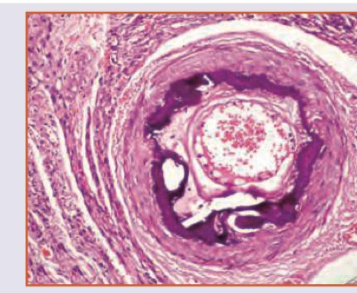
Which is true about the image shown?

Explanation: ***CMV*** - **Cytomegalovirus (CMV)** infection is very common in solid organ transplant recipients, particularly in the period between **1 to 6 months post-transplant**, known as the **intermediate period** [1]. - This timing is due to the cumulative effect of **immunosuppression** compromising the patient's ability to control latent viral shedding or newly acquired infection. *EBV* - **Epstein-Barr virus (EBV)** infection is also common in transplant recipients, but it is more significantly associated with the development of **post-transplant lymphoproliferative disorder (PTLD)**, rather than being the *most likely* general infection at 4 months [2], [3]. - While EBV can occur, CMV is typically more prevalent as a symptomatic viral infection in the intermediate post-transplant period [1]. *Candida* - **Candida** infections (fungal) are more common in the **early post-transplant period** (within the first month), often associated with surgical complications, indwelling catheters, or broad-spectrum antibiotic use [1]. - While possible, it is less likely to be the *most common* infection at 4 months compared to CMV. *Histoplasma* - **Histoplasma** infections are a **systemic fungal infection** that is typically seen in transplant patients who have been exposed to endemic areas. - It is not a common opportunistic infection universally seen in transplant recipients at 4 months post-transplant but rather depends on geographical exposure and specific risk factors.
Explanation: ***Minutes to hours*** - **Hyperacute rejection** is a rapidly occurring complication post-transplant, characterized by its onset within minutes to hours after **organ reperfusion** [1]. - This type of rejection is mediated by pre-formed **recipient antibodies** that recognize donor antigens, leading to immediate graft damage [1]. *12 hours* - While plausible, 12 hours is a bit too broad as **hyperacute rejection** primarily begins much sooner, typically within the first few hours [1]. - This timeframe might overlap with the initial stages of **acute cellular rejection**, which typically occurs days to weeks later [1]. *24 hours* - **Hyperacute rejection** is almost always observed and causes graft failure well before the 24-hour mark, if it is going to happen. - Rejection occurring within this extended period is more indicative of **accelerated acute rejection** rather than true hyperacute rejection. *6 hours* - While hyperacute rejection certainly can occur within 6 hours, "minutes to hours" better captures the immediate onset, often within seconds or minutes [1]. - Some cases of **hyperacute rejection** can be so rapid that the 6-hour mark would be considered a late presentation. **References:** [1] Kumar V, Abbas AK, et al.. Robbins and Cotran Pathologic Basis of Disease. 9th ed. Diseases of the Immune System, pp. 241-242.
Explanation: ***Sepsis*** - **Infection** is the leading cause of morbidity and mortality after intestinal transplantation, making **sepsis** the most common complication. - The immunocompromised state due to immunosuppressive therapy and the inherent bacterial load of the gastrointestinal tract contribute significantly to the high risk of severe infections. *Intestinal obstruction* - While intestinal obstruction can occur post-transplant due to adhesions or strictures, it is **less common** than infectious complications. - It typically manifests later and may require surgical intervention but doesn't have the same high frequency as sepsis. *Graft versus host disease* - **Graft-versus-host disease (GVHD)** is a significant complication in intestinal transplantation, but it is **not the most common**. - Its incidence varies, and while serious, it does not surpass the overall frequency of infectious complications and sepsis. *Intestinal necrosis* - **Intestinal necrosis** (e.g., due to infarction or severe rejection) is a severe complication but is **less frequent** than sepsis. - It is often a consequence of vascular compromise or overwhelming rejection, leading to graft failure or perforation.
Explanation: ***CMV*** - **Cytomegalovirus (CMV)** is the most common viral infection in solid organ transplant recipients, often reactivating in immunosuppressed patients [1]. - It can cause a wide range of clinical syndromes including **fever**, **leukopenia**, **hepatitis**, **pneumonitis**, and **gastroenteritis**, and is a significant cause of morbidity and mortality [1]. *EBV* - **Epstein-Barr virus (EBV)** is also common in transplant recipients but is most notably associated with **post-transplant lymphoproliferative disorder (PTLD)**, a serious complication [1]. - While present, it is not as frequently the cause of symptomatic infection as CMV in the immediate post-transplant period. *HSV* - **Herpes simplex virus (HSV)** infections can occur, manifesting as mucocutaneous lesions or, less commonly, severe systemic disease in transplant patients. - However, its incidence and severity are generally lower compared to CMV in the overall transplant population. *HPV* - **Human papillomavirus (HPV)** infections are typically associated with **warts** and increased risk of **malignancies** (e.g., anogenital cancers) in immunosuppressed individuals, including transplant recipients. - While important for long-term surveillance, HPV does not represent the most common acute infection post-transplant.
Explanation: ***Renal artery thrombosis*** - **Renal artery thrombosis** is generally associated with conditions like **atherosclerosis**, atrial fibrillation, or vasculitis, not directly with chronic pancreatitis. - While chronic pancreatitis can lead to systemic complications, direct renal arterial clotting is an atypical and **uncommon sequela**. *Pancreatic pseudocyst* - **Pancreatic pseudocysts** are common complications of chronic pancreatitis, occurring when fluid collections around the pancreas become walled off by fibrous tissue [1]. - They can cause pain, obstruction, and even rupture if left untreated [2]. *Splenic vein thrombosis* - **Splenic vein thrombosis** can result from inflammation and compression of the splenic vein by the diseased pancreatic tissue in chronic pancreatitis [1]. - This can lead to **splenomegaly** and **gastric varices** due to increased pressure in the portal system. *Pancreatic fistula* - A **pancreatic fistula** occurs when pancreatic fluid leaks from the gland, often forming a connection to another organ or the skin [2]. - This is a well-recognized complication of both acute and chronic pancreatitis, usually due to ductal disruption.
Explanation: ***Cytomegalovirus*** - **CMV infection** is very common and a frequent opportunistic infection in **immunosuppressed solid organ transplant recipients**, especially within the first few months post-transplant [1]. - **CMV pneumonitis**, characterized by diffuse interstitial pneumonia and fever, is a classic presentation of CMV disease in this patient population [1]. *Varicella zoster virus* - While VZV can cause serious infections in immunosuppressed individuals, **pneumonia due to VZV** is typically part of a disseminated disease and less common than CMV pneumonitis in transplant recipients. - **Cutaneous vesicular lesions** would usually precede or accompany VZV pneumonia, which are not mentioned here. *Herpes simplex virus* - HSV can cause severe mucocutaneous infections in immunocompromised patients, but **HSV pneumonia** is rare and usually manifests as tracheobronchitis or a focal necrotizing pneumonia, not typically diffuse interstitial. - **Esophagitis or encephalitis** are more common serious manifestations of HSV in this population than primary pneumonitis. *Epstein-barr virus* - EBV is primarily associated with **post-transplant lymphoproliferative disorder (PTLD)** in transplant recipients, which can involve the lungs. - While PTLD can manifest with fever and pulmonary infiltrates, **diffuse interstitial pneumonia** solely due to primary EBV infection is less characteristic than for CMV.
Explanation: ***Serum lipase*** - The symptoms of **epigastric pain**, **vomiting**, and **guarding** in a chronic alcoholic patient are highly suggestive of **acute pancreatitis** [1]. - **Serum lipase** is a highly specific and sensitive marker for acute pancreatitis and is the initial diagnostic test of choice. *Alcohol breath test* - An alcohol breath test would indicate current alcohol intoxication but would not help in diagnosing the underlying cause of the patient's severe abdominal pain. - While relevant to his history, it will not guide immediate management of his acute symptoms. *Upper GI endoscopy* - **Upper GI endoscopy** is an invasive procedure and is typically reserved for investigating upper gastrointestinal bleeding or structural abnormalities of the esophagus, stomach, or duodenum, often after initial diagnostic tests. - It is not the initial test for suspected acute pancreatitis. *CECT* - **CECT (Contrast-Enhanced Computed Tomography)** of the abdomen is useful for assessing the severity and complications of pancreatitis, and for confirming the diagnosis if serum lipase is equivocal, but it is not the first-line diagnostic test [1]. - It is generally performed after initial laboratory tests confirm suspicion of pancreatitis, or if complications are suspected [1].
Explanation: ***Naïve B cells*** - Chronic Lymphocytic Leukaemia (CLL) and Small Lymphocytic Lymphoma (SLL) originate from **CD5-positive B lymphocytes** arrested in a mature but **naïve differentiation stage** [1]. - These cells express both **B-cell markers (CD19, CD20, CD23)** and a T-cell marker (CD5), which is characteristic of the clone [4]. *Mature B cells* - While CLL/SLL are derived from B cells, they are specifically from **naïve, not fully mature, B cells**. - **Other B-cell lymphomas** like follicular lymphoma or mantle cell lymphoma originate from distinct stages of mature B-cell differentiation [2]. *Progenitor T cells* - **Progenitor T cells** are the cells of origin for **T-cell acute lymphoblastic leukaemia (T-ALL)**, not CLL/SLL [3]. - T-ALL involves immature T lymphocytes and presents with different clinical and immunophenotypic features [3]. *Mature T cells* - **Mature T cells** can give rise to various **peripheral T-cell lymphomas**, like peripheral T-cell lymphoma, not otherwise specified (PTCL-NOS) or cutaneous T-cell lymphoma (Mycosis Fungoides). - These are distinct from CLL/SLL, which is a B-cell neoplasm [4]. **References:** [1] Kumar V, Abbas AK, et al.. Robbins and Cotran Pathologic Basis of Disease. 9th ed. Diseases of White Blood Cells, Lymph Nodes, Spleen, and Thymus, pp. 596-598. [2] Kumar V, Abbas AK, et al.. Robbins and Cotran Pathologic Basis of Disease. 9th ed. Diseases of White Blood Cells, Lymph Nodes, Spleen, and Thymus, pp. 610-612. [3] Kumar V, Abbas AK, et al.. Robbins and Cotran Pathologic Basis of Disease. 9th ed. Diseases of White Blood Cells, Lymph Nodes, Spleen, and Thymus, pp. 598-599. [4] Kumar V, Abbas AK, et al.. Robbins and Cotran Pathologic Basis of Disease. 9th ed. Diseases of White Blood Cells, Lymph Nodes, Spleen, and Thymus, p. 598.
Explanation: ***Gp IIb/IIIa*** - Glanzmann thrombasthenia is a **rare, inherited bleeding disorder** characterized by a defect or deficiency in the **glycoprotein IIb/IIIa (Gp IIb/IIIa) complex** on the platelet surface [1]. - This complex is crucial for platelet aggregation as it acts as the receptor for **fibrinogen**, which links activated platelets together [1]. *Gp VI* - **Glycoprotein VI (Gp VI)** is a collagen receptor on platelets, important for initial **platelet adhesion and activation** at sites of vascular injury. - Defects in Gp VI are associated with milder bleeding disorders, not Glanzmann thrombasthenia. *Thromboxane A2* - **Thromboxane A2 (TXA2)** is a potent **vasoconstrictor** and **platelet aggregator** synthesized by platelets. - Disorders in TXA2 synthesis or response, such as aspirin-induced platelet dysfunction, cause bleeding but are biochemically distinct from Glanzmann thrombasthenia. *Gp Ia/IIa* - The **glycoprotein Ia/IIa (Gp Ia/IIa) complex** (also known as integrin ̡2̢1) is another **collagen receptor** on platelets, mediating platelet adhesion to collagen. - Defects in Gp Ia/IIa lead to a different type of mild bleeding disorder, affecting initial adhesion rather than aggregation. **References:** [1] Kumar V, Abbas AK, et al.. Robbins and Cotran Pathologic Basis of Disease. 9th ed. Red Blood Cell and Bleeding Disorders, pp. 668-669.
Explanation: ***Monckeberg's Arteriosclerosis*** - This condition involves **calcification of the media of muscular arteries**, typically in individuals over 50 years old. - It is characterized by **ring-like calcifications** in the vessel wall, which can be seen on imaging or histology, and is usually not clinically significant as it does not narrow the lumen. *Hyaline Arteriosclerosis* - This type is characterized by **homogeneous, pink, hyaline thickening** of the walls of arterioles, with narrowing of the lumen. - It is typically seen in **benign hypertension** and **diabetes mellitus**, affecting small arteries and arterioles. *Hyperplastic Arteriosclerosis* - This condition is associated with **malignant hypertension** and is characterized by **concentric, laminated thickening** of the arteriole walls, often described as "onion-skinning." [1] - It involves proliferation of smooth muscle cells and reduplication of the basement membrane, leading to severe luminal narrowing [1]. *Fibrinoid necrosis* - This is a form of **necrosis** seen in the walls of blood vessels, characterized by deposition of **fibrin-like material** that stains intensely eosinophilic. - It is typically associated with **malignant hypertension** [2], **vasculitis**, or immune-mediated vascular damage, and is not a primary form of arteriosclerosis. **References:** [1] Kumar V, Abbas AK, et al.. Robbins and Cotran Pathologic Basis of Disease. 9th ed. The Kidney, p. 945. [2] Cross SS. Underwood's Pathology: A Clinical Approach. 6th ed. Common Clinical Problems From Cardiovascular Disease, pp. 276-277.
Get full access to all questions, explanations, and performance tracking.
Start For Free