A 55-year-old white woman has had recurrent episodes of alcohol-induced pancreatitis. Despite abstinence, the patient develops postprandial abdominal pain, bloating, weight loss despite good appetite, and bulky, foul-smelling stools. Kidney, ureter, bladder (KUB) x-ray shows pancreatic calcifications. In this patient, you should expect to find which of the following?
Cells central to the production of pancreatic fibrosis are?
Pancreatitis is a common complication of which one of the following?
Which of the following is not a recognized complication of chronic pancreatitis?
Which is not a component of Ranson's criteria for acute pancreatitis?
A 55-year-old man presents with intermittent epigastric pain, relieved by eating, and worsened by fasting. What is the most likely cause?
A chronic alcoholic patient came to emergency with severe pain in epigastrium and multiple episodes of vomiting. On examination, guarding was present in upper epigastrium. Chest X-ray was normal. What is the next best step?
In cystic fibrosis, which of the following structures is affected in the pancreas?
Type of necrosis in pancreatitis-
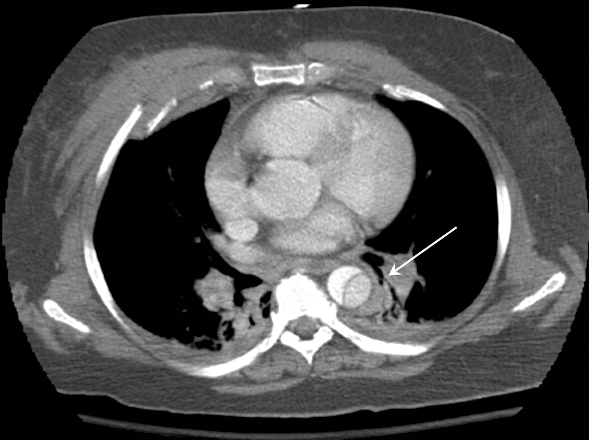
The CT thorax image shows:

Explanation: Diabetes mellitus - Chronic pancreatitis, especially due to recurrent alcohol-induced episodes, often leads to the destruction of pancreatic islet cells, resulting in impaired insulin production and consequently, diabetes mellitus [1]. - The combination of pancreatic calcifications and symptoms like weight loss despite good appetite, and malabsorption due to pancreatic insufficiency, makes diabetes a strong expected complication [1]. Malabsorption of fat-soluble vitamins D and K - While chronic pancreatitis often causes steatorrhea and malabsorption of fat-soluble vitamins (A, D, E, K), the question asks what one should expect to find, and diabetes mellitus is a more direct and universally expected consequence of widespread pancreatic damage from recurrent pancreatitis [1]. - The symptoms described, such as bulky, foul-smelling stools, are indicative of fat malabsorption, which leads to deficiencies in fat-soluble vitamins, but the direct mention of diabetes mellitus reflects a more advanced stage of pancreatic destruction [2]. Positive fecal occult blood test - A positive fecal occult blood test suggests gastrointestinal bleeding, which is not a direct or typical consequence of chronic pancreatitis itself. - While complications like peptic ulcers or pancreatic cancer (a long-term risk of chronic pancreatitis) could cause GI bleeding, it's not an expected finding directly associated with the pancreatitis symptoms described. Courvoisier sign - Courvoisier sign (a palpable, non-tender gallbladder with jaundice) is typically associated with obstruction of the common bile duct due to a malignancy in the head of the pancreas or other periampullary tumors. - It is not a characteristic finding in uncomplicated chronic pancreatitis, especially without mention of jaundice.
Explanation: ***Stellate cells*** - **Pancreatic stellate cells** (PSCs) play a crucial role in the development of **pancreatic fibrosis** by producing and secreting extracellular matrix components. - Upon activation by injury or inflammation, PSCs transform into myofibroblast-like cells, leading to increased **collagen deposition** and scarring. *Alpha cells* - **Alpha cells** are endocrine cells in the pancreatic islets responsible for producing and secreting **glucagon**, which raises blood glucose levels. - They are not directly involved in the **fibrotic process** of the pancreas. *Beta cells* - **Beta cells** are endocrine cells in the pancreatic islets that produce and secrete **insulin**, which lowers blood glucose levels. - While dysfunction or death of beta cells is central to diabetes, they are not primarily responsible for **pancreatic fibrosis**. *Acinar cells* - **Acinar cells** are exocrine cells of the pancreas that produce and secrete **digestive enzymes** into the pancreatic duct. - While injury to acinar cells can lead to inflammation (e.g., pancreatitis), they are not the primary drivers of **collagen synthesis** and **fibrosis**.
Explanation: ***Didanosine (ddI)*** - **Didanosine (ddI)** is a nucleoside reverse transcriptase inhibitor (NRTI) known for causing dose-dependent **pancreatitis** as a significant adverse effect. - Patients on didanosine require monitoring for symptoms and elevated **amylase/lipase** levels. *Zidovudine* - **Zidovudine** (AZT) is an NRTI primarily associated with **bone marrow suppression** (anemia, neutropenia) and myopathy. - While it can cause lactic acidosis, **pancreatitis** is not its most common or dose-limiting side effect. *Zalcitabine* - **Zalcitabine** (ddC) is an NRTI whose primary dose-limiting toxicity is **peripheral neuropathy**, particularly in the extremities. - **Pancreatitis** is a less common adverse effect compared to didanosine. *Stavudine* - **Stavudine** (d4T) is an NRTI frequently associated with **peripheral neuropathy** and **lipoatrophy** (loss of subcutaneous fat). - Although it can also contribute to lactic acidosis, **pancreatitis** is not its characteristic or most common side effect.
Explanation: ***Renal artery thrombosis*** - **Renal artery thrombosis** is generally associated with conditions like **atherosclerosis**, atrial fibrillation, or vasculitis, not directly with chronic pancreatitis. - While chronic pancreatitis can lead to systemic complications, direct renal arterial clotting is an atypical and **uncommon sequela**. *Pancreatic pseudocyst* - **Pancreatic pseudocysts** are common complications of chronic pancreatitis, occurring when fluid collections around the pancreas become walled off by fibrous tissue [1]. - They can cause pain, obstruction, and even rupture if left untreated [2]. *Splenic vein thrombosis* - **Splenic vein thrombosis** can result from inflammation and compression of the splenic vein by the diseased pancreatic tissue in chronic pancreatitis [1]. - This can lead to **splenomegaly** and **gastric varices** due to increased pressure in the portal system. *Pancreatic fistula* - A **pancreatic fistula** occurs when pancreatic fluid leaks from the gland, often forming a connection to another organ or the skin [2]. - This is a well-recognized complication of both acute and chronic pancreatitis, usually due to ductal disruption.
Explanation: ***Serum lipase*** - **Serum lipase** is not a component of Ranson's criteria. While it is a crucial diagnostic marker for acute pancreatitis, Ranson's criteria focus on other clinical and laboratory values for predicting severity. - The criteria were developed before widespread availability and use of lipase as a primary diagnostic marker for pancreatitis. *Age* - **Age** is a component of Ranson's criteria, specifically "Age > 55 years" for admission and initial assessment [1]. - Older age is associated with increased severity and mortality in acute pancreatitis due to decreased physiologic reserve [1]. *Base deficit* - **Base deficit** is a component of Ranson's criteria, specifically "Base deficit > 4 mEq/L" after 48 hours. - A significant base deficit indicates **metabolic acidosis**, which is a marker of severe systemic inflammation and organ dysfunction in acute pancreatitis. *Blood glucose* - **Blood glucose** is a component of Ranson's criteria, specifically "Blood glucose > 200 mg/dL (11.1 mmol/L)" for admission and initial assessment. - Elevated blood glucose can reflect the severity of pancreatic inflammation and insult to the **islet cells**, or systemic stress response.
Explanation: ### Peptic ulcer disease - The classic presentation of **duodenal ulcers**, a common type of peptic ulcer, includes epigastric pain that is **relieved by eating** and **worsens with fasting** [1]. - This pattern is due to the buffering effect of food on gastric acid and the increased acid secretion during fasting, which irritates the ulcer. *Cholelithiasis* - Characterized by **biliary colic**, which is typically severe, intermittent right upper quadrant pain, often radiating to the back or shoulder, and frequently triggered by fatty meals. - Pain relief with eating is not a typical feature, and it does not usually worsen with fasting. *Chronic pancreatitis* - Presents with persistent or recurrent **epigastric pain** that often **radiates to the back** [2], and can be worsened by eating fatty foods. - The pain is usually not relieved by eating, and symptoms like steatorrhea and diabetes development are common later in the disease [2]. *Gastroesophageal reflux disease (GERD)* - Primarily causes **heartburn** (burning sensation behind the sternum) and **regurgitation**, which often worsen after meals, when lying down, or bending over. - Pain is typically not relieved by eating, nor does it characteristically worsen with fasting; instead, it is often associated with acid reflux.
Explanation: ***Serum lipase*** - The symptoms of **epigastric pain**, **vomiting**, and **guarding** in a chronic alcoholic patient are highly suggestive of **acute pancreatitis** [1]. - **Serum lipase** is a highly specific and sensitive marker for acute pancreatitis and is the initial diagnostic test of choice. *Alcohol breath test* - An alcohol breath test would indicate current alcohol intoxication but would not help in diagnosing the underlying cause of the patient's severe abdominal pain. - While relevant to his history, it will not guide immediate management of his acute symptoms. *Upper GI endoscopy* - **Upper GI endoscopy** is an invasive procedure and is typically reserved for investigating upper gastrointestinal bleeding or structural abnormalities of the esophagus, stomach, or duodenum, often after initial diagnostic tests. - It is not the initial test for suspected acute pancreatitis. *CECT* - **CECT (Contrast-Enhanced Computed Tomography)** of the abdomen is useful for assessing the severity and complications of pancreatitis, and for confirming the diagnosis if serum lipase is equivocal, but it is not the first-line diagnostic test [1]. - It is generally performed after initial laboratory tests confirm suspicion of pancreatitis, or if complications are suspected [1].
Explanation: ***Pancreatic ducts*** - In cystic fibrosis, the **CFTR protein** dysfunction leads to thick, viscous secretions that obstruct the **pancreatic ducts** [2]. - This obstruction prevents digestive enzymes from reaching the intestine, causing **malabsorption** and progressive pancreatic damage [2]. *Acinar cells* - While pancreatic acinar cells are responsible for producing digestive enzymes, they are not directly dysfunctional in cystic fibrosis. - Their function is secondarily impaired due to the **blockage of the ducts** that carry their secretions [2]. *Islets of Langerhans* - The **islets of Langerhans** contain endocrine cells (e.g., insulin-producing beta cells) and are generally unaffected early in cystic fibrosis [1]. - Long-standing inflammation and fibrosis in severe cases can eventually impair islet function, leading to **CF-related diabetes** [1]. *Stromal tissue* - Stromal tissue (supporting connective tissue) is not the primary site of pathology in cystic fibrosis. - While chronic inflammation may lead to **fibrosis** of stromal tissue over time, the initial and primary defect is in the **ductal obstruction**, not in the stroma itself. **References:** [1] Kumar V, Abbas AK, et al.. Robbins and Cotran Pathologic Basis of Disease. 9th ed. The Pancreas, pp. 893-895. [2] Kumar V, Abbas AK, et al.. Robbins and Cotran Pathologic Basis of Disease. 9th ed. The Gastrointestinal Tract, p. 789.
Explanation: ***Fat*** - In pancreatitis, the release of **lipases** from damaged pancreatic cells leads to the breakdown of fat cells, resulting in the formation of **fatty acids** and **glycerol** [1]. - These fatty acids then combine with calcium to form **calcium soaps**, which appear as white, chalky deposits and signify **fat necrosis** [1]. *Coagulative* - This type of necrosis typically occurs due to **ischemia** (lack of blood supply) in solid organs, preserving the outline of the cells for a period [1]. - While ischemia can play a role in severe pancreatitis, the primary and distinctive type of necrosis in this condition is not coagulative. *Caseous* - **Caseous necrosis** is characteristic of **tuberculosis** and certain fungal infections, where the tissue has a crumbly, cheese-like appearance [1]. - It involves a combination of liquefactive and coagulative necrosis, but it is not seen in pancreatitis. *Fibrinoid* - **Fibrinoid necrosis** is often associated with **immune-mediated vascular damage**, such as in cases of **vasculitis** or **malignant hypertension** [2]. - It involves the deposition of immune complexes and fibrin in arterial walls, which is not the primary necrotic process in pancreatitis. **References:** [1] Kumar V, Abbas AK, et al.. Robbins and Cotran Pathologic Basis of Disease. 9th ed. Cellular Responses to Stress and Toxic Insults: Adaptation, Injury, and Death, pp. 53-55. [2] Cross SS. Underwood's Pathology: A Clinical Approach. 6th ed. (Basic Pathology) introduces the student to key general principles of pathology, both as a medical science and as a clinical activity with a vital role in patient care. Part 2 (Disease Mechanisms) provides fundamental knowledge about the cellular and molecular processes involved in diseases, providing the rationale for their treatment. Part 3 (Systematic Pathology) deals in detail with specific diseases, with emphasis on the clinically important aspects., pp. 103-104.
Explanation: ***Ascending aortic dissection*** - The CT image shows a **classic intimal flap** separating the true and false lumens in the ascending aorta, which is the hallmark feature of an aortic dissection. - This represents a **Stanford Type A dissection** involving the ascending aorta, which is a life-threatening emergency requiring **immediate surgical intervention** due to high risk of complications including rupture, cardiac tamponade, and acute aortic regurgitation. - The presence of the intimal flap creating two distinct channels (true and false lumens) is pathognomonic for dissection. *Descending aortic dissection* - While the intimal flap is characteristic of dissection, the image specifically shows involvement of the **ascending aorta** (proximal to the left subclavian artery), not the descending thoracic aorta. - Descending aortic dissections (Stanford Type B) are typically managed **medically** with blood pressure control, unlike ascending dissections which require surgery. *Aortic aneurysm* - An **aortic aneurysm** represents focal dilatation of the aortic wall (>50% increase in diameter) without separation of the intimal layers. - While aneurysms can be a risk factor for dissection, the key finding here is the **intimal flap dividing the lumen**, which defines dissection rather than simple aneurysmal dilatation. - The image does not show the uniform circumferential enlargement typical of aneurysms. *Aortic coarctation* - **Aortic coarctation** is a congenital narrowing of the aorta, typically located at the aortic isthmus (near the ligamentum arteriosum), distal to the left subclavian artery. - CT would show focal narrowing with pre-stenotic dilatation and collateral vessel formation, not an intimal flap. - This is a completely different pathology without the characteristic dissection flap seen in this image.
Get full access to all questions, explanations, and performance tracking.
Start For Free