Which of the following represents a secondary brain injury mechanism?
Post contusional syndrome includes:
The earliest manifestation of increased intracranial pressure following head injury is:
A 43-year-old man presents to the emergency department after falling down a flight of stairs and landing on his head. He did not lose consciousness. He complains of severe headache, marked decreased acuity in hearing in the left ear, and a "runny nose" since the fall. On physical examination, he is found to have a left-sided Battle's sign (an ecchymosis in the area of the left mastoid process) and hemotympanum. He has a constant dripping of a clear, watery fluid through his nose. Findings on his neurologic examination, other than the hearing loss, are completely normal. X-ray studies will reveal which of the following?
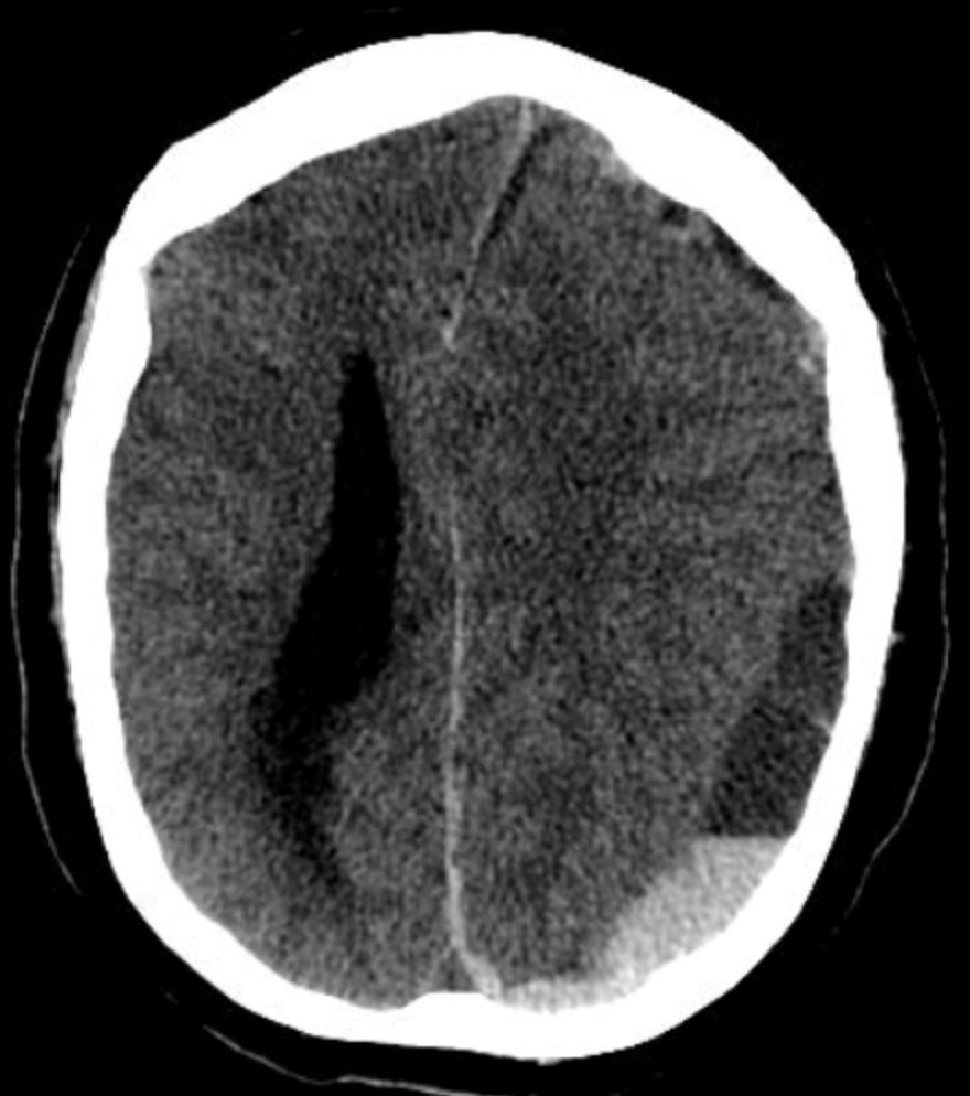
A polytrauma patient's CT brain shows a crescent-shaped extra-axial collection with a concave inner margin. What is the most likely diagnosis?

Glasgow coma scale -moderate includes
What is the primary pathological mechanism in classical Guillain-Barré syndrome affecting the peripheral nervous system?
A child presented with microcephaly, hepatomegaly and periventricular calcification. What is the best specimen for diagnosis of CMV by PCR?
Investigation of choice for leptomeningeal carcinomatosis:
Treatment of Neurocysticercosis includes all of the following except -
Explanation: ***Inflammatory response*** - **Inflammatory response** is a classic **secondary brain injury mechanism** that occurs hours to days after initial trauma through delayed activation of **microglia**, **cytokine release**, and **neuroinflammation**. - It represents a **cascade of cellular processes** that develops after the primary injury, causing additional neuronal damage through **blood-brain barrier disruption**, **cerebral edema**, and **cellular apoptosis**. *Immediate axonal shearing* - **Immediate axonal shearing** is a **primary brain injury mechanism** that occurs at the exact moment of trauma due to **rotational and acceleration forces**. - It represents **direct mechanical damage** to axons during impact, not a delayed secondary process that develops after the initial injury. *Contusion at impact site* - **Contusion at impact site** is a **primary brain injury** resulting from **direct mechanical trauma** to brain tissue at the point of impact. - It occurs **immediately upon trauma** through direct tissue damage and hemorrhage, rather than through delayed secondary mechanisms. *Skull fracture* - **Skull fracture** is a **primary injury** that results from **direct mechanical force** applied to the skull during the traumatic event. - It represents **immediate structural damage** occurring at the moment of impact, not a secondary process that develops over time after initial trauma.
Explanation: ***Headache*** - **Headache** is the **most common and characteristic symptom** of **post-concussion syndrome (PCS)**, present in up to 90% of cases. - Typically described as tension-type or migraine-like headaches that persist for weeks to months after mild traumatic brain injury. - This is a **core diagnostic feature** of PCS according to ICD-10 (F07.2) and DSM-5 criteria. - Among the given options, this is the **most definitive symptom** of post-concussion syndrome. *Delirium* - **Delirium** is an acute confusional state with fluctuating consciousness, impaired attention, and cognitive dysfunction. - This is **NOT a feature of post-concussion syndrome**, which involves persistent symptoms in clear consciousness. - Delirium may occur immediately after severe traumatic brain injury but is not part of the chronic post-concussional syndrome picture. - Post-concussion syndrome involves cognitive difficulties (memory, concentration) but not delirium. *Nausea & vomiting* - **Nausea** can occur as part of post-concussion syndrome, particularly when associated with vestibular dysfunction or migraine-like headaches. - However, it is **less characteristic and less persistent** than headache, and is not present in all cases. - While recognized in ICD-10 criteria for PCS, nausea is not as defining or universal as headache. - Vomiting is less common in chronic PCS compared to acute concussion. *All of the options* - This is incorrect because **delirium is NOT a feature of post-concussion syndrome**. - While headache is the hallmark symptom and nausea can occur, delirium represents acute brain dysfunction, not the chronic syndrome. - PCS is characterized by persistent somatic (headache, dizziness), cognitive (concentration, memory problems), and psychological (irritability, anxiety) symptoms in clear consciousness.
Explanation: ***Altered mental status*** - **Altered mental status** (e.g., confusion, irritability, drowsiness) is often the earliest sign of increased intracranial pressure (ICP) due to its profound effect on global brain function. - This change reflects the **brain's reduced perfusion** and metabolic compromise as pressure within the rigid skull rises. *Hemiparesis* - **Hemiparesis** indicates focal neurological deficits, usually resulting from direct injury or significant pressure on specific motor pathways, which typically manifest later than global mental status changes. - It suggests a more advanced stage of neurological compromise or a localized mass effect. *Ipsilateral pupillary dilatation* - **Ipsilateral pupillary dilatation** is a classic sign of uncal herniation, where the temporal lobe compresses the **oculomotor nerve** (CN III) on the same side. - While critical, it is generally a *late and ominous sign* of significantly elevated ICP, indicating severe brainstem compression. *Contralateral pupillary dilatation* - **Contralateral pupillary dilatation** is highly unusual in the context of typical uncal herniation, which almost always causes *ipsilateral* signs due to direct compression. - Its presence would suggest atypical herniation patterns or other causes of pupillary asymmetry.
Explanation: ***A temporal bone fracture with CSF rhinorrhea*** - The combination of **Battle's sign**, **hemotympanum**, unilateral hearing loss, and clear nasal discharge after head trauma strongly indicates a **temporal bone fracture**. - **CSF rhinorrhea** refers to cerebrospinal fluid leaking from the nose due to a skull base fracture involving the temporal bone, typically affecting the petrous part. - The CSF can reach the nasal cavity via the **eustachian tube** or through fracture lines extending to the middle ear and mastoid air cells. *Occipital bone fracture* - While occipital fractures are possible with head trauma, they do not directly explain the specific findings of **hemotympanum** or unilateral hearing loss. - An occipital fracture would typically cause symptoms related to damage to the **brainstem** or **cerebellum**, depending on the extent. *A skull-base fracture with a mucocele* - A **mucocele** is a cyst filled with mucus, usually resulting from obstruction of a sinus ostium, and is not an acute traumatic finding. - While a skull-base fracture is present, the presence of a mucocele does not fit the acute injury presentation. *A fracture of the cribriform plate with a CSF leak into the paranasal sinuses* - A **cribriform plate fracture** would result in CSF rhinorrhea, but it typically causes CSF to leak directly from the anterior cranial fossa into the nasal cavity. - It would not explain the **hemotympanum**, Battle's sign, or unilateral hearing loss, which are characteristic of **temporal bone injury**.
Explanation: ***SDH*** - The image shows a **crescent-shaped collection** of hemorrhage with a concave inner margin, consistent with a **subdural hematoma** (SDH). - SDHs result from the tearing of **bridging veins** and typically conform to the brain's surface, crossing suture lines but not limited by bony sutures. *EDH* - An **epidural hematoma (EDH)** characteristically appears as a **lenticular** or **biconvex** shape on CT, not crescent-shaped. - EDHs are typically caused by arterial bleeding, often from the **middle meningeal artery**, and are limited by cranial sutures. *Contusion* - A **contusion** is brain tissue bruising that appears as **heterogeneous areas** of hemorrhage and edema within the brain parenchyma itself. - It would not manifest as a distinct extra-axial collection with a smooth, concave margin. *Diffuse axonal injury* - **Diffuse axonal injury (DAI)** involves widespread microscopic damage to axons, often at the gray-white matter junction. - It may appear as *punctate hemorrhages* or **small lesions** at these junctions on CT, but often the CT can be normal, and it would not present as a large extra-axial collection.
Explanation: ***9-12*** - A **Glasgow Coma Scale (GCS)** score between 9 and 12 is classified as **moderate head injury** or **moderate coma** [1]. - This range indicates significant impairment of consciousness, often requiring medical intervention and close monitoring. *<3* - A GCS score of **less than 3** is not clinically possible as the minimum score achievable is 3 (1 for each component: Eye, Verbal, Motor response). - This option is technically inaccurate based on the GCS scoring system. *>12* - A GCS score of **greater than 12** typically falls into the category of **mild head injury** (13-15) [1]. - Patients in this range usually have a better prognosis and less severe neurological deficits. *3-8* - A GCS score between **3 and 8** is indicative of **severe head injury** or **severe coma** [1]. - Patients in this range often require immediate critical care, including intubation and ventilatory support due to compromised airway reflexes.
Explanation: ***It causes demyelination of peripheral nerves.*** - Classical Guillain-Barré syndrome (AIDP - Acute Inflammatory Demyelinating Polyneuropathy) is an autoimmune disorder where the immune system attacks the **myelin sheath** surrounding peripheral nerves. - This **demyelination** impairs nerve signal conduction, leading to weakness and paralysis. - AIDP represents the most common form of GBS in Western countries (~85-90% of cases). *It blocks neurotransmitter release.* - Conditions like **Lambert-Eaton myasthenic syndrome** primarily involve antibodies targeting presynaptic voltage-gated calcium channels, thereby reducing neurotransmitter release. - While GBS affects nerve conduction, its primary mechanism is not the blockage of neurotransmitter release at the synapse. *It inhibits muscle contraction.* - Inhibition of muscle contraction is a downstream effect of impaired nerve innervation, but the fundamental problem in GBS is with the **nerve itself**, not the muscle's ability to contract directly. - Conditions like **myasthenia gravis** directly affect neuromuscular transmission by blocking acetylcholine receptors on muscle fibers. *It leads to axonal degeneration.* - While **axonal variants** of GBS exist (AMAN - Acute Motor Axonal Neuropathy; AMSAN - Acute Motor-Sensory Axonal Neuropathy), particularly common in Asia, the **classical and most common form** is characterized by **primary demyelination** (AIDP). - Pure axonal degeneration as a primary pathology is seen in specific GBS variants, not the classical presentation. - Secondary axonal damage can occur in severe or prolonged cases.
Explanation: ***Urine*** - **Urine** is the most sensitive and commonly used specimen for diagnosing **congenital CMV infection** via PCR, especially in neonates, due to high viral shedding in urine. - A positive urine CMV PCR within the first 2-3 weeks of life is highly indicative of **congenital CMV**, which can cause symptoms like **microcephaly**, **hepatomegaly**, and **periventricular calcifications**. *CSF* - While CMV can be detected in **CSF** in congenital infections, particularly in symptomatic cases with neurological involvement, it is less sensitive than urine for initial diagnosis. - **CSF PCR** is typically reserved for evaluating central nervous system involvement and may not detect systemic infection as reliably as urine. *Blood* - **Blood PCR** for CMV can be positive in congenital infection, but it can also be positive in postnatal CMV acquisition or maternal viremia without congenital transmission. - The presence of viral DNA in blood is transient, and its sensitivity for diagnosing congenital infection is generally lower than that of urine. *Liver biopsy* - **Liver biopsy** is an invasive procedure and is not the primary diagnostic method for CMV infection, although histological examination can reveal characteristic viral inclusions if there is significant hepatic involvement. - It carries risks and is typically performed only when other diagnostic methods are inconclusive or when assessing the extent of liver damage.
Explanation: ***Gd enhanced MRI*** - **Gadolinium-enhanced MRI** is the investigation of choice for **leptomeningeal carcinomatosis** as it can visualize the subtle nodular or linear enhancement along the leptomeninges, indicating tumor dissemination. - It offers superior **soft tissue contrast** and spatial resolution compared to CT, enabling detection of small lesions and accurate mapping of disease extent. *CT scan* - A **CT scan** has limited sensitivity for detecting leptomeningeal involvement due to poor contrast resolution of soft tissues and the dura/arachnoid spaces. - It might show hydrocephalus or large tumor deposits, but subtle leptomeningeal enhancement is often missed. *SPECT* - **Single photon emission computed tomography (SPECT)** is primarily used for functional imaging and is not the investigation of choice for anatomical visualization of leptomeningeal carcinomatosis. - Its resolution is too low to detect the fine structural changes associated with leptomeningeal spread. *PET* - **Positron emission tomography (PET)**, often combined with CT, identifies metabolically active tumor cells and can detect diffuse metastatic disease. - While useful for overall cancer staging and identifying primary lesions, it is less effective than gadolinium-enhanced MRI for directly visualizing the morphology and enhancement patterns of leptomeningeal carcinomatosis due to limited spatial resolution in the CSF spaces.
Explanation: ***Niclosamide***- **Niclosamide** is an oral anthelmintic primarily used to treat **intestinal tapeworm infections** by inhibiting **oxidative phosphorylation** in the parasites.- It has **poor systemic absorption** and therefore is **not effective** against **neurocysticercosis**, which involves cysts in the brain parenchyma requiring drugs with good CNS penetration.*Praziquantel*- **Praziquantel** is an orally administered anthelmintic that increases the **calcium permeability** of the parasite's cell membrane, leading to paralysis and death.- It is used in the treatment of **neurocysticercosis**, particularly for **viable parenchymal cysts** and as an alternative to albendazole [2].*Albendazole*- **Albendazole** is a broad-spectrum anthelmintic that works by inhibiting **tubulin polymerization**, causing disruption of **parasite metabolism** and glucose uptake.- It is considered the **first-line treatment** for **parenchymal neurocysticercosis** due to its excellent penetration into the central nervous system and proven efficacy.*Corticosteroids*- **Corticosteroids** (such as **dexamethasone** or **prednisolone**) are used as **adjunct therapy** in neurocysticercosis management.- They help reduce **inflammation and edema** associated with parasite death, preventing complications like seizures and increased intracranial pressure during anthelmintic treatment [1].
Get full access to all questions, explanations, and performance tracking.
Start For Free