Which of the following most likely causes a communicating (nonobstructive) hydrocephalus?
Arnold Chiari malformation is characterized by all of the following except:
When should folic acid supplementation start in order to be effective in preventing neural tube defects?
Which of the following is the MOST accurate test for detecting neural tube defects?
A 10 year old child presented with headache, vomiting, gait instability and diplopia. On examination he had papilledema and gait ataxia. The most probable diagnosis is –
Meningomyelocele with progressive hydrocephalus is commonly seen in
Porencephaly refers to -
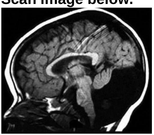
Identify the condition shown in the CT scan image.

A newborn baby presented with a failure to pass meconium in the immediate postnatal period. The pediatrician also notices visible yet ineffective peristalsis, and abdominal distention. A radiological contrast enema demonstrated a narrow conical segment and a dilated proximal bowel. A diagnosis of Hirschsprung disease was made. Which of the following is a cause of the condition in the patient?
A muscle biopsy shows type 1 fiber atrophy and increased internal nuclei on histology. Which additional finding on light microscopy would be most specific for myotonic dystrophy?
Explanation: ***Blockage of the arachnoid granulations*** - **Communicating hydrocephalus** occurs when CSF flow from the ventricles is unobstructed, but its **reabsorption** into the venous system is impaired [1] - **Arachnoid granulations** (pacchionian bodies) are responsible for reabsorbing CSF into the dural venous sinuses [1] - Blockage (e.g., due to **subarachnoid hemorrhage**, chronic **meningitis**, or venous thrombosis) prevents proper reabsorption, leading to CSF accumulation [2] - This is the **direct pathophysiologic mechanism** of communicating hydrocephalus *Tuberculous meningitis* - This can cause **communicating hydrocephalus** through inflammation and fibrosis of the **basilar meninges**, which obstructs the arachnoid granulations and impairs CSF reabsorption - It is a clinically important cause, especially in endemic regions - However, it works through the mechanism of arachnoid granulation blockage, making it an indirect cause - Less commonly, it can cause obstructive hydrocephalus if inflammatory exudates block the basal cisterns [2] *Stenosis of the duct of Sylvius* - Also known as **aqueductal stenosis**, this is a classic cause of **non-communicating (obstructive) hydrocephalus** - It blocks the flow of CSF from the third to the fourth ventricle, leading to dilation of the lateral and third ventricles - CSF cannot communicate between the ventricular system and subarachnoid space *Ependymoma of the fourth ventricle* - An **ependymoma** in this location causes **non-communicating (obstructive) hydrocephalus** [2] - The tumor physically blocks the outflow of CSF from the fourth ventricle through the foramina of Luschka and Magendie into the subarachnoid space - Ependymomas are the most common posterior fossa tumor in children **References:** [1] Kumar V, Abbas AK, et al.. Robbins and Cotran Pathologic Basis of Disease. 9th ed. Peripheral Nerves and Skeletal Muscles, pp. 1256-1257. [2] Cross SS. Underwood's Pathology: A Clinical Approach. 6th ed. Common Clinical Manifestations Of Central And Peripheral Nervous System Disease, pp. 703-704.
Explanation: Hypoplasia of cerebellar vermis - **Hypoplasia of the cerebellar vermis** is characteristic of **Dandy-Walker malformation**, not Arnold-Chiari malformation. - In Arnold-Chiari malformation, the cerebellar tonsils are displaced, but the vermis itself is typically not hypoplastic [2]. *Herniation of cerebellum* - **Type I Chiari malformation** is defined by the **caudal displacement of the cerebellar tonsils** through the foramen magnum [1]. - This herniation can lead to compression of the brainstem and spinal cord [1], [3]. *Flattened base of skull* - A flattened skull base, or **platybasia**, is often associated with Chiari malformation, particularly **Type I**. - This anatomical anomaly can **reduce the posterior cranial fossa volume**, contributing to cerebellar herniation [2]. *Syringomyelia* - **Syringomyelia**, the formation of a fluid-filled cyst within the spinal cord, is a **common complication** of Chiari I malformation [1]. - It results from cerebrospinal fluid flow obstruction caused by the cerebellar tonsil herniation [1].
Explanation: ***At least one month before conception*** - **Neural tube closure** occurs very early in pregnancy, typically between days 21 and 28 after conception, often before a woman even knows she is pregnant. - Adequate **folate levels** are crucial during this critical period, making preconception supplementation essential for prevention. *As soon as pregnancy is diagnosed* - By the time pregnancy is diagnosed, often several weeks after conception, the **neural tube has already closed**, or the critical window for its closure has passed. - Starting supplementation at this point would be too late to prevent most **neural tube defects**. *Before the beginning of 2nd trimester* - The second trimester begins at week 13 of pregnancy, which is far too late to prevent **neural tube defects**, as the neural tube has already formed and closed in the first month. - This timing is not aligned with the critical developmental period for the neural tube. *At least 1 week before conception* - While closer to the correct timing, one week before conception may not be sufficient to build up optimal **folate levels** in the body, particularly in women with lower baseline intake or increased needs. - Most guidelines recommend **at least one month** to ensure adequate saturation and effectiveness.
Explanation: ***USG (Ultrasound)*** - **High-resolution ultrasound** is the **gold standard and most accurate imaging modality** for detecting **neural tube defects (NTDs)** due to its ability to directly visualize anatomical structures of the fetus. - **Diagnostic accuracy**: Detection rate >95% for anencephaly and 80-90% for open spina bifida with targeted anomaly scan at 18-20 weeks. - Can identify specific features such as **lemon sign** (frontal bone scalloping), **banana sign** (cerebellar compression), direct visualization of **spina bifida**, **anencephaly**, or **encephalocele**. - **Non-invasive, safe, and widely available**, making it the primary diagnostic tool in clinical practice. *Amniocentesis* - **Amniocentesis** measures **alpha-fetoprotein (AFP)** and **acetylcholinesterase (AChE)** in amniotic fluid, which are elevated in open NTDs. - While highly accurate as a **confirmatory test** (near 99% sensitivity with both markers), it is **invasive** with risk of miscarriage (0.1-0.3%). - Used primarily when ultrasound findings are **equivocal** or for **biochemical confirmation**, not as the first-line diagnostic test. - In modern practice, ultrasound has largely replaced amniocentesis for NTD diagnosis due to superior imaging technology. *Chromosomal analysis* - **Chromosomal analysis** (karyotyping) detects **chromosomal abnormalities** like trisomies (Down syndrome, Edwards syndrome). - NTDs are **structural malformations**, not chromosomal abnormalities, though some chromosomal disorders may have associated structural defects. - Does not directly diagnose NTDs. *Placentography* - **Placentography** is used to localize the **placenta** in cases of suspected **placenta previa** or for guiding invasive procedures. - Provides no information about **fetal anatomy** and is not used for detecting NTDs.
Explanation: ***Midline posterior fossa tumour*** - The combination of **headache, vomiting, papilledema (signs of increased intracranial pressure)**, **gait instability, and ataxia** strongly suggests a **midline posterior fossa tumor** in a child. These tumors often obstruct CSF flow, leading to hydrocephalus and cerebellar symptoms. - Common tumors in this location in children include **medulloblastoma** and **pilocytic astrocytoma**, which frequently present with these symptoms due to their proximity to the **fourth ventricle** and **cerebellum**. *Suprasellar tumour* - **Suprasellar tumors** typically present with **visual field deficits** (e.g., bitemporal hemianopia) due to compression of the optic chiasm, and/or **endocrine dysfunction** (e.g., growth delay, diabetes insipidus). - While they can cause hydrocephalus and increased intracranial pressure if large, the prominent **gait instability and ataxia** point away from a primary suprasellar lesion as the most likely cause. *Hydrocephalus* - **Hydrocephalus** itself explains the **increased intracranial pressure (headache, vomiting, papilledema)** and sometimes **gait instability (ataxia)**. - However, hydrocephalus is usually a *consequence* of an underlying obstruction, and in a child presenting acutely with cerebellar dysfunction, a **tumor blocking CSF flow in the posterior fossa** is the most probable underlying cause, not hydrocephalus as the primary diagnosis. *Brain stem tumour* - **Brain stem tumors** typically cause **cranial nerve deficits** (e.g., facial weakness, dysphagia), **long tract signs (hemiparesis)**, and often **multiple types of ataxia**, alongside signs of increased intracranial pressure if they obstruct CSF flow. - While gait instability and diplopia can occur, the overall picture of prominent **gait ataxia** and papilledema without other focal cranial nerve signs makes a primary midline posterior fossa tumor compressing the cerebellum and fourth ventricle more likely.
Explanation: ***Arnold Chiari II*** - **Arnold Chiari II malformation** is characterized by the downward displacement of the **cerebellar vermis and tonsils**, along with the brainstem, through the foramen magnum. - This malformation is almost always associated with **meningomyelocele** and often leads to **hydrocephalus** due to obstruction of CSF flow at the level of the foramen magnum and aqueductal stenosis. *Vein of Galen malformation* - A **Vein of Galen malformation** is an arteriovenous malformation located in the brain, which can cause high-output cardiac failure in neonates. - It can lead to hydrocephalus due to venous congestion but is not typically associated with **meningomyelocele**. *Dandy-Walker malformation* - **Dandy-Walker malformation** involves a hypoplastic or absent **cerebellar vermis**, cystic dilation of the fourth ventricle, and an enlarged posterior fossa. - While it often presents with hydrocephalus, it is not directly associated with **meningomyelocele**. *Choroid plexus papilloma* - A **Choroid plexus papilloma** is a rare, benign tumor that typically causes **hydrocephalus** due to **overproduction of CSF**. - It is not associated with **meningomyelocele** or Chiari malformations.
Explanation: ***Vascular lesions due to degenerative vessel disease and head injury*** - **Porencephaly** refers to cysts or cavities within the brain parenchyma, which may communicate with the ventricular system or subarachnoid space. - Porencephaly can be **congenital** (developmental) or **acquired** (destructive). This option describes **acquired porencephaly**, which results from destructive processes such as **ischemic or hemorrhagic strokes**, **perinatal hypoxic-ischemic injury**, and **traumatic brain injury** [1]. - These vascular insults and trauma cause tissue necrosis and subsequent cyst formation, which is the hallmark of porencephalic cavities. *Neural tube defects* - These are **congenital malformations** resulting from incomplete closure of the neural tube during early embryonic development (weeks 3-4). - Examples include **spina bifida** and **anencephaly**, which represent failures of neural tube closure rather than destructive cystic lesions in formed brain tissue. *Fetal alcohol syndrome* - This condition results from **maternal alcohol consumption** during pregnancy and causes a spectrum of physical, neurodevelopmental, and behavioral abnormalities. - While it may cause brain structural abnormalities (microcephaly, corpus callosum abnormalities), it does not typically manifest as focal **porencephalic cysts**. *Dandy-Walker syndrome* - This is a **congenital posterior fossa malformation** characterized by hypoplasia/agenesis of the cerebellar vermis, cystic dilatation of the fourth ventricle, and enlarged posterior fossa. - It represents a developmental anomaly of the cerebellum, not a destructive cystic lesion of the cerebral hemispheres. **References:** [1] Kumar V, Abbas AK, et al.. Robbins and Cotran Pathologic Basis of Disease. 9th ed. The Central Nervous System, pp. 1260-1261.
Explanation: ***Dandy-Walker malformation*** - The image shows an enlarged posterior fossa with **cystic dilation of the fourth ventricle** and **absence/hypoplasia of the cerebellar vermis**, which are classic features of Dandy-Walker malformation. - The elevated tentorium and upward displacement of the transverse sinuses are also characteristic, contributing to the distinct appearance. *Cerebellar vermis hypoplasia* - While cerebellar vermis hypoplasia is a component of Dandy-Walker malformation, it is not the sole, defining feature. - Dandy-Walker also includes cystic dilation of the fourth ventricle and an enlarged posterior fossa, which are evident in the image and go beyond isolated vermis hypoplasia. *Mega cisterna magna* - A **mega cisterna magna** is a benign enlargement of the cisterna magna, which is the space between the cerebellum and the medulla oblongata. - Unlike in Dandy-Walker malformation, a mega cisterna magna usually does not involve displacement of the tentorium or hypoplasia of the cerebellar vermis, and the fourth ventricle is typically normal in shape and size. *None of the options* - The image clearly displays the diagnostic hallmarks of Dandy-Walker malformation, making this option incorrect.
Explanation: ***Failure of migration of neural crest cells*** - Hirschsprung disease is characterized by the **absence of ganglion cells** (specifically **Auerbach's and Meissner's plexuses**) in the distal bowel. - This aganglionosis results from the **failure of neural crest cells to migrate** completely into the intestinal wall during embryonic development. *Persistence of embryonic structures in the bowel wall* - This mechanism is associated with conditions like **Meckel's diverticulum**, where a remnant of the **vitelline duct** persists. - It does not explain the absence of ganglion cells or the functional obstruction seen in Hirschsprung disease. *Congenital obstruction due to external factors* - This would involve conditions such as an **annular pancreas**, **bands**, or **malrotation with volvulus**, creating a physical barrier. - Hirschsprung disease is a **functional obstruction** due to neuromuscular dysfunction, not an external compression or blockage. *Abnormal peristalsis due to neural dysfunction* - While there is abnormal peristalsis, the underlying cause is not just **"neural dysfunction"** in a general sense, but specifically the **absence of entire ganglion cell plexuses** within the bowel wall. - This option is too broad and doesn't pinpoint the precise developmental defect.
Explanation: ***Ring fibers*** - The presence of **ring fibers** (annular fibers with peripherally arranged myofibrils forming a ring around the fiber core) is a **classic and highly specific** histological finding for **myotonic dystrophy type 1 (DM1)**. - Ring fibers are seen in **25-50% of cases** and result from abnormal myofibrillar architecture with peripheral displacement of myofibrils. - Combined with type 1 fiber atrophy and increased internal nuclei, ring fibers provide strong confirmation of myotonic dystrophy [1]. *Nemaline rods* - **Nemaline rods** (rod bodies) are characteristic protein aggregates composed of Z-line material found in **nemaline myopathy**, a distinct congenital muscle disorder. - They appear as dark red structures on Gomori trichrome stain and are not a feature of myotonic dystrophy. *Ragged red fibers* - **Ragged red fibers** are a hallmark of **mitochondrial myopathies**, indicating abnormal subsarcolemmal accumulation of mitochondria seen on modified Gomori trichrome stain. - These are definitively associated with primary mitochondrial dysfunction (e.g., MELAS, MERRF), not myotonic dystrophy. *Central cores* - **Central cores** are areas within muscle fibers devoid of oxidative enzyme activity, characteristic of **central core disease** (autosomal dominant RYR1 mutations). - This finding is not expected in myotonic dystrophy and represents a different congenital myopathy. **References:** [1] Cross SS. Underwood's Pathology: A Clinical Approach. 6th ed. Common Clinical Manifestations Of Central And Peripheral Nervous System Disease, pp. 732-733.
Get full access to all questions, explanations, and performance tracking.
Start For Free