What is not seen in tuberous sclerosis?
Which of the following statements about meningiomas is true?
Which of the following is the most frequent primary malignant tumor of the CNS?
A patient presents with headache, confusion, and a diagnosis of a brain tumor. The family history reveals brain and kidney tumors. What is the most likely diagnosis?
Which one of the following is the most common CNS tumor associated with type I neurofibromatosis?
Most common tumor in the part of the brain shown (arrow) among children is
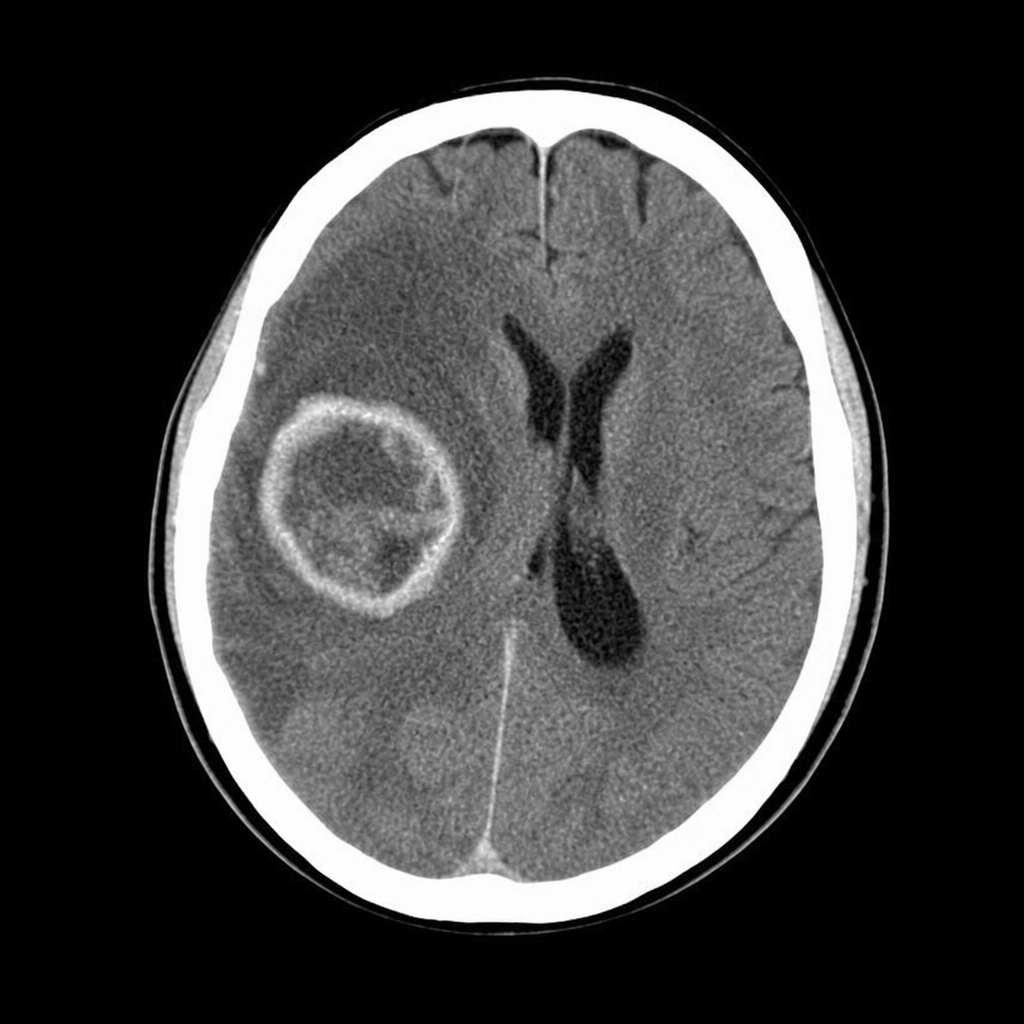
Most common site for medulloblastoma is-
Most common supratentorial tumor in children
Which of the following CNS tumor shows increased growth during pregnancy?
CNS tumor seen in Von Hippel Lindau syndrome is:
Explanation: ***Ependymoma*** - **Ependymomas** are primary central nervous system tumors that arise from **ependymal cells**, typically found in the ventricles and spinal cord. - While they are brain tumors, they are **not characteristic lesions** associated with **Tuberous Sclerosis Complex (TSC)**. *Giant Cell Astrocytoma* - **Subependymal giant cell astrocytomas (SEGAs)** are a classic feature of **Tuberous Sclerosis Complex (TSC)**, often growing within the ventricles. - They can cause **hydrocephalus** and are a significant source of morbidity in TSC patients. *White matter migration lines* - **Radial glial migration lines**, or **cerebral white matter lesions**, are due to abnormalities in neuronal migration during development and are a common finding in **Tuberous Sclerosis Complex (TSC)**. - These are typically seen as **linear hypdensities** on neuroimaging. *Sub-ependymal nodules* - **Subependymal nodules (SENs)** are small, benign lesions located just beneath the ependyma of the lateral ventricles in **Tuberous Sclerosis Complex (TSC)**. - They are a diagnostic hallmark of TSC and can sometimes develop into **Subependymal Giant Cell Astrocytomas (SEGAs)**.
Explanation: ***95% cure rate following treatment*** - Meningiomas generally have a **high cure rate of approximately 95%** following surgical resection, especially when they are completely excised [1]. - They are typically **benign tumors**, resulting in favorable outcomes with appropriate management [1]. *Arise from arachnoid layer* - Meningiomas actually arise from **meningothelial cells** of the **arachnoid layer**, but this statement does not fully explain their pathogenesis. - This mischaracterization does not provide an accurate understanding of the tumor's origin and biology. *50% are malignant* - Most meningiomas are benign; only a small percentage, about **1-5%**, are classified as malignant. - Thus, stating that **50% are malignant** significantly overestimates the incidence of aggressive forms. *More common in men* - Meningiomas are more prevalent in **women**, especially those aged between 30-70 years, with a female-to-male ratio of approximately **3:1**. - This option is incorrect as it misrepresents the demographic distribution of the disease. **References:** [1] Kumar V, Abbas AK, et al.. Robbins and Cotran Pathologic Basis of Disease. 9th ed. The Central Nervous System, pp. 1316-1317.
Explanation: ***Glioblastoma multiforme*** - **Glioblastoma multiforme (GBM)** is the most common and aggressive primary malignant brain tumor in adults [1]. - It is a **grade IV astrocytoma**, characterized by rapid growth, necrosis, and microvascular proliferation [1]. *Oligodendroglioma* - **Oligodendrogliomas** are primary glial tumors but are less common than GBM. - They typically have a more indolent course than GBM and are often characterized by **IDH mutations** and **1p/19q co-deletion**. *Medulloblastoma* - **Medulloblastoma** is the most common malignant brain tumor in children, but it is rare in adults [2]. - It arises in the **cerebellum** and is a type of embryonal tumor [2]. *Meningioma* - **Meningiomas** are the most common primary brain tumors overall, but they are typically **benign** and originate from the meninges. - While they can be symptomatic due to compression, they are not primarily malignant in the way GBM is. **References:** [1] Kumar V, Abbas AK, et al.. Robbins and Cotran Pathologic Basis of Disease. 9th ed. The Central Nervous System, pp. 1308-1310. [2] Cross SS. Underwood's Pathology: A Clinical Approach. 6th ed. Common Clinical Manifestations Of Central And Peripheral Nervous System Disease, pp. 725-726.
Explanation: ***VHL syndrome*** - **Von Hippel-Lindau (VHL) syndrome** is an inherited disorder characterized by the growth of tumors and cysts in various parts of the body, including the **brain (hemangioblastomas)** and **kidneys (renal cell carcinoma)**. - The presentation of a brain tumor, kidney tumors, and a positive family history for both organs strongly points to VHL syndrome. *Neurofibromatosis* - **Neurofibromatosis (NF)** typically presents with **cafe-au-lait spots**, neurofibromas, optic gliomas, and Lisch nodules. - While it involves brain tumors, kidney tumors are not a primary feature of NF. *Li-Fraumeni syndrome* - **Li-Fraumeni syndrome** is associated with an increased risk of various cancers, including **sarcomas**, **breast cancer**, **adrenocortical carcinomas**, and **leukemia**. - While brain tumors can occur, the specific combination of brain and kidney tumors with a clear family history is less characteristic of Li-Fraumeni than VHL syndrome. *Churg-Strauss syndrome* - **Churg-Strauss syndrome (Eosinophilic Granulomatosis with Polyangiitis)** is a systemic vasculitis characterized by **asthma**, **eosinophilia**, and **granulomatous inflammation**. - It does not involve the development of brain or kidney tumors.
Explanation: ***Optic nerve glioma*** - **Optic nerve gliomas** are the most frequently encountered central nervous system tumors in patients with **Type 1 neurofibromatosis (NF1)**, occurring in about 15% of individuals. - They are typically low-grade **astrocytomas** and can cause vision loss and proptosis depending on their size and location. *Meningioma* - While more common in **Type 2 neurofibromatosis (NF2)**, meningiomas can occur in NF1, but are not the most common CNS tumor. - Meningiomas are tumors that arise from the **meninges**, the membranes surrounding the brain and spinal cord. *Acoustic schwannoma* - **Bilateral acoustic schwannomas (vestibular schwannomas)** are the hallmark feature of **Type 2 neurofibromatosis (NF2)**, not NF1 [1]. - These tumors arise from the Schwann cells of the **vestibulocochlear nerve** and can cause hearing loss and balance issues [1]. *Low grade astrocytoma* - While optic nerve gliomas are a type of low-grade astrocytoma, this option is too general; **optic nerve glioma** is the specific and most common presentation in NF1. - Other forms of low-grade astrocytomas can occur in NF1 but are not as universally characteristic as optic nerve gliomas.
Explanation: ***Astrocytoma*** - The image shows a **brainstem lesion**, which is a common location for astrocytomas in children. - **Pilocytic astrocytoma** is the most common brain tumor in children, often presenting in the cerebellum, brainstem, or optic pathways. *DNET (Dysembryoplastic Neuroepithelial Tumor)* - DNETs are typically found in the **temporal lobe** and are associated with a history of seizures. - While benign, their characteristic location and symptoms do not align with a brainstem mass as the primary finding. *Ependymoma* - Ependymomas frequently arise in the **fourth ventricle** in children, often causing hydrocephalus. - Although they can occur in the brainstem, astrocytomas are generally more common in this specific region in pediatric cases. *Medulloblastoma* - Medulloblastomas are nearly always located in the **cerebellum** (infratentorial region) and are known for their aggressive nature and propensity for CSF dissemination. - The lesion shown appears to be within the brainstem, making medulloblastoma less likely given its typical cerebellar location.
Explanation: ***Cerebellum*** - **Medulloblastoma** is a highly malignant primary brain tumor that characteristically arises in the **cerebellum** [1]. - It is the most common malignant brain tumor in children, typically originating from the **roof of the fourth ventricle**. *Pituitary* - The **pituitary gland** is mostly associated with **adenomas**, which are benign tumors arising from anterior pituitary cells. - Tumors like **craniopharyngiomas** can also be found in the sellar region, but medulloblastomas do not originate here. *Cerebrum* - The **cerebrum** is the most common site for **gliomas** (e.g., glioblastoma multiforme) and metastatic tumors in adults. - Medulloblastoma specifically originates from primitive neuroectodermal cells in the posterior fossa [1]. *Pineal gland* - The **pineal gland** is associated with **pinealomas** (e.g., pineoblastoma, pineocytoma) and **germinomas** [2]. - These are distinct from medulloblastomas in their cellular origin and typical anatomical location.
Explanation: ***Craniopharyngioma*** - **Craniopharyngiomas** are the **most common supratentorial tumors in children**, accounting for approximately 5-10% of all pediatric intracranial tumors. - They arise from **Rathke's pouch remnants** near the pituitary gland and hypothalamus in the **sellar/suprasellar region**. - Present with **visual disturbances, endocrine dysfunction, and increased intracranial pressure**. - Bimodal age distribution with peaks at **5-14 years** and 50-74 years. *Astrocytoma* - While **astrocytomas** are the **most common primary CNS tumors in children overall**, they are predominantly located in the **posterior fossa (infratentorial)**, particularly as **cerebellar astrocytomas** [1], [2]. - In the **supratentorial compartment specifically**, astrocytomas are less common than craniopharyngiomas in the pediatric population. - They arise from **astrocytes** and range from low-grade (pilocytic) to high-grade (glioblastoma) [1]. *Oligodendroglioma* - **Oligodendrogliomas** are **rare in children** and more prevalent in adults (peak 40-50 years). - They originate from **oligodendrocytes** and are typically **slow-growing tumors**. - Account for only 1-2% of pediatric brain tumors. *Meningioma* - **Meningiomas** are **very rare in children** (less than 2% of pediatric brain tumors) and are primarily tumors of adulthood. - They arise from **arachnoid cap cells** of the meninges and show increasing incidence with age. - When they occur in children, they are often associated with **neurofibromatosis type 2** [2]. **References:** [1] Cross SS. Underwood's Pathology: A Clinical Approach. 6th ed. Common Clinical Manifestations Of Central And Peripheral Nervous System Disease, pp. 725-726. [2] Kumar V, Abbas AK, et al.. Robbins and Cotran Pathologic Basis of Disease. 9th ed. The Central Nervous System, pp. 1319-1320.
Explanation: ***Meningioma*** - **Meningiomas** often express receptors for **estrogen** and **progesterone**, leading to increased growth rates during the **estrogen-rich environment of pregnancy**. - This tumor's growth during pregnancy can exacerbate neurological symptoms or lead to the discovery of previously asymptomatic meningiomas. *Oligodendroglioma* - **Oligodendrogliomas** are a type of glioma that do not typically show an increased growth rate in response to hormonal changes during pregnancy. - Their growth is generally independent of **sex hormones**, and their progression is determined by other genetic and molecular factors. *Craniopharyngioma* - **Craniopharyngiomas** are benign epithelial tumors that arise from Rathke's pouch remnants and primarily affect children, though they can occur in adults. - Their growth is not significantly influenced by **hormonal changes** related to pregnancy. *Glioblastoma Multiforme* - **Glioblastoma multiforme (GBM)** is a highly aggressive and fast-growing primary brain tumor, but its growth is not directly stimulated by the hormonal changes of pregnancy. - While GBM can rapidly progress during pregnancy, this is due to its inherent aggressive nature rather than hormonal effects on tumor cells.
Explanation: ***Cerebellar hemangioblastoma*** - **Cerebellar hemangioblastomas** are characteristic CNS tumors associated with **Von Hippel-Lindau syndrome** due to mutations in the VHL gene [1]. - Patients typically present with symptoms related to raised **intracranial pressure** or focal neurological deficits depending on the tumor's size and location. *Meningioma* - Meningiomas are **benign tumors** arising from the meninges and are generally not a primary feature of **Von Hippel-Lindau syndrome** [1]. - They are more commonly associated with **Neurofibromatosis type 2** if linked to a genetic syndrome [1]. *Glioma* - Gliomas, which include astrocytomas, oligodendrogliomas, and ependymomas, are a diverse group of brain tumors that are not typically or specifically linked to **Von Hippel-Lindau syndrome** [1]. - While central nervous system tumors, they lack the specific association seen with **hemangioblastomas** in VHL. *CNS lymphoma* - **CNS lymphoma** is primarily associated with **immunocompromised states**, such as HIV/AIDS, or occurs as a primary brain tumor in older adults [1]. - It has no known direct association with **Von Hippel-Lindau syndrome** [1]. **References:** [1] Cross SS. Underwood's Pathology: A Clinical Approach. 6th ed. Common Clinical Manifestations Of Central And Peripheral Nervous System Disease, pp. 724-728.
Get full access to all questions, explanations, and performance tracking.
Start For Free