Which one of the following is an antiapoptotic protein or gene?
All of the following are chromosomal breakage syndromes except?
Inheritance pattern of Familial Hypercholesterolemia is:
What is the CD marker for histiocytosis?
All are factors of poor wound healing EXCEPT?
Deletion of chromosome 11 is associated with which of the following conditions?
A normal parent has two siblings with osteogenesis imperfecta. What is the most likely pattern of inheritance?
A tissue preparation is experimentally subjected to a hypoxic environment. The cells in this tissue begin to swell, and chromatin begins to clump in cell nuclei. ATPases are activated, and ATP production decreases. Which of the following ions accumulating in mitochondria and the cytosol contributes most to these findings and to eventual cell death?
Which of the following is an execution caspase?
BRAF V600E mutations are seen in all of the following except?
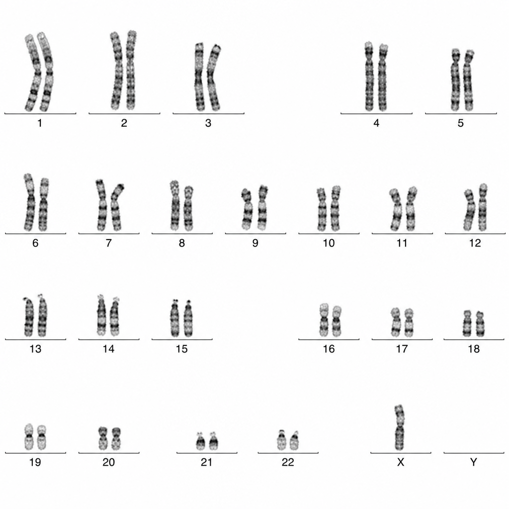
Explanation: **Explanation:** The regulation of apoptosis (programmed cell death) is primarily governed by the **BCL-2 family of proteins**, which act as a rheostat to determine cell survival. These proteins are categorized into three functional groups based on their BCL-homology (BH) domains: 1. **Anti-apoptotic (Pro-survival):** These proteins, including **BCL-2**, BCL-XL, and MCL-1, reside in the outer mitochondrial membrane [3]. They prevent the leakage of Cytochrome C by inhibiting the formation of mitochondrial pores. BCL-2 specifically binds to and neutralizes pro-apoptotic proteins [1]. 2. **Pro-apoptotic (Effectors):** These include **BAX** and **BAK**. Upon activation, they oligomerize to form pores in the mitochondrial membrane (MOMP - Mitochondrial Outer Membrane Permeabilization), leading to the release of Cytochrome C and activation of the caspase cascade [3]. 3. **Pro-apoptotic (BH3-only sensors):** These include **BIM**, BID, and BAD. They act as "stress sensors" that neutralize anti-apoptotic proteins and directly activate BAX/BAK. **Analysis of Options:** * **BCL-2 (Correct):** It is the prototype anti-apoptotic protein. Overexpression (e.g., in Follicular Lymphoma via t(14;18)) leads to increased cell survival and oncogenesis [1]. * **BAX & BAK (Incorrect):** These are the "executioner" pro-apoptotic proteins that create mitochondrial channels [3]. * **BIM (Incorrect):** This is a BH3-only protein that promotes apoptosis in response to growth factor withdrawal. **High-Yield Clinical Pearls for NEET-PG:** * **Follicular Lymphoma:** Characterized by **t(14;18)**, which translocates the BCL-2 gene to the IgH locus, causing BCL-2 overexpression [1], [2]. * **Venetoclax:** A BH3-mimetic drug that inhibits BCL-2, used in the treatment of CLL and AML. * **Guardian of the Genome:** p53 induces apoptosis by upregulating BAX [3]. **References:** [1] Kumar V, Abbas AK, et al.. Robbins and Cotran Pathologic Basis of Disease. 9th ed. Neoplasia, pp. 310-311. [2] Kumar V, Abbas AK, et al.. Robbins and Cotran Pathologic Basis of Disease. 9th ed. Diseases of White Blood Cells, Lymph Nodes, Spleen, and Thymus, pp. 602-604. [3] Kumar V, Abbas AK, et al.. Robbins and Cotran Pathologic Basis of Disease. 9th ed. Neoplasia, p. 310.
Explanation: The correct answer is **Ehlers-Danlos syndrome (EDS)** because it is a disorder of **collagen synthesis**, not a defect in DNA repair [1]. Chromosomal breakage syndromes are a group of genetic disorders characterized by chromosomal instability, high rates of breakage, and a significantly increased risk of malignancy. **Why Ehlers-Danlos Syndrome is the exception:** EDS is a heterogeneous group of connective tissue disorders caused by defects in the synthesis or structure of fibrillar collagen (e.g., mutations in *COL5A1*, *COL3A1*) [1]. It manifests clinically as skin hyperextensibility, joint hypermobility, and tissue fragility [1]. It does not involve defects in DNA repair mechanisms or chromosomal instability. **Analysis of Incorrect Options (Chromosomal Breakage Syndromes):** * **Fanconi’s Anaemia:** An autosomal recessive disorder caused by defects in the **FANC gene complex**, which is responsible for repairing DNA interstrand cross-links [2]. It presents with pancytopenia, thumb deformities, and a high risk of AML. * **Ataxia Telangiectasia:** Caused by a mutation in the **ATM gene**, which codes for a protein kinase that senses DNA double-strand breaks [2]. It presents with cerebellar ataxia, oculocutaneous telangiectasia, and immunodeficiency. * **Bloom Syndrome:** Caused by a mutation in the **BLM gene** (recQ helicase family), leading to defective DNA helicase activity [2]. Clinical features include "butterfly" facial rash, growth retardation, and "sister chromatid exchanges." **High-Yield Clinical Pearls for NEET-PG:** * **Xeroderma Pigmentosum** is another classic breakage syndrome caused by defective **Nucleotide Excision Repair (NER)** [2]. * **Nijmegen Breakage Syndrome** (NBN gene) is often grouped here; it involves defective double-strand break repair. * **Diagnostic Test:** Fanconi’s anaemia is diagnosed using the **Diepoxybutane (DEB) test** or Mitomycin C test to induce chromosomal breaks in vitro. **References:** [1] Kumar V, Abbas AK, et al.. Robbins and Cotran Pathologic Basis of Disease. 9th ed. Genetic Disorders, pp. 154-155. [2] Kumar V, Abbas AK, et al.. Robbins and Cotran Pathologic Basis of Disease. 9th ed. Neoplasia, pp. 322-323.
Explanation: **Explanation:** **Familial Hypercholesterolemia (FH)** is a classic example of an **Autosomal Dominant (AD)** disorder [3]. It is primarily caused by mutations in the **LDLR gene**, which encodes the Low-Density Lipoprotein (LDL) receptor [1]. 1. **Why Autosomal Dominant is Correct:** In FH, a mutation in a single allele (heterozygous state) is sufficient to cause a significant clinical phenotype [3]. Heterozygotes have a 50% reduction in functional LDL receptors, leading to a two-to-threefold increase in plasma cholesterol levels from birth. Homozygotes (inheriting two mutant alleles) are much more severely affected, often presenting with myocardial infarction before age 20, demonstrating a "gene dosage effect" common in AD conditions [2]. 2. **Why Other Options are Incorrect:** * **Autosomal Recessive:** While a rare form (ARH) exists due to mutations in the *LDLRAP1* protein, the classic and most common form of FH follows a dominant pattern where carriers are clinically symptomatic [3]. * **X-linked Dominant/Recessive:** The LDLR gene is located on **Chromosome 19** (an autosome), not on the sex chromosomes. Therefore, the inheritance is independent of the patient's biological sex. **NEET-PG High-Yield Pearls:** * **Key Mutations:** Most commonly the **LDLR gene**; other mutations include **ApoB-100** (ligand for LDLR) and **PCSK9** (which degrades LDLR) [1]. * **Clinical Triad:** Hypercholesterolemia, Tendon Xanthomas (especially Achilles tendon), and premature Coronary Artery Disease (CAD). * **Pathophysiology:** Impaired hepatic uptake of LDL leads to increased plasma LDL and increased scavenger receptor-mediated uptake by macrophages (forming foam cells). [1] * **Treatment:** Statins are the mainstay; PCSK9 inhibitors are used for refractory cases. **References:** [1] Kumar V, Abbas AK, et al.. Robbins and Cotran Pathologic Basis of Disease. 9th ed. Genetic Disorders, pp. 157-159. [2] Kumar V, Abbas AK, et al.. Robbins and Cotran Pathologic Basis of Disease. 9th ed. Genetic Disorders, pp. 149-150. [3] Cross SS. Underwood's Pathology: A Clinical Approach. 6th ed. (Basic Pathology) introduces the student to key general principles of pathology, both as a medical science and as a clinical activity with a vital role in patient care. Part 2 (Disease Mechanisms) provides fundamental knowledge about the cellular and molecular processes involved in diseases, providing the rationale for their treatment. Part 3 (Systematic Pathology) deals in detail with specific diseases, with emphasis on the clinically important aspects., pp. 53-54.
Explanation: **Langerhans Cell Histiocytosis (LCH)** is a proliferative disorder of Langerhans cells, which are specialized dendritic cells [1]. In pathology, identifying these cells requires specific immunohistochemical (IHC) markers. **Why CD1a is the correct answer:** CD1a is the most specific and widely used diagnostic marker for Langerhans cells. It is a protein involved in presenting lipid antigens to T-cells. In the context of LCH, the presence of **CD1a** and **S100** on IHC, along with the identification of **Birbeck granules** (tennis-racket shaped organelles) on electron microscopy, confirms the diagnosis [1]. **Analysis of incorrect options:** * **CD1b, CD1c, and CD1d:** While these are also members of the CD1 family involved in lipid antigen presentation, they are not used as diagnostic markers for histiocytosis. CD1b and CD1c are expressed on various subsets of dendritic cells and B-cells, while CD1d is primarily involved in presenting lipids to NKT cells. They lack the diagnostic specificity required for LCH. **High-Yield Clinical Pearls for NEET-PG:** * **Langerin (CD207):** This is the most specific marker for LCH as it is directly associated with the formation of Birbeck granules [1]. * **BRAF V600E Mutation:** Found in approximately 50% of LCH cases; it is a common "hot topic" in molecular pathology [1]. * **Classic Triad (Hand-Schüller-Christian disease):** Calvarial bone defects, diabetes insipidus, and exophthalmos. * **Letterer-Siwe Disease:** The aggressive, multisystem form of LCH seen in infants (<2 years). **References:** [1] Kumar V, Abbas AK, et al.. Robbins and Cotran Pathologic Basis of Disease. 9th ed. Diseases of White Blood Cells, Lymph Nodes, Spleen, and Thymus, pp. 629-630.
Explanation: **Explanation:** Wound healing is a complex biological process divided into inflammatory, proliferative, and remodeling phases [1]. Factors affecting this process are categorized into local and systemic factors [1]. **Why "Young Age" is the correct answer:** Age is a significant systemic factor in wound healing. **Young age** is generally associated with a robust immune response, rapid cell proliferation, and efficient collagen synthesis, leading to **faster and better wound healing**. In contrast, advanced age (old age) is a factor for poor wound healing due to reduced vascularity, slower re-epithelialization, and comorbid conditions like diabetes. **Analysis of Incorrect Options:** * **Infection:** This is the **single most important local cause** of delayed wound healing [1]. It prolongs the inflammatory phase and causes persistent tissue injury [1]. * **Zinc Deficiency:** Zinc is a vital cofactor for **DNA polymerase and RNA polymerase**, which are essential for cell proliferation. It is also required for **matrix metalloproteinases (MMPs)** involved in wound remodeling. Its deficiency significantly impairs epithelialization and collagen synthesis. * **Vitamin D Deficiency:** While Vitamin C (proline hydroxylation) is the most famous vitamin linked to healing [1], recent studies and clinical pathology confirm that Vitamin D is crucial for the production of **antimicrobial peptides** (like cathelicidin) and the modulation of keratinocytes. Deficiency impairs the early stages of wound repair and increases infection risk. **High-Yield Clinical Pearls for NEET-PG:** * **Most important systemic factor:** Diabetes Mellitus [1]. * **Most important local factor:** Infection [1]. * **Glucocorticoids:** Impair healing by inhibiting TGF-̢ and reducing collagen synthesis (resulting in weak scars) [1]. * **Vitamin C:** Essential for the hydroxylation of proline and lysine; deficiency leads to **Scurvy** and wound dehiscence [1]. * **Keloid vs. Hypertrophic Scar:** Keloids extend beyond the boundaries of the original wound and do not regress, whereas hypertrophic scars stay within the boundaries. **References:** [1] Kumar V, Abbas AK, et al.. Robbins and Cotran Pathologic Basis of Disease. 9th ed. Inflammation and Repair, pp. 116-117.
Explanation: **Explanation:** The correct answer is **Wilms tumor (Nephroblastoma)**. This association is rooted in the molecular pathology of the **WT1 gene**, a tumor suppressor gene located on **chromosome 11p13** [1]. Deletions or mutations in this region lead to the development of Wilms tumor, either sporadically or as part of syndromic complexes like **WAGR syndrome** (Wilms tumor, Aniridia, Genitourinary anomalies, and intellectual disability/Range of developmental delays) [1]. Another locus on chromosome 11, **11p15.5 (WT2 gene)**, is associated with **Beckwith-Wiedemann Syndrome**, which also predisposes to Wilms tumor. **Analysis of Incorrect Options:** * **Neuroblastoma:** Characterized primarily by **N-myc amplification** and deletions of the short arm of **chromosome 1 (1p36)**. * **Retinoblastoma:** Associated with the **RB1 gene** located on **chromosome 13q14** [2]. It follows Knudson’s "two-hit" hypothesis [2]. * **Osteosarcoma:** Most commonly associated with mutations in the **RB1 gene (13q14)** and the **TP53 gene (17p13)**. Patients with hereditary retinoblastoma have a significantly increased risk of developing osteosarcoma later in life. **High-Yield Clinical Pearls for NEET-PG:** * **WAGR Syndrome:** Caused by a microdeletion on 11p13 involving both the *WT1* and *PAX6* (aniridia) genes [1]. * **Denys-Drash Syndrome:** Associated with *WT1* mutations, characterized by gonadal dysgenesis and early-onset nephropathy [1]. * **Beckwith-Wiedemann Syndrome (BWS):** Involves genomic imprinting defects at 11p15.5; look for macroglossia, hemihyperplasia, and omphalocele in the clinical stem. * **Histology:** Wilms tumor typically shows a "triphasic" pattern: blastemal, stromal, and epithelial cells. **References:** [1] Kumar V, Abbas AK, et al.. Robbins and Cotran Pathologic Basis of Disease. 9th ed. Diseases of Infancy and Childhood, pp. 487-488. [2] Kumar V, Abbas AK, et al.. Robbins and Cotran Pathologic Basis of Disease. 9th ed. Neoplasia, p. 300.
Explanation: ### Explanation The correct answer is **Germline mosaicism (D)**. **Understanding the Concept:** In this scenario, phenotypically normal parents have multiple children affected by **Osteogenesis Imperfecta (OI)**, which is typically an autosomal dominant (AD) disorder [1]. If a parent were a carrier of a somatic mutation, they would show symptoms. If it were a *de novo* mutation, it would rarely occur twice in the same family. **Germline mosaicism** occurs when a mutation happens post-zygotically during the parent's embryonic development, affecting only a subset of germ cells (sperm or eggs) but not the somatic cells. Consequently, the parent remains clinically healthy (normal phenotype) but can pass the mutant allele to multiple offspring. This is a classic "exception" to Mendelian inheritance patterns frequently tested in NEET-PG. **Why other options are incorrect:** * **Mutation (A):** While a *de novo* mutation explains a single affected child from normal parents, the occurrence of **two** affected siblings makes a single random mutation statistically improbable. * **Anticipation (B):** This refers to the increasing severity or earlier onset of a disease in successive generations (e.g., Huntington’s disease, Fragile X). It is associated with trinucleotide repeat expansions, not the recurrence of AD traits in normal parents [2]. * **Genomic imprinting (C):** This involves differential gene expression depending on whether the allele is inherited from the mother or father (e.g., Prader-Willi/Angelman syndromes). It does not explain the recurrence of a structural bone defect like OI in this context. **High-Yield Clinical Pearls for NEET-PG:** * **Osteogenesis Imperfecta:** Most commonly due to mutations in **COL1A1** or **COL1A2** (Type I Collagen) [3]. * **Suspect Germline Mosaicism:** Whenever two or more offspring are affected by an **Autosomal Dominant** or **X-linked** disorder, but both parents are phenotypically and genotypically (in blood tests) normal. * **Common Examples:** Osteogenesis Imperfecta, Duchenne Muscular Dystrophy (DMD), and Achondroplasia. **References:** [1] Cross SS. Underwood's Pathology: A Clinical Approach. 6th ed. (Basic Pathology) introduces the student to key general principles of pathology, both as a medical science and as a clinical activity with a vital role in patient care. Part 2 (Disease Mechanisms) provides fundamental knowledge about the cellular and molecular processes involved in diseases, providing the rationale for their treatment. Part 3 (Systematic Pathology) deals in detail with specific diseases, with emphasis on the clinically important aspects., pp. 53-54. [2] Kumar V, Abbas AK, et al.. Robbins and Cotran Pathologic Basis of Disease. 9th ed. Genetic Disorders, pp. 149-150. [3] Kumar V, Abbas AK, et al.. Robbins and Cotran Pathologic Basis of Disease. 9th ed. Bones, Joints, and Soft Tissue Tumors, p. 1188.
Explanation: ### Explanation The correct answer is **A. Ca2+**. **Mechanism of Cell Injury:** In a hypoxic environment, the failure of oxidative phosphorylation leads to a depletion of **ATP**. This triggers a cascade of events: 1. **Failure of the Na+/K+ pump:** Leads to intracellular accumulation of Na+ and water (causing the cell swelling mentioned in the question). 2. **Failure of the Ca2+ pump:** Normally, cytosolic calcium is maintained at very low levels. ATP depletion causes an influx of Ca2+ from the extracellular space and its release from intracellular stores (mitochondria and endoplasmic reticulum) [3]. **Why Ca2+ is the key driver:** Increased cytosolic calcium acts as a "second messenger" that activates several potentially lethal enzymes [3]: * **ATPases:** Further deplete ATP stores. * **Phospholipases:** Cause membrane damage. * **Proteases:** Breakdown cytoskeleton and membrane proteins [4]. * **Endonucleases:** Responsible for the **chromatin clumping** and DNA fragmentation. * **Mitochondrial Permeability Transition (MPT):** High mitochondrial Ca2+ leads to the opening of the MPT pore, resulting in the loss of membrane potential and release of pro-apoptotic proteins (like Cytochrome c), ultimately leading to cell death [2], [4]. **Why other options are incorrect:** * **B (Cl-) & C (HCO3-):** While ion shifts occur during swelling, they are passive consequences of osmotic changes and do not possess the enzymatic-activating properties that drive irreversible injury. * **D (K+):** Under hypoxic conditions, K+ typically **leaves** the cell (efflux) due to the failure of the Na+/K+ ATPase, rather than accumulating in the cytosol. **NEET-PG High-Yield Pearls:** * **Reversible Injury:** Characterized by cellular swelling (hydropic change) and fatty change [1]. * **Irreversible Injury:** Characterized by **severe mitochondrial damage**, extensive plasma membrane damage, and lysosomal rupture [4]. * **Point of No Return:** The massive influx of calcium is considered a hallmark of the transition from reversible to irreversible cell injury [3]. **References:** [1] Kumar V, Abbas AK, et al.. Robbins and Cotran Pathologic Basis of Disease. 9th ed. Cellular Responses to Stress and Toxic Insults: Adaptation, Injury, and Death, pp. 61-62. [2] Cross SS. Underwood's Pathology: A Clinical Approach. 6th ed. (Basic Pathology) introduces the student to key general principles of pathology, both as a medical science and as a clinical activity with a vital role in patient care. Part 2 (Disease Mechanisms) provides fundamental knowledge about the cellular and molecular processes involved in diseases, providing the rationale for their treatment. Part 3 (Systematic Pathology) deals in detail with specific diseases, with emphasis on the clinically important aspects., pp. 102-103. [3] Kumar V, Abbas AK, et al.. Robbins and Cotran Pathologic Basis of Disease. 9th ed. Cellular Responses to Stress and Toxic Insults: Adaptation, Injury, and Death, pp. 57-59. [4] Kumar V, Abbas AK, et al.. Robbins and Cotran Pathologic Basis of Disease. 9th ed. Cellular Responses to Stress and Toxic Insults: Adaptation, Injury, and Death, pp. 60-61.
Explanation: Apoptosis (programmed cell death) is mediated by a family of cysteine proteases called **Caspases** [1]. These are broadly categorized into **Initiator caspases** and **Executioner (Effector) caspases**. **1. Why Caspase 3 is Correct:** Caspase 3 is the primary **Executioner Caspase**. Once activated by initiator caspases, executioners (Caspases 3, 6, and 7) cleave structural proteins and activate nucleases (like CAD - Caspase Activated DNase), leading to the characteristic morphological changes of apoptosis, such as DNA fragmentation and cell shrinkage. **2. Analysis of Incorrect Options:** * **Caspase 8:** This is an **Initiator Caspase** for the **Extrinsic (Death Receptor) Pathway** [1]. It is activated by the binding of ligands like FasL to the Fas receptor. * **Caspase 9:** This is an **Initiator Caspase** for the **Intrinsic (Mitochondrial) Pathway** [1]. It is activated when Cytochrome C is released into the cytosol and forms the "Apoptosome" complex. * **Caspase 5:** This belongs to the **Inflammatory Caspase** group (along with Caspases 1, 4, and 11), which are involved in pyroptosis and cytokine processing rather than the standard apoptotic cascade [1]. **High-Yield Clinical Pearls for NEET-PG:** * **Initiator Caspases:** 8, 9, 10. * **Executioner Caspases:** 3, 6, 7. * **Intrinsic Pathway Marker:** Cytochrome C release from mitochondria [1]. * **Extrinsic Pathway Marker:** Activation of Caspase 8 (which can also cross-talk with the intrinsic pathway via the protein **Bid**). * **DNA Laddering:** A hallmark of apoptosis caused by internucleosomal cleavage of DNA by Caspase-activated nucleases. **References:** [1] Kumar V, Abbas AK, et al.. Robbins and Cotran Pathologic Basis of Disease. 9th ed. Cellular Responses to Stress and Toxic Insults: Adaptation, Injury, and Death, pp. 64-69.
Explanation: **Explanation:** The **BRAF V600E mutation** is a gain-of-function mutation in the *BRAF* oncogene, leading to constitutive activation of the MAPK/ERK signaling pathway, which promotes cellular proliferation. **Why Grade II Astrocytoma is the correct answer:** Diffuse gliomas, such as **Grade II Astrocytomas** (e.g., Diffuse Astrocytoma, IDH-mutant), are characterized by mutations in **IDH1 or IDH2** genes. BRAF V600E mutations are notably absent in these low-grade diffuse gliomas. In contrast, BRAF mutations (specifically the *KIAA1549-BRAF* fusion) are the hallmark of **Pilocytic Astrocytoma (Grade I)**, while the **BRAF V600E** point mutation is frequently seen in **Pleomorphic Xanthoastrocytoma (PXA)** and Gangliogliomas. **Analysis of Incorrect Options:** * **Langerhans Cell Histiocytosis (LCH):** Approximately 50-60% of LCH cases harbor the BRAF V600E mutation, making it a key diagnostic and therapeutic target [1]. * **Erdheim-Chester Disease (ECD):** This rare non-Langerhans cell histiocytosis shows BRAF V600E mutations in about 50% of patients, similar to LCH. * **Ovarian Carcinoma:** BRAF V600E mutations are specifically associated with **Low-grade Serous Carcinoma** of the ovary and its precursor, serous borderline tumors. (Note: High-grade serous carcinomas typically harbor *TP53* mutations instead). **High-Yield Clinical Pearls for NEET-PG:** * **Mnemonic for BRAF V600E:** "**M**y **B**est **H**airy **P**ill **C**an **L**ift **E**veryone" * **M**elanoma (50%) * **B**orderline Ovarian Tumor * **H**airy Cell Leukemia (100%—pathognomonic) [1] * **P**apillary Thyroid Carcinoma * **C**olorectal Carcinoma (MSI-H subtype) * **L**angerhans Cell Histiocytosis [1] * **E**rdheim-Chester Disease * **Targeted Therapy:** Vemurafenib and Dabrafenib are BRAF inhibitors used in treating these malignancies. **References:** [1] Kumar V, Abbas AK, et al.. Robbins and Cotran Pathologic Basis of Disease. 9th ed. Diseases of White Blood Cells, Lymph Nodes, Spleen, and Thymus, pp. 629-630.
Principles of Molecular Pathology
Practice Questions
DNA and RNA Analysis Techniques
Practice Questions
Cytogenetics
Practice Questions
Polymerase Chain Reaction Applications
Practice Questions
Next-Generation Sequencing
Practice Questions
Molecular Diagnosis of Infectious Diseases
Practice Questions
Molecular Oncology
Practice Questions
Pharmacogenomics
Practice Questions
Genetic Counseling and Risk Assessment
Practice Questions
Molecular Diagnostics Quality Control
Practice Questions
Get full access to all questions, explanations, and performance tracking.
Start For Free