
Molecular Pathology — MCQs

On this page
What is the rapid method for chromosome identification in intersex conditions?
What is true about Klinefelter syndrome?
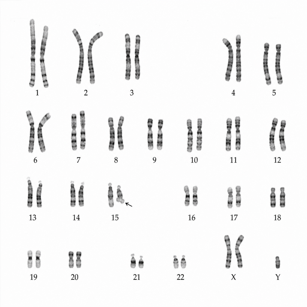
The following karyotype is seen in which of the following syndromes?
Mutation in the gene encoding Dystrophin is known to cause all the following except?
Trinucleotide repeat expansion causes all of the following conditions EXCEPT:
Which of the following is a true statement regarding autosomal dominant disorders?
Jumping genes (transposons) are primarily involved in which of the following biological processes?
What is the chromosomal abnormality associated with 100% recurrence of disease in Down's syndrome?
Which of the following is not associated with Down syndrome?
Which of the following is a polygenic disorder?
Practice by Chapter
Principles of Molecular Pathology
Practice Questions
DNA and RNA Analysis Techniques
Practice Questions
Cytogenetics
Practice Questions
Polymerase Chain Reaction Applications
Practice Questions
Next-Generation Sequencing
Practice Questions
Molecular Diagnosis of Infectious Diseases
Practice Questions
Molecular Oncology
Practice Questions
Pharmacogenomics
Practice Questions
Genetic Counseling and Risk Assessment
Practice Questions
Molecular Diagnostics Quality Control
Practice Questions
Want unlimited practice?
Get full access to all questions, explanations, and performance tracking.
Scan to download app