
Molecular Pathology — MCQs

On this page
Gene instability associated with malignancy is seen in which of the following conditions?
Barr body is absent in which individuals?
Which of the following is a polygenic disorder?
Which of the following conditions is not inherited in an autosomal dominant manner?
Best sample for karyotyping is?
Karyotyping is done in which phase of the cell cycle?
Which genetic mutation is most commonly associated with male breast carcinoma?
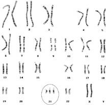
The given karyotype is seen in which of the following syndromes?
In breast cancer, Her-2/Neu promotes tumorigenesis by what mechanism?
Which of the following disorders is most commonly associated with multifactorial inheritance?
Practice by Chapter
Principles of Molecular Pathology
Practice Questions
DNA and RNA Analysis Techniques
Practice Questions
Cytogenetics
Practice Questions
Polymerase Chain Reaction Applications
Practice Questions
Next-Generation Sequencing
Practice Questions
Molecular Diagnosis of Infectious Diseases
Practice Questions
Molecular Oncology
Practice Questions
Pharmacogenomics
Practice Questions
Genetic Counseling and Risk Assessment
Practice Questions
Molecular Diagnostics Quality Control
Practice Questions
Want unlimited practice?
Get full access to all questions, explanations, and performance tracking.
Scan to download app