
Molecular Pathology — MCQs

On this page
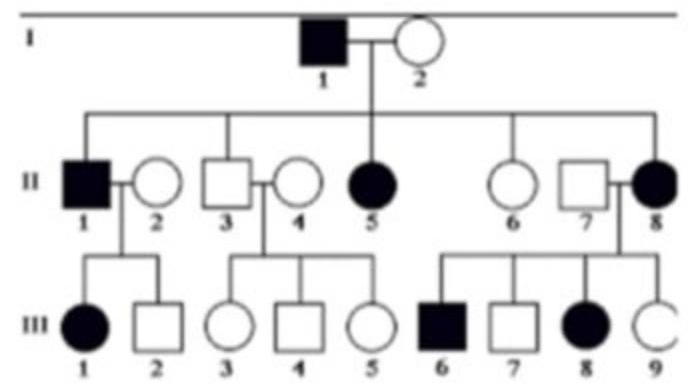
A family has a history of retinitis pigmentosa. The patient's pedigree chart shows multiple generations affected, with both males and females exhibiting the disease. What is the most likely pattern of inheritance?
Which molecular technique is used to detect chromosomal translocations in cancers such as chronic myelogenous leukemia?
A 45-year-old female is suspected of having a specific balanced translocation based on clinical presentation. Which of the following tests would be most appropriate for rapid confirmation and detailed analysis of the suspected chromosomal rearrangement?
Which molecular test is preferred for detecting HER2/neu amplification in breast cancer?
What is the effect of defective mismatch repair genes in Lynch syndrome on DNA replication fidelity and cancer risk?
In the context of molecular diagnostics, which method is the best for detecting chromosomal translocations?
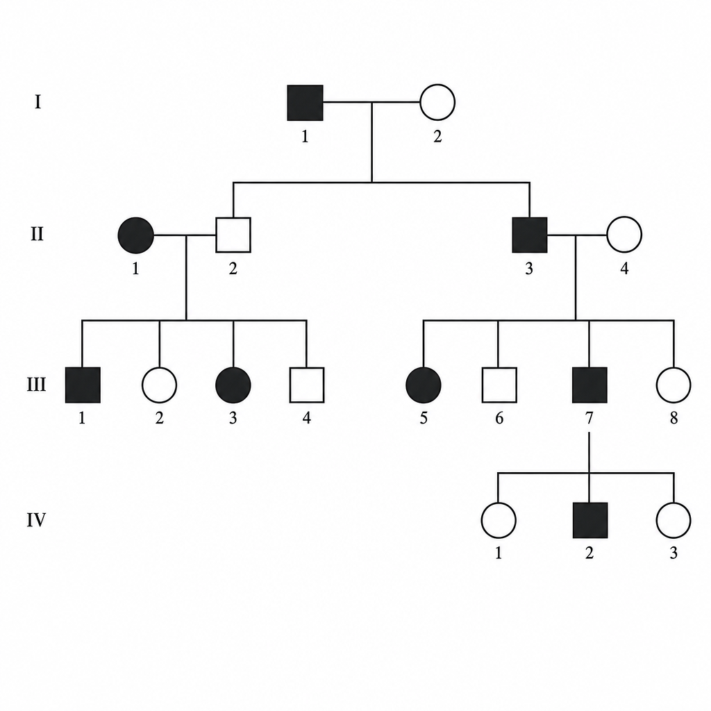
25-year-old man presents for a routine physical examination. The patient is tall and on examination, he was found to have an early diastolic murmur. His family pedigree is given below (image attached). Which of the following is the mode of inheritance by which the disease is likely to be transmitted?
Which one of the following is an autosomal recessive disorder?
Which single gene disorder does not follow Mendelian inheritance?
In Turner's syndrome, which of the following is NOT typically observed?
Practice by Chapter
Principles of Molecular Pathology
Practice Questions
DNA and RNA Analysis Techniques
Practice Questions
Cytogenetics
Practice Questions
Polymerase Chain Reaction Applications
Practice Questions
Next-Generation Sequencing
Practice Questions
Molecular Diagnosis of Infectious Diseases
Practice Questions
Molecular Oncology
Practice Questions
Pharmacogenomics
Practice Questions
Genetic Counseling and Risk Assessment
Practice Questions
Molecular Diagnostics Quality Control
Practice Questions
Want unlimited practice?
Get full access to all questions, explanations, and performance tracking.
Scan to download app