Which of the following syndromes are caused due to genomic imprinting? I. Rubinstein Taybi syndrome II. Prader-Willi syndrome III. Angelman syndrome IV. Edward syndrome Select the correct answer using the code given below :
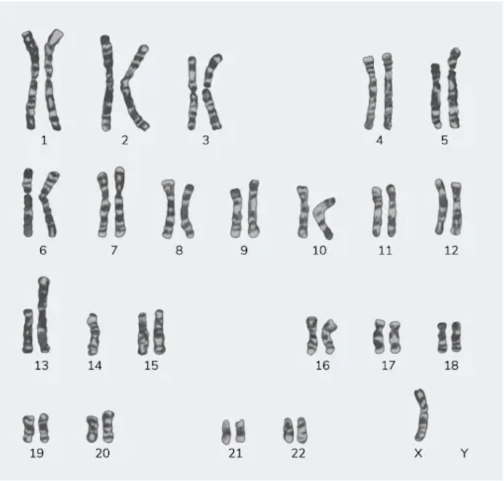
A 23-year-old female with a height of 4 feet has a karyotype as shown in the image below. Which among the following indicates the correct etiology?

Identify the gene commonly involved in the condition shown in the image?

Which PCR technique is best suited for identifying a syndrome with multiple causative agents?
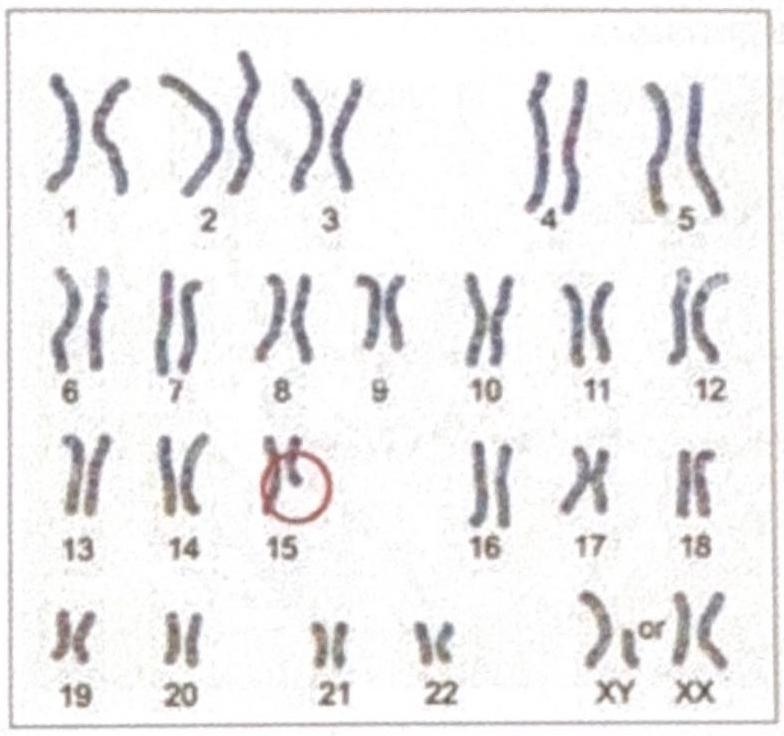
The diagrammatic representation of the karyotype of an individual indicates a specific genetic abnormality. What is the diagnosis?

HNPCC has defect in which
Which of the following is associated with defect in mismatch repair
Best method for the detection of mutations with low allele frequency is:
What is the interpretation of the given pedigree chart?

Fluorescence in situ hybridization (FISH) is required in which of the following interpretations of Her2/neu?
Explanation: ***II and III (Correct Answer)*** - **Prader-Willi syndrome** and **Angelman syndrome** are classic examples of disorders caused by **genomic imprinting** defects on chromosome 15 [1]. - **Prader-Willi syndrome** results from the loss of paternal 15q11-q13 expression, while **Angelman syndrome** results from the loss of maternal 15q11-q13 expression [1]. - Both conditions demonstrate parent-of-origin effects, where the same chromosomal region causes different phenotypes depending on whether the mutation is inherited from the mother or father [1]. *II and IV (Incorrect)* - While Prader-Willi syndrome is linked to genomic imprinting, **Edward syndrome** (Trisomy 18) is caused by a chromosomal abnormality (an extra copy of chromosome 18), not genomic imprinting. - Edward syndrome presents with distinct clinical features like **micrognathia** and **rocker-bottom feet**, different from imprinting disorders. *I and IV (Incorrect)* - **Rubinstein-Taybi syndrome** is caused by mutations in the **CREBBP** gene or deletion of 16p13.3, which are not related to genomic imprinting. - **Edward syndrome** is a chromosomal aneuploidy (Trisomy 18), not a disorder of genomic imprinting. *I and III (Incorrect)* - **Rubinstein-Taybi syndrome** is a genetic disorder caused by mutations in the CREBBP or EP300 genes, and it is not associated with genomic imprinting. - Only **Angelman syndrome** among these two options is caused by genomic imprinting. **References:** [1] Kumar V, Abbas AK, et al.. Robbins and Cotran Pathologic Basis of Disease. 9th ed. Genetic Disorders, pp. 181-182.
Explanation: **Turner syndrome** - The **karyotype shows 45,X**, meaning there is only one X chromosome and no second sex chromosome (Y or another X). This absence of a full second sex chromosome is the defining genetic characteristic of **Turner syndrome** [1]. - The clinical presentation of a **23-year-old female with a height of 4 feet (short stature)** is a classic sign of Turner syndrome, which results from the partial or complete monosomy of the X chromosome. Short stature in these patients is specifically linked to the haploinsufficiency of the SHOX gene [1]. *Klinefelter's syndrome* - This syndrome is characterized by the presence of an **extra X chromosome in males**, leading to a karyotype typically 47,XXY [2]. - While individuals with Klinefelter's syndrome may also have a variety of physical and developmental challenges, the patient's biological sex (female) and the specific karyotype shown **(45,X)** do not align with this condition. *Down syndrome* - Down syndrome is caused by a **trisomy of chromosome 21**, meaning there are three copies of chromosome 21 instead of the usual two [2]. - The provided karyotype clearly shows **two copies of chromosome 21** and a sex chromosome abnormality (45,X), making Down syndrome an incorrect diagnosis [1]. *Edward's syndrome* - Edward's syndrome is characterized by a **trisomy of chromosome 18**, meaning there are three copies of chromosome 18 [1]. - The presented karyotype shows **two copies of chromosome 18** and an abnormality in the sex chromosomes, ruling out Edward's syndrome. **References:** [1] Kumar V, Abbas AK, et al.. Robbins and Cotran Pathologic Basis of Disease. 9th ed. Genetic Disorders, pp. 171-177. [2] Cross SS. Underwood's Pathology: A Clinical Approach. 6th ed. (Basic Pathology) introduces the student to key general principles of pathology, both as a medical science and as a clinical activity with a vital role in patient care. Part 2 (Disease Mechanisms) provides fundamental knowledge about the cellular and molecular processes involved in diseases, providing the rationale for their treatment. Part 3 (Systematic Pathology) deals in detail with specific diseases, with emphasis on the clinically important aspects., pp. 92-93.
Explanation: ***BRAF V600E*** - The image displays cells with **Langerhans cell morphology**, including folded nuclei and abundant pale cytoplasm, which are characteristic of **Langerhans cell histiocytosis (LCH)** [1]. - The **BRAF V600E mutation** is the most common genetic alteration found in LCH, present in about 50-60% of cases and activating the MAPK pathway [1]. *RAS* - **RAS mutations** are frequently seen in various cancers, including colorectal adenocarcinoma, pancreatic adenocarcinoma, and non-small cell lung cancer. - While RAS pathway activation can occur in LCH, a direct RAS mutation is not the most common genetic driver; rather, downstream effectors like BRAF V600E are more prominent [1]. *RET* - **RET mutations** are primarily associated with **medullary thyroid carcinoma** (in both sporadic and inherited forms like MEN 2A and MEN 2B) and can also be found in certain types of lung cancer. - They are not a characteristic genetic alteration for Langerhans cell histiocytosis. *P53* - The **TP53 gene** encodes the tumor suppressor protein p53, and mutations in this gene are among the most frequent genetic alterations across a wide spectrum of human cancers. - Although p53 plays a critical role in cell cycle regulation and apoptosis, it is not a primary or common driver mutation specifically associated with Langerhans cell histiocytosis [1]. **References:** [1] Kumar V, Abbas AK, et al.. Robbins and Cotran Pathologic Basis of Disease. 9th ed. Diseases of White Blood Cells, Lymph Nodes, Spleen, and Thymus, pp. 629-630.
Explanation: ***Multiplex PCR*** - **Multiplex PCR** allows for the simultaneous amplification of **multiple DNA targets** in a single reaction, making it ideal for identifying syndromes with numerous potential causative agents. - This method uses **multiple primer pairs** in one reaction tube, each designed to amplify a specific target sequence, thus efficiently detecting various pathogens or genetic markers. *RT-PCR* - **Reverse Transcription PCR (RT-PCR)** is used to detect **RNA targets** by first converting RNA into cDNA, which is then amplified. - While useful for RNA viruses or gene expression studies, it is not primarily designed for simultaneous detection of multiple diverse causative agents in the same way as multiplex PCR. *Nested PCR* - **Nested PCR** uses two sets of primers in sequential reactions to **increase sensitivity and specificity** by reducing non-specific binding. - This technique is generally employed to detect very low copies of a specific target or to overcome issues with non-specific amplification, rather than for identifying multiple different agents concurrently. *Conventional PCR* - **Conventional PCR** amplifies a **single specific DNA target** using one pair of primers per reaction. [1] - It requires separate reactions for each potential causative agent, making it inefficient and labor-intensive when testing for a syndrome with multiple etiologies. **References:** [1] Cross SS. Underwood's Pathology: A Clinical Approach. 6th ed. (Basic Pathology) introduces the student to key general principles of pathology, both as a medical science and as a clinical activity with a vital role in patient care. Part 2 (Disease Mechanisms) provides fundamental knowledge about the cellular and molecular processes involved in diseases, providing the rationale for their treatment. Part 3 (Systematic Pathology) deals in detail with specific diseases, with emphasis on the clinically important aspects., pp. 56-57.
Explanation: ***Prader-Willi syndrome*** - The karyotype shows an abnormality on **chromosome 15**, which is consistent with Prader-Willi syndrome caused by **deletion of 15q11-q13** inherited from the **paternal** chromosome or **maternal uniparental disomy**. - While PWS deletions are typically **submicroscopic**, larger deletions can occasionally be visible on standard karyotyping, particularly when they represent **class I deletions** that are more extensive and involve additional chromosomal material beyond the typical PWS critical region. *Angelman syndrome* - Although Angelman syndrome also involves **chromosome 15q11-q13 deletion**, it results from **maternal** deletion or **paternal uniparental disomy**, and presents with distinctly different clinical features. - Clinical presentation includes **severe intellectual disability**, **ataxia**, **seizures**, **absent speech**, and **inappropriate laughter** (happy demeanor), which differs significantly from the PWS phenotype. *DiGeorge syndrome* - DiGeorge syndrome is caused by **deletion of chromosome 22q11.2**, not chromosome 15 as shown in the karyotype. - Clinical features include **cardiac defects** (conotruncal abnormalities), **thymic hypoplasia**, **parathyroid hypoplasia** (hypocalcemia), **cleft palate**, and characteristic facial features (CATCH-22 syndrome). *Cri du Chat syndrome* - This syndrome results from **deletion of chromosome 5p** (short arm of chromosome 5), not chromosome 15 as indicated in the karyotype. - Characteristic features include **high-pitched cry** resembling a cat's meow in infancy, **intellectual disability**, **microcephaly**, and **distinctive facial features**.
Explanation: ***Mismatch repair gene*** - **HNPCC (hereditary non-polyposis colorectal cancer)**, also known as Lynch syndrome, is caused by inherited mutations in genes responsible for **DNA mismatch repair** [1]. - These genes, such as **MLH1, MSH2, MSH6, and PMS2**, normally correct errors that occur during DNA replication, preventing the accumulation of mutations. *Base pair excision* - **Base excision repair** is a distinct DNA repair pathway that primarily fixes small base lesions, such as damaged or modified bases. - This mechanism is not primarily implicated in the development of HNPCC. *Point mutation* - A **point mutation** refers to a single nucleotide change in a DNA sequence, which can be the *result* of a defective repair mechanism but is not the defect itself. - While mismatch repair defects lead to an increased rate of point mutations, the underlying *defect* in HNPCC is in the repair system, not in the mutation type. *Nucleotide excision* - **Nucleotide excision repair** is a major pathway for removing bulky, helix-distorting DNA lesions, such as those caused by UV radiation. - Defects in this pathway are associated with conditions like **xeroderma pigmentosum**, not HNPCC. **References:** [1] Kumar V, Abbas AK, et al.. Robbins and Cotran Pathologic Basis of Disease. 9th ed. The Gastrointestinal Tract, p. 817.
Explanation: ***Hereditary HNPCC*** - **Hereditary Nonpolyposis Colorectal Cancer (HNPCC)**, also known as Lynch syndrome, is caused by inherited mutations in **DNA mismatch repair (MMR) genes** [1]. - Defective MMR leads to an accumulation of **mutations** in microsatellite regions, increasing the risk of colorectal and other cancers [1]. *MUTYH Associated Polyposis* - This condition is associated with mutations in the **MUTYH gene**, which plays a role in **base excision repair**, not mismatch repair [1]. - It leads to an increased risk of colorectal polyps and cancer, but through a different DNA repair pathway. *Bloom Disorder* - Bloom syndrome is caused by mutations in the **BLM gene**, which encodes a DNA helicase involved in **DNA replication** and repair. - It results in genomic instability, increased cancer risk, and characteristic growth retardation and photosensitivity, distinct from mismatch repair defects. *SCID* - **Severe Combined Immunodeficiency (SCID)** refers to a group of genetic disorders that impair the development and function of **T and B lymphocytes**. - While some forms involve defects in DNA repair enzymes vital for V(D)J recombination (**e.g., RAG enzymes, Artemis**), SCID is primarily an immune disorder and not directly associated with the mismatch repair pathway in the context of cancer predisposition like HNPCC. **References:** [1] Kumar V, Abbas AK, et al.. Robbins and Cotran Pathologic Basis of Disease. 9th ed. The Gastrointestinal Tract, p. 817.
Explanation: ***Droplet digital PCR*** - **Droplet digital PCR (ddPCR)** offers superior sensitivity for detecting **low allele frequency mutations** by partitioning the sample into thousands of individual reactions. - This compartmentalization allows for the direct quantification of target DNA molecules without relying on a standard curve, making it highly accurate for rare mutation detection. *FISH* - **Fluorescence in situ hybridization (FISH)** primarily detects **chromosomal abnormalities** like translocations, deletions, or amplifications, rather than single-nucleotide variants or small indels with low allele frequencies [2]. - It visualizes genetic changes at a **cytogenetic level** on an intracellular basis, not typically for quantifying rare DNA mutations in a heterogeneous sample. *Sanger sequencing* - **Sanger sequencing** is the gold standard for **sequencing individual DNA fragments** but has a detection limit of around 15-20% for allele frequency, making it unsuitable for very low allele frequency mutations [1]. - It struggles to reliably detect minor alleles when they are present in a small proportion of the total DNA pool. *Nested PCR* - **Nested PCR** increases the sensitivity and specificity of amplification by using two sets of primers in a sequential manner but does not inherently provide the **quantification capability** or the same level of **low allele frequency detection** as ddPCR processes. - While sensitive for detecting target sequences, it is not designed for precise quantification of rare mutations in a background of wild-type sequences. **References:** [1] Kumar V, Abbas AK, et al.. Robbins and Cotran Pathologic Basis of Disease. 9th ed. Genetic Disorders, p. 185. [2] Kumar V, Abbas AK, et al.. Robbins and Cotran Pathologic Basis of Disease. 9th ed. Genetic Disorders, pp. 185-186.
Explanation: ***Autosomal dominant*** - The trait appears in **every generation**, indicated by affected individuals in the first, second, and third generations [1]. - Both males and females are affected relatively equally, and affected individuals transmit the trait to approximately half of their offspring, consistent with **vertical transmission** [1]. - **Male-to-male transmission** is present, which definitively rules out X-linked inheritance patterns [1]. *Incomplete penetrance* - This pattern would typically show some individuals who carry the disease-causing genotype but **do not express the phenotype** (unaffected). - In this pedigree, all individuals who are expected to express the trait based on the clear dominant pattern are indeed affected. *Autosomal recessive* - This mode of inheritance would typically show **skipped generations**, meaning affected individuals would often have unaffected parents who are carriers [1]. - In this chart, every affected individual has at least one affected parent, ruling out an autosomal recessive pattern [1]. X-linked dominant - In X-linked dominant inheritance, **no male-to-male transmission** would be observed, as fathers pass their X chromosome only to daughters. - The presence of affected males transmitting the trait to male offspring rules out this inheritance pattern. **References:** [1] Kumar V, Abbas AK, et al.. Robbins and Cotran Pathologic Basis of Disease. 9th ed. Genetic Disorders, pp. 148-151.
Explanation: ***Correct: 2+*** A **Her2/neu immunohistochemistry (IHC) score of 2+** is considered **equivocal**, meaning it's uncertain whether Her2/neu is overexpressed. In such cases, **Fluorescence In Situ Hybridization (FISH)** is required to determine the amplification status of the *HER2* gene, which guides treatment decisions regarding anti-HER2 therapy (trastuzumab) [1], [2]. The 2+ score shows incomplete and weak to moderate membrane staining in >10% of tumor cells, necessitating gene amplification confirmation. *Incorrect: All of the options* While FISH is crucial for equivocal interpretations, it is **not required for all** possible Her2/neu IHC results [2]. Some scores (1+ and 3+) definitively indicate Her2/neu status without requiring confirmatory testing. Routinely performing FISH for all IHC scores would be unnecessary and costly. *Incorrect: 1+* An IHC score of **1+** indicates **no Her2/neu overexpression** (faint/barely perceptible incomplete membrane staining in >10% of tumor cells). In this situation, the patient is considered **Her2-negative**, and FISH testing is **not required** as the result is clearly negative. *Incorrect: 3+* An IHC score of **3+** indicates **clear Her2/neu overexpression** (strong, complete membrane staining in >10% of tumor cells) [1]. Patients with an IHC 3+ score are considered **Her2-positive**, and typically **FISH testing is not required** to confirm this result, as the overexpression is unequivocal [2]. **References:** [1] Cross SS. Underwood's Pathology: A Clinical Approach. 6th ed. (Basic Pathology) introduces the student to key general principles of pathology, both as a medical science and as a clinical activity with a vital role in patient care. Part 2 (Disease Mechanisms) provides fundamental knowledge about the cellular and molecular processes involved in diseases, providing the rationale for their treatment. Part 3 (Systematic Pathology) deals in detail with specific diseases, with emphasis on the clinically important aspects., pp. 256-259. [2] Kumar V, Abbas AK, et al.. Robbins and Cotran Pathologic Basis of Disease. 9th ed. The Breast, pp. 1064-1066.
Principles of Molecular Pathology
Practice Questions
DNA and RNA Analysis Techniques
Practice Questions
Cytogenetics
Practice Questions
Polymerase Chain Reaction Applications
Practice Questions
Next-Generation Sequencing
Practice Questions
Molecular Diagnosis of Infectious Diseases
Practice Questions
Molecular Oncology
Practice Questions
Pharmacogenomics
Practice Questions
Genetic Counseling and Risk Assessment
Practice Questions
Molecular Diagnostics Quality Control
Practice Questions
Get full access to all questions, explanations, and performance tracking.
Start For Free