
Molecular Pathology — MCQs

On this page
A couple has two children affected with tuberous sclerosis. On detailed clinical and laboratory evaluation, including molecular studies, both parents are normal. Which one of the following explains the presence of two affected children in this family?
The most common chromosomal syndrome is
Which of the following genetic aberrations is not a causative mechanism in cases of Prader-Willi syndrome?
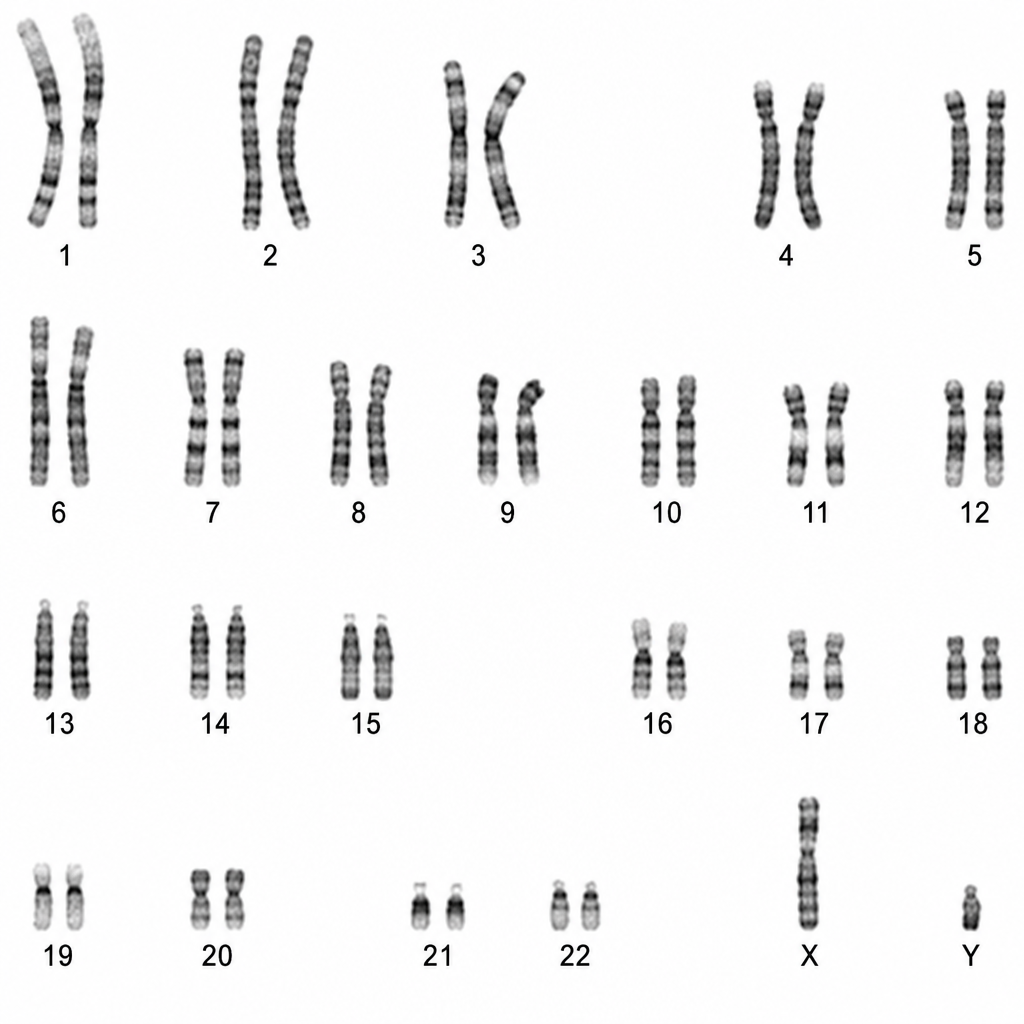
A patient's karyotype shows the following. What clinical abnormality is expected?
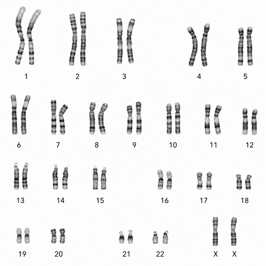
A specific karyotype is observed in a female patient. Which of the following syndromes is associated with this karyotype?
Karyotyping can be used to diagnose which of the following diseases?
What is the trinucleotide repeat seen in Fragile X syndrome?
Which of the following statements is true regarding ataxia telangiectasia?
Klinefelter syndrome is diagnosed by?
22q11 deletion leading to Di George syndrome is associated with which of the following?
Practice by Chapter
Principles of Molecular Pathology
Practice Questions
DNA and RNA Analysis Techniques
Practice Questions
Cytogenetics
Practice Questions
Polymerase Chain Reaction Applications
Practice Questions
Next-Generation Sequencing
Practice Questions
Molecular Diagnosis of Infectious Diseases
Practice Questions
Molecular Oncology
Practice Questions
Pharmacogenomics
Practice Questions
Genetic Counseling and Risk Assessment
Practice Questions
Molecular Diagnostics Quality Control
Practice Questions
Want unlimited practice?
Get full access to all questions, explanations, and performance tracking.
Scan to download app