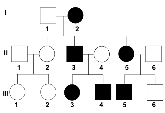
Analyze the provided pedigree chart to identify the underlying genetic disease.

What is the most common tumor in females with tuberous sclerosis?
What is a Leiden mutation?
All of the following are intermediate filaments except?
What is the gene involved in GIST?
A patient died of Alzheimer's disease. At autopsy, the heart contains a yellow-brown, finely granular pigment. What is the most likely cause of this pigment?
Which of the following is a sign of chronic inflammation?
What is the function of the FMR1 protein?
Ring sideroblasts in myelodysplastic syndrome are associated with mutations in which gene?
Which stain is used for tissue fats?
Explanation: ***Crouzon syndrome*** - Shows **autosomal dominant inheritance** pattern with multiple generations affected, both sexes involved, and approximately **50% of offspring** affected. - Exhibits **father-to-son transmission**, which is characteristic of autosomal dominant conditions and excludes X-linked inheritance. *Sickle cell anaemia* - Follows **autosomal recessive inheritance** where typically only one generation is affected with unaffected parents having affected children. - Would require **both parents to be carriers** and show a **25% recurrence risk** in offspring, not the pattern seen in the pedigree. *Diabetes insipidus* - Most forms are **acquired** rather than inherited, and when hereditary, usually show **autosomal dominant** or **X-linked recessive** patterns. - **Nephrogenic diabetes insipidus** (X-linked) would show male predominance with no father-to-son transmission, inconsistent with this pedigree. *Hunter syndrome* - Exhibits **X-linked recessive inheritance** with affected males and carrier females, showing a distinctive pattern. - Would demonstrate **no father-to-son transmission** and predominantly affect males through carrier mothers, unlike the pedigree shown.
Explanation: **Tuberous Sclerosis Complex (TSC)** is an autosomal dominant neurocutaneous syndrome caused by mutations in the **TSC1 (Hamartin)** or **TSC2 (Tuberin)** genes [1]. These proteins normally inhibit the mTOR pathway; their loss leads to uncontrolled cell growth and hamartoma formation across multiple organs [1]. **Why Angiomyolipoma (AML) is correct:** Renal Angiomyolipoma is the most common mesenchymal tumor associated with TSC, occurring in approximately **70-80% of patients** [1]. While AMLs occur in both genders, they are significantly more frequent and larger in **females** due to the presence of estrogen receptors on the tumor cells. These are benign triphasic tumors composed of abnormal blood vessels, smooth muscle, and adipose tissue. **Analysis of Incorrect Options:** * **Pulmonary Lymphangioleiomyomatosis (LAM):** This is a cystic lung disease seen almost exclusively in females with TSC [1]. However, its prevalence (approx. 30-40%) is lower than that of renal AMLs. * **Rhabdomyosarcoma:** This is a malignant skeletal muscle tumor. TSC is associated with **Cardiac Rhabdomyomas** (the most common fetal cardiac tumor), not rhabdomyosarcomas [1]. * **Optic Glioma:** This is a classic feature of **Neurofibromatosis Type 1 (NF1)**, not Tuberous Sclerosis [2]. TSC is instead associated with Retinal Astrocytic Hamartomas (Phakomas). **High-Yield Clinical Pearls for NEET-PG:** * **Vogt’s Triad:** Adenoma sebaceum (facial angiofibromas), mental retardation, and seizures (seen in only 30% of cases) [1]. * **Dermatological markers:** Ash-leaf spots (earliest sign), Shagreen patches (connective tissue nevi), and Periungual fibromas (Koenen tumors). * **CNS findings:** Subependymal Giant Cell Astrocytoma (SEGA) and cortical tubers [1]. * **Management:** mTOR inhibitors like **Everolimus** or Sirolimus are used to reduce the size of SEGA and renal AMLs. **References:** [1] Kumar V, Abbas AK, et al.. Robbins and Cotran Pathologic Basis of Disease. 9th ed. The Central Nervous System, pp. 1318-1319. [2] Cross SS. Underwood's Pathology: A Clinical Approach. 6th ed. Common Clinical Manifestations Of Central And Peripheral Nervous System Disease, pp. 724-725.
Explanation: **Factor V Leiden** is the most common inherited cause of hypercoagulability (thrombophilia) [1], [2]. ### **Why the Correct Answer is Right** The Factor V Leiden mutation is a specific **point mutation** (specifically a **mis-sense mutation**) where there is a substitution of **Guanine by Adenine (G→A)** at nucleotide 1691. This genetic change results in the replacement of the amino acid **Arginine with Glutamine** at position 506 (Arg506Gln) [2]. * **Mechanism:** This mutation occurs at the cleavage site where **Activated Protein C (APC)** normally inactivates Factor Va. The structural change renders Factor Va resistant to degradation by APC, leading to a pro-thrombotic state [2]. ### **Why Other Options are Wrong** * **Frame shift mutation:** These involve insertions or deletions of nucleotides (not in multiples of three), shifting the reading frame [3]. Example: Tay-Sachs disease. * **Tri-nucleotide repeat mutation:** These involve expansions of specific three-base sequences. Example: Huntington’s disease or Fragile X syndrome. * **Non-sense mutation:** This occurs when a point mutation creates a premature stop codon, leading to a truncated protein. Example: Some forms of Beta-thalassemia. ### **High-Yield Clinical Pearls for NEET-PG** * **Inheritance:** Autosomal Dominant. * **Clinical Presentation:** Recurrent Deep Vein Thrombosis (DVT) and pulmonary embolism [2]. It is also associated with pregnancy complications like recurrent miscarriages. * **Diagnosis:** Suspect in patients with "Activated Protein C Resistance" (APCR) [2]. Confirmed by PCR-based genetic testing. * **Key Fact:** Heterozygotes have a 5-10 fold increased risk of venous thrombosis, while homozygotes have an 80-fold increased risk. **References:** [1] "Cross SS. Underwood's Pathology: A Clinical Approach. 6th ed. Common Clinical Problems From Cardiovascular Disease, pp. 281-282." [2] "Kumar V, Abbas AK, et al.. Robbins and Cotran Pathologic Basis of Disease. 9th ed. Hemodynamic Disorders, Thromboembolic Disease, and Shock, pp. 133-134." [3] "Cross SS. Underwood's Pathology: A Clinical Approach. 6th ed. (Basic Pathology) introduces the student to key general principles of pathology, both as a medical science and as a clinical activity with a vital role in patient care. Part 2 (Disease Mechanisms) provides fundamental knowledge about the cellular and molecular processes involved in diseases, providing the rationale for their treatment. Part 3 (Systematic Pathology) deals in detail with specific diseases, with emphasis on the clinically important aspects., pp. 57-58."
Explanation: **Explanation:** The cytoskeleton of a cell consists of three main components: microfilaments (actin), microtubules (tubulin), and **intermediate filaments (IFs)**. Intermediate filaments provide mechanical strength to cells and are categorized into several types based on their tissue distribution. **Why Cadherin is the correct answer:** **Cadherins** are not intermediate filaments; they are **transmembrane cell adhesion molecules**. They play a critical role in cell-cell junctions (like desmosomes and adherens junctions) by mediating calcium-dependent adhesion [1]. While cadherins often link to the cytoskeleton internally, they are structurally distinct from the IF family. **Analysis of incorrect options:** * **Lamin (Option A):** These are Type V intermediate filaments located within the nucleus (nuclear lamina). They provide structural support to the nuclear envelope and regulate DNA replication. * **Vimentin (Option C):** A Type III intermediate filament found in cells of **mesenchymal origin** (e.g., fibroblasts, endothelium, smooth muscle). It is a classic IHC marker for sarcomas. * **Desmin (Option D):** Also a Type III intermediate filament, specifically found in **all types of muscle cells** (skeletal, cardiac, and smooth). It is used as an IHC marker to identify myogenic tumors (e.g., rhabdomyosarcoma). **High-Yield Clinical Pearls for NEET-PG:** * **Cytokeratin:** IF for epithelial cells (Marker for Carcinomas). * **GFAP:** IF for glial cells (Marker for Astrocytomas). * **Neurofilaments:** IF for neurons (Marker for Neuroblastoma/Pheochromocytoma). * **Mallory Hyaline:** These are inclusions of pre-keratin intermediate filaments found in alcoholic liver disease. * **Diagnostic Tip:** If a question asks for a marker of "mesenchymal origin," always look for **Vimentin**. **References:** [1] Kumar V, Abbas AK, et al.. Robbins and Cotran Pathologic Basis of Disease. 9th ed. With Illustrations By, pp. 23-24.
Explanation: **Explanation:** **Gastrointestinal Stromal Tumor (GIST)** is the most common mesenchymal tumor of the gastrointestinal tract. The correct answer is **C-KIT** because approximately 85-90% of GISTs are driven by oncogenic gain-of-function mutations in the **c-KIT gene** (which encodes the CD117 receptor tyrosine kinase) [1]. This mutation leads to constitutive activation of the kinase signaling pathway, resulting in uncontrolled cell proliferation [1]. **Analysis of Options:** * **C-KIT (CD117):** This is the hallmark diagnostic marker for GIST. In cases where c-KIT is negative, mutations in **PDGFRA** (Platelet-Derived Growth Factor Receptor Alpha) are often found [1]. * **BRCA-1 & BRCA-2:** These are tumor suppressor genes involved in DNA repair. Mutations are primarily associated with hereditary **Breast and Ovarian Cancer** syndromes, not mesenchymal GI tumors. * **p53:** Known as the "Guardian of the Genome," this is the most commonly mutated gene in human cancers (e.g., Li-Fraumeni syndrome). While it may be involved in the progression of many tumors, it is not the primary driver or diagnostic marker for GIST. **High-Yield Clinical Pearls for NEET-PG:** * **Origin:** GISTs arise from the **Interstitial Cells of Cajal (ICC)**, the pacemakers of the gut [1]. * **Most Common Site:** Stomach (60%), followed by the small intestine. * **Markers:** **CD117** (most specific) and **DOG1** (Discovered on GIST-1). * **Treatment:** The discovery of the c-KIT mutation led to the use of **Imatinib mesylate** (a tyrosine kinase inhibitor), which has revolutionized the prognosis of unresectable or metastatic GIST [1]. **References:** [1] Kumar V, Abbas AK, et al.. Robbins and Cotran Pathologic Basis of Disease. 9th ed. The Gastrointestinal Tract, pp. 782-784.
Explanation: **Explanation:** The correct answer is **B. Lipochrome (Lipofuscin)**. The description of a **yellow-brown, finely granular pigment** in the heart of an elderly patient (implied by Alzheimer's disease) is the classic presentation of **Lipofuscin**, also known as "wear and tear" or "aging" pigment [1]. 1. **Why it is correct:** Lipofuscin is an insoluble pigment composed of polymers of lipids and phospholipids complexed with protein [1]. It is derived through the **peroxidation of polyunsaturated lipids** of subcellular membranes. It is not harmful to the cell itself but serves as a hallmark of aging and free radical injury. In the heart, extensive accumulation leads to a condition known as **"Brown Atrophy."** 2. **Why the other options are incorrect:** * **A. Hemosiderin:** This is a golden-yellow to brown, granular pigment derived from hemoglobin (iron). While it looks similar, it is associated with iron overload (hemosiderosis) and would typically stain positive with **Prussian Blue**, unlike lipofuscin. * **C. Glycogen:** Glycogen is a clear/white intracellular inclusion on H&E staining (appearing as empty vacuoles) [1]. It requires a **PAS stain** for visualization and is not yellow-brown. * **D. Fat:** Intracellular fat (steatosis) appears as clear, sharply demarcated vacuoles on H&E. **High-Yield Clinical Pearls for NEET-PG:** * **Staining:** Lipofuscin is **PAS positive** and can be visualized with Sudan Black B (though it is not a true fat). * **Location:** Most commonly seen in **perikaryal (around the nucleus)** locations in permanent cells like **myocytes and neurons** [1]. * **Mechanism:** It is a sign of **autophagy**; the pigment represents undigested material persisting in residual bodies within lysosomes [1]. **References:** [1] Kumar V, Abbas AK, et al.. Robbins and Cotran Pathologic Basis of Disease. 9th ed. Cellular Responses to Stress and Toxic Insults: Adaptation, Injury, and Death, p. 75.
Explanation: **Explanation:** Chronic inflammation is a prolonged process (weeks to months) characterized by the simultaneous occurrence of inflammation, tissue injury, and attempts at repair. [1] **Why Angiogenesis is Correct:** Angiogenesis (the formation of new blood vessels) is a hallmark of the **repair phase** of chronic inflammation. [1] It is mediated by growth factors like VEGF and FGF, which stimulate endothelial cell proliferation to supply oxygen and nutrients to the developing granulation tissue. [1] Along with mononuclear cell infiltration (macrophages, lymphocytes) and fibrosis, angiogenesis constitutes the classic morphological triad of chronic inflammation. [1] **Analysis of Incorrect Options:** * **Purulent Exudate:** This is a feature of **acute inflammation**, specifically "suppurative inflammation." It consists of neutrophils, liquefied debris, and edema fluid (pus), typically seen in pyogenic bacterial infections. * **Edema:** This is one of the cardinal signs of **acute inflammation**. It results from increased vascular permeability and vasodilation (exudation), occurring within minutes to hours of injury. * **Induration:** While induration (hardening of tissue) can occur in chronic states, it is specifically the clinical hallmark of a **Type IV Hypersensitivity reaction** (e.g., a positive Tuberculin/Mantoux test) rather than a general pathological sign of all chronic inflammatory processes. **NEET-PG High-Yield Pearls:** * **Cells of Chronic Inflammation:** Macrophages are the "dominant" cells. [1] * **Granulomatous Inflammation:** A specific subtype of chronic inflammation characterized by epithelioid histiocytes (activated macrophages). * **Key Growth Factor:** **VEGF** is the most important mediator for angiogenesis in chronic inflammation and wound healing. [1] * **Morphological Triad:** 1. Infiltration with mononuclear cells; 2. Tissue destruction; 3. Healing by connective tissue replacement (Angiogenesis + Fibrosis). [1] **References:** [1] Kumar V, Abbas AK, et al.. Robbins and Cotran Pathologic Basis of Disease. 9th ed. Inflammation and Repair, pp. 103-119.
Explanation: **Explanation:** The **FMR1 gene** (Fragile X Mental Retardation 1) is located on the long arm of the X chromosome (Xq27.3). The hallmark of Fragile X Syndrome is the expansion of a **CGG trinucleotide repeat** within the 5' untranslated region (UTR) of this gene [1]. In a normal individual, there are 6–54 repeats; however, in affected individuals, this expands to a "full mutation" (>200 repeats), leading to hypermethylation of the promoter and transcriptional silencing of the gene [1]. **Analysis of Options:** * **Option D (Correct):** The FMR1 gene contains (5′-CGG-3′)n repeat segments. The expansion of these repeats is the primary molecular mechanism behind the disease [1]. * **Option A (Incorrect):** In Fragile X Syndrome, the FMR1 protein (FMRP) is **absent or significantly reduced** due to gene silencing [1]. It is not "expressed" in the disease state; its absence causes the pathology. * **Option B (Incorrect):** While FMRP is widely expressed, it is most abundantly found in the **brain and testes** (not ovaries) [1]. This explains the clinical presentation of intellectual disability and macro-orchidism. * **Option C (Incorrect):** Loss of FMRP is **sufficient** to cause the syndrome. FMRP is an RNA-binding protein that regulates the translation of specific mRNAs at the synapse; its absence disrupts synaptic plasticity [1]. **High-Yield Clinical Pearls for NEET-PG:** * **Inheritance:** X-linked dominant with variable expressivity (Anticipation occurs). * **Clinical Triad:** Long face with large mandible, large everted ears, and **Macro-orchidism** (post-pubertal). * **Premutation (55–200 repeats):** Associated with **FXTAS** (Fragile X-associated Tremor/Ataxia Syndrome) and **POI** (Primary Ovarian Insufficiency). * **Diagnosis:** PCR (for small repeats) and **Southern Blot** (to detect full mutations and methylation status). **References:** [1] Kumar V, Abbas AK, et al.. Robbins and Cotran Pathologic Basis of Disease. 9th ed. Genetic Disorders, pp. 179-181.
Explanation: **Explanation:** **Correct Answer: D. SF3B1** The presence of **ring sideroblasts** (erythroblasts with iron-laden mitochondria encircling the nucleus) is a hallmark of specific subtypes of Myelodysplastic Syndrome (MDS). The **SF3B1** (*Splicing Factor 3b Subunit 1*) gene mutation is highly specific for MDS with ring sideroblasts (MDS-RS). This gene encodes a core component of the RNA splicing machinery (spliceosome) [1]. Mutations lead to aberrant splicing of iron transporters (like *ABCB7*), causing mitochondrial iron accumulation. In the revised WHO/ICC classifications, the presence of an SF3B1 mutation allows for the diagnosis of MDS-RS even if the ring sideroblast count is as low as 5% (compared to the usual 15% threshold). **Incorrect Options:** * **A. ASXL1:** An epigenetic regulator. Mutations are common in MDS and AML but are associated with a **poor prognosis** and do not specifically correlate with ring sideroblasts [1]. * **B. EZH2:** Part of the Polycomb Repressive Complex 2 (PRC2). Mutations are linked to myelofibrosis and MDS but signify an adverse prognosis rather than a specific morphological feature [1]. * **C. TET2:** One of the most common mutations in MDS and Clonal Hematopoiesis of Indeterminate Potential (CHIP). It involves DNA demethylation; while common, it is not specific to the sideroblastic phenotype [1]. **High-Yield Clinical Pearls for NEET-PG:** * **Stain for Ring Sideroblasts:** Perls’ Prussian Blue stain. * **Definition:** ≥5 granules covering at least 1/3rd of the nuclear circumference. * **Prognostic Value:** SF3B1 mutations in MDS are generally associated with a **favorable prognosis** and lower risk of transformation to AML. * **Treatment:** Luspatercept is a newer agent used specifically for anemia in MDS-RS patients. **References:** [1] Kumar V, Abbas AK, et al.. Robbins and Cotran Pathologic Basis of Disease. 9th ed. Diseases of White Blood Cells, Lymph Nodes, Spleen, and Thymus, pp. 622-624.
Explanation: **Explanation:** **Sudan IV** is the correct answer because it belongs to a group of lipid-soluble dyes (including Sudan III and Oil Red O) used to demonstrate **neutral fats, triglycerides, and lipids** in tissue sections. These stains work on the principle of physical solubility: the dye is more soluble in the lipid droplets than in the solvent (usually alcohol or propylene glycol), causing it to migrate into the fat and stain it a brilliant red-orange. *Note:* For lipid staining, tissues must be processed as **frozen sections** because routine paraffin embedding involves organic solvents (like xylene) that dissolve fats [1]. **Analysis of Incorrect Options:** * **A. PAS (Periodic Acid-Schiff):** Used primarily to detect **glycogen**, mucopolysaccharides, and basement membranes [2]. It stains these structures magenta. * **B. Prussian Blue (Perl’s Stain):** Used to detect **ferric iron** (hemosiderin). It is the gold standard for diagnosing conditions like hemochromatosis or sideroblastic anemia. * **C. Alcian Blue:** Used to identify **acidic mucopolysaccharides** (mucin). It is frequently used to diagnose Barrett’s esophagus (staining goblet cells blue). **NEET-PG High-Yield Pearls:** * **Oil Red O:** The most commonly used stain for lipids in modern pathology (superior to Sudan IV). * **Osmium Tetroxide:** A unique stain that colors lipids **black** and is also used as a fixative in electron microscopy. * **Sudan Black B:** Stains phospholipids and is used in hematopathology to differentiate Acute Myeloid Leukemia (AML) from ALL. **References:** [1] Cross SS. Underwood's Pathology: A Clinical Approach. 6th ed. (Basic Pathology) introduces the student to key general principles of pathology, both as a medical science and as a clinical activity with a vital role in patient care. Part 2 (Disease Mechanisms) provides fundamental knowledge about the cellular and molecular processes involved in diseases, providing the rationale for their treatment. Part 3 (Systematic Pathology) deals in detail with specific diseases, with emphasis on the clinically important aspects., pp. 25-26. [2] Kumar V, Abbas AK, et al.. Robbins and Cotran Pathologic Basis of Disease. 9th ed. Cellular Responses to Stress and Toxic Insults: Adaptation, Injury, and Death, p. 75.
Principles of Molecular Pathology
Practice Questions
DNA and RNA Analysis Techniques
Practice Questions
Cytogenetics
Practice Questions
Polymerase Chain Reaction Applications
Practice Questions
Next-Generation Sequencing
Practice Questions
Molecular Diagnosis of Infectious Diseases
Practice Questions
Molecular Oncology
Practice Questions
Pharmacogenomics
Practice Questions
Genetic Counseling and Risk Assessment
Practice Questions
Molecular Diagnostics Quality Control
Practice Questions
Get full access to all questions, explanations, and performance tracking.
Start For Free