'Onion skin' fibrosis of bile duct is seen in?
What is the most common carcinoma associated with cirrhosis?
Which of the following statements about fibrolamellar carcinoma is TRUE?
The liver biopsy in acute hepatitis due to hepatitis B virus is likely to show all of the following, except?
NCPF is due to:
Mallory's degeneration seen in alcoholic liver disease is a form of which type of degeneration?
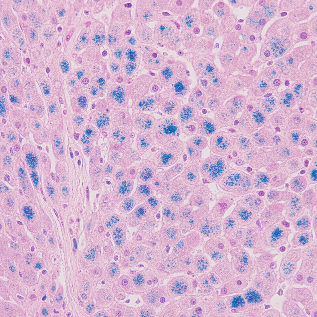
What is the most common site of metastasis in cholangiocarcinoma?
Which of the following is NOT exclusively produced by the liver?
Which of the following is NOT a feature of liver damage?
Liver biopsy of an icteric patient shows intensely eosinophilic objects called as Councilman bodies. What is the likely diagnosis of this patient?
Explanation: **Explanation:** **Primary Sclerosing Cholangitis (PSC)** is the correct answer. The hallmark pathological feature of PSC is **periductal "onion-skin" fibrosis** [2]. This represents concentric layers of fibrous tissue depositing around medium and small-sized intrahepatic bile ducts. Over time, this progressive fibrosis leads to the obliteration of the duct lumen, leaving behind a solid fibrous scar known as a **"tombstone scar."** Radiologically, this alternating segment of stenosis and dilation creates a characteristic "beaded appearance" on ERCP/MRCP [1]. **Analysis of Incorrect Options:** * **Primary Biliary Cirrhosis (PBC):** Characterized by the **"Florid Duct Lesion,"** which involves granulomatous destruction of small intrahepatic bile ducts. It does not typically show concentric onion-skin fibrosis. * **Extrahepatic Biliary Atresia/Fibrosis:** Presents with neonatal jaundice due to complete obstruction of the extrahepatic biliary tree. Histology shows bile duct proliferation and portal tract edema, not the specific onion-skin pattern. * **Congenital Hepatic Fibrosis:** Part of the fibrocystic liver disease spectrum (associated with ARPKD). It features broad bands of mature collagenous tissue and malformed, dilated bile ducts (ductal plate malformation). **High-Yield Clinical Pearls for NEET-PG:** * **Association:** Strongly associated with **Ulcerative Colitis** (approx. 70% of PSC patients have UC) [1], [2]. * **Antibody:** **p-ANCA** is positive in about 60-80% of cases. * **Gender:** More common in **males** (unlike PBC, which is more common in females) [1]. * **Risk:** Significant increased risk of **Cholangiocarcinoma**. * **Imaging Gold Standard:** MRCP/ERCP showing "beading" of the biliary tree [1]. **References:** [1] Kumar V, Abbas AK, et al.. Robbins and Cotran Pathologic Basis of Disease. 9th ed. Liver and Gallbladder, pp. 865-866. [2] Cross SS. Underwood's Pathology: A Clinical Approach. 6th ed. Common Clinical Problems From Liver And Biliary System Disease, pp. 393-394.
Explanation: **Explanation:** **Hepatocellular Carcinoma (HCC)** is the most common primary malignancy of the liver and is strongly associated with **cirrhosis** [1], [2]. In approximately 80-90% of cases, HCC develops within a cirrhotic liver. The underlying mechanism involves chronic inflammation, repeated cycles of hepatocyte death, and compensatory regeneration, which leads to genomic instability and the formation of dysplastic nodules that eventually progress to carcinoma [4]. Chronic Hepatitis B and C, alcoholic liver disease, and Non-Alcoholic Steatohepatitis (NASH) are the leading causes of the preceding cirrhosis [1]. **Analysis of Incorrect Options:** * **Fibrolamellar Carcinoma:** This is a distinct variant of HCC that typically occurs in **young adults (20-40 years)** and, crucially, is **not associated with cirrhosis** or HBV/HCV infection. It has a better prognosis and presents as a single large scirrhous tumor with fibrous bands. * **Hepatoblastoma:** This is the most common liver tumor of **childhood** (usually occurring before age 3). It is associated with FAP and Beckwith-Wiedemann syndrome, not adult-onset cirrhosis. * **Pancreatic Cancer:** While it can metastasize to the liver, it is a primary malignancy of the pancreas and does not arise from cirrhotic liver tissue [3]. **High-Yield Clinical Pearls for NEET-PG:** * **Tumor Marker:** Serum **Alpha-fetoprotein (AFP)** is the most common screening marker, though it can be normal in 30% of cases [2]. * **Radiology:** HCC shows a characteristic **"Wash-in"** (arterial enhancement) and **"Wash-out"** (venous phase) pattern on Triphasic CT [4]. * **Aflatoxin B1:** Exposure (from *Aspergillus flavus*) is a major risk factor for HCC, often causing a specific mutation in the **p53 gene (codon 249)** [1]. **References:** [1] Kumar V, Abbas AK, et al.. Robbins and Cotran Pathologic Basis of Disease. 9th ed. Liver and Gallbladder, pp. 876-877. [2] Cross SS. Underwood's Pathology: A Clinical Approach. 6th ed. Common Clinical Problems From Liver And Biliary System Disease, pp. 399-400. [3] Cross SS. Underwood's Pathology: A Clinical Approach. 6th ed. Common Clinical Problems From Liver And Biliary System Disease, pp. 398-399. [4] Kumar V, Abbas AK, et al.. Robbins and Cotran Pathologic Basis of Disease. 9th ed. Liver and Gallbladder, pp. 878-879.
Explanation: **Explanation:** Fibrolamellar Carcinoma (FLC) is a distinct variant of Hepatocellular Carcinoma (HCC) that differs significantly from the conventional type in terms of demographics and clinical behavior. **1. Why Option D is Correct:** Fibrolamellar carcinoma generally carries a **better prognosis** compared to conventional HCC. This is primarily because it typically arises in a **non-cirrhotic liver** [1], allowing for more aggressive surgical resection. Additionally, patients are usually younger and have better physiological reserves. **2. Why the other options are incorrect:** * **Option A (Diffuse in nature):** FLC usually presents as a **single, large, well-circumscribed hard mass** (scirrhous tumor) with a characteristic central stellate scar. It is not diffuse. * **Option B (Occurs after 60 years):** FLC typically affects **young adults** (usually between 20 and 40 years of age). Conventional HCC is more common in older populations. * **Option C (Cirrhosis as a feature):** Unlike conventional HCC, FLC is **not associated with cirrhosis** [1], HBV, or HCV infection. This is a hallmark diagnostic feature. **3. High-Yield Clinical Pearls for NEET-PG:** * **Morphology:** Characterized by large, polygonal cells with abundant eosinophilic cytoplasm (due to mitochondria) and prominent nucleoli, separated by **parallel (lamellar) bundles of collagen**. * **Tumor Marker:** Serum **Alpha-fetoprotein (AFP) levels are usually normal**. * **Molecular Hallmark:** A characteristic recurrent **DNAJB1-PRKACA gene fusion** is found in almost all cases. * **Radiology:** Often shows a central calcified scar (unlike the non-calcified scar of Focal Nodular Hyperplasia). **References:** [1] Cross SS. Underwood's Pathology: A Clinical Approach. 6th ed. Common Clinical Problems From Liver And Biliary System Disease, pp. 399-400.
Explanation: The key to answering this question lies in distinguishing between **acute** and **chronic** viral hepatitis. **Why "Ground glass hepatocytes" is the correct answer:** Ground glass hepatocytes are a hallmark of **Chronic Hepatitis B**, not acute infection [1]. This appearance is caused by the massive accumulation of HBsAg (Hepatitis B surface Antigen) within the smooth endoplasmic reticulum of the hepatocyte cytoplasm [2]. It takes time for this viral antigen to accumulate to a level visible by light microscopy; therefore, it is absent in the acute phase. On staining, these cells appear granular and eosinophilic and are positive for **Shikata’s orcein stain** [3]. **Explanation of incorrect options (Features of Acute Hepatitis):** * **Ballooning change (Option A):** This is a form of reversible cell injury where hepatocytes swell due to the accumulation of water (hydropic degeneration). It is a classic feature of acute viral injury [4]. * **Focal or spotty necrosis (Option B):** In acute hepatitis, individual or small clusters of hepatocytes undergo necrosis [4]. This is often accompanied by an inflammatory infiltrate. * **Acidophil bodies (Option D):** Also known as **Councilman bodies**, these represent hepatocytes undergoing apoptosis [4]. They appear as small, shrunken, deeply eosinophilic (pink) rounded bodies. They are characteristic of acute viral hepatitis. **High-Yield Clinical Pearls for NEET-PG:** * **Councilman bodies** are seen in both Viral Hepatitis and Yellow Fever. * **Bridging necrosis** (portal-to-portal or portal-to-central) indicates a more severe form of hepatitis and carries a risk of progression to cirrhosis. * **Interface hepatitis** (formerly "piecemeal necrosis") is the hallmark of Chronic Active Hepatitis. * **Mallory-Denk bodies** (cytokeratin filaments) are typically associated with Alcoholic Liver Disease, not viral hepatitis. **References:** [1] Kumar V, Abbas AK, et al.. Robbins and Cotran Pathologic Basis of Disease. 9th ed. Liver and Gallbladder, pp. 843-844. [2] Kumar V, Abbas AK, et al.. Robbins and Cotran Pathologic Basis of Disease. 9th ed. Liver and Gallbladder, p. 844. [3] Kumar V, Abbas AK, et al.. Robbins and Cotran Pathologic Basis of Disease. 9th ed. Liver and Gallbladder, pp. 844-845. [4] Cross SS. Underwood's Pathology: A Clinical Approach. 6th ed. Common Clinical Problems From Liver And Biliary System Disease, pp. 386-387.
Explanation: **Explanation:** **Non-Cirrhotic Portal Fibrosis (NCPF)** is a clinical syndrome characterized by portal hypertension, splenomegaly, and variceal bleeding in the absence of cirrhosis or extrahepatic portal vein obstruction. **1. Why the correct answer is right:** The primary pathology in NCPF involves **obliterative venopathy** of the small portal vein branches. Because these changes occur within the liver but *before* the blood reaches the hepatic sinusoids, it is classified as an **intrahepatic pre-sinusoidal obstruction** [1]. The portal pressure is elevated, but the Wedged Hepatic Venous Pressure (WHVP)—which reflects sinusoidal pressure—remains normal or near-normal. **2. Why the incorrect options are wrong:** * **Extrahepatic obstruction:** This refers to conditions like Extrahepatic Portal Vein Obstruction (EHPVO), where the blockage is in the main portal vein outside the liver [1]. * **Intrahepatic sinusoidal obstruction:** This is the hallmark of **Cirrhosis**, where the resistance to blood flow occurs within the sinusoids due to fibrosis and nodule formation [1]. * **Intrahepatic post-sinusoidal obstruction:** This occurs in **Sinusoidal Obstruction Syndrome (SOS)** (formerly Veno-occlusive disease), where the terminal hepatic venules are affected. **High-Yield Clinical Pearls for NEET-PG:** * **Commonest Presentation:** Massive hematemesis due to esophageal varices in a young/middle-aged patient. * **Liver Function Tests (LFTs):** Usually normal (unlike cirrhosis). * **Biopsy Findings:** Portal fibrosis and "narrowing/obliteration" of portal vein branches; **no cirrhosis** (no regenerative nodules). * **Schistosomiasis:** Another classic cause of intrahepatic pre-sinusoidal portal hypertension (due to eggs lodging in portal triads) [1]. * **Idiopathic Portal Hypertension (IPH):** The term used in Japan and the West for the same clinical entity as NCPF. **References:** [1] Kumar V, Abbas AK, et al.. Robbins and Cotran Pathologic Basis of Disease. 9th ed. Liver and Gallbladder, pp. 832-835.
Explanation: **Explanation:** **Mallory-Denk bodies** (Mallory’s hyaline) are a hallmark finding in alcoholic hepatitis [1]. They represent **Hyaline degeneration**, which refers to an intra-cytoplasmic accumulation of proteinaceous material that appears homogeneous, glassy, and eosinophilic (pink) on H&E staining [2]. 1. **Why Hyaline Degeneration is Correct:** Mallory bodies are composed of tangled intermediate filaments, specifically **cytokeratins 8 and 18**, complexed with other proteins like ubiquitin and heat shock proteins. This protein aggregation occurs due to hepatocyte injury, leading to the characteristic "rope-like" eosinophilic inclusions around the nucleus [1]. 2. **Why Other Options are Incorrect:** * **Amyloid degeneration:** This involves the extracellular deposition of misfolded fibrillar proteins (beta-pleated sheets). Mallory bodies are intracellular. * **Hydropic degeneration:** Also known as cloudy swelling, this is caused by an influx of water into the cell due to failure of Na+/K+ pumps. It results in a pale, swollen cytoplasm, not dense protein aggregates. * **Fatty degeneration (Steatosis):** This is the accumulation of triglycerides within hepatocytes (clear vacuoles) [1]. While common in alcoholic liver disease, it is distinct from the proteinaceous Mallory bodies. **High-Yield NEET-PG Pearls:** * **Stain:** Mallory bodies are highlighted by **Ubiquitin** immunohistochemical stains. * **Not Pathognomonic:** Although classic for Alcohol, they are also seen in **NASH** (Non-alcoholic steatohepatitis) [3], **Wilson disease**, **Primary Biliary Cholangitis**, and **Indian Childhood Cirrhosis**. * **Morphology:** They are typically described as "eosinophilic rope-like" inclusions. **References:** [1] Cross SS. Underwood's Pathology: A Clinical Approach. 6th ed. Common Clinical Problems From Liver And Biliary System Disease, pp. 389-390. [2] Cross SS. Underwood's Pathology: A Clinical Approach. 6th ed. Common Clinical Problems From Liver And Biliary System Disease, pp. 388-389. [3] Kumar V, Abbas AK, et al.. Robbins and Cotran Pathologic Basis of Disease. 9th ed. Liver and Gallbladder, pp. 852-854.
Explanation: **Explanation:** Cholangiocarcinoma (CCA) is a malignant tumor arising from the epithelial lining of the bile ducts. Understanding its spread is crucial for NEET-PG. **1. Why Liver is the Correct Answer:** Cholangiocarcinoma, particularly the intrahepatic and perihilar (Klatskin tumor) types, primarily spreads via **direct extension** and **portal venous drainage**. Because the bile ducts are anatomically embedded within or in close proximity to the hepatic parenchyma, the **liver** is the most common site for both direct invasion and metastatic seeding [1]. Intrahepatic spread often presents as satellite nodules within the liver. **2. Analysis of Incorrect Options:** * **B. Bones:** While CCA can metastasize to the bone, it is a late-stage occurrence and significantly less common than hepatic or lymphatic spread. * **C. Lung:** The lungs are the most common site for **extra-abdominal** distant metastasis, but they occur less frequently than intra-abdominal spread to the liver and regional lymph nodes. * **D. Pancreas:** While distal cholangiocarcinomas can involve the head of the pancreas via direct extension due to anatomical proximity, it is not considered a primary site of distant metastasis. **3. High-Yield Clinical Pearls for NEET-PG:** * **Risk Factors:** Primary Sclerosing Cholangitis (most common in the West), *Clonorchis sinensis* (liver fluke), and Choledochal cysts. * **Tumor Marker:** **CA 19-9** is the most commonly used marker for monitoring. * **Morphology:** Most are well-to-moderately differentiated **adenocarcinomas** with a dense fibrous stroma (desmoplasia) [1]. * **Klatskin Tumor:** A specific type of hilar cholangiocarcinoma occurring at the confluence of the right and left hepatic ducts. **References:** [1] Kumar V, Abbas AK, et al.. Robbins and Cotran Pathologic Basis of Disease. 9th ed. Liver and Gallbladder, pp. 880-881.
Explanation: **Explanation:** The liver is the primary metabolic factory of the body, responsible for synthesizing the majority of plasma proteins. However, the production of **Globulins** is the exception to this rule. **1. Why Globulin is the correct answer:** Globulins are divided into alpha, beta, and gamma fractions. While alpha and beta globulins are synthesized in the liver, **Gamma-globulins (Immunoglobulins)** are produced by **plasma cells** (derived from B-lymphocytes) in the lymphoid tissue. Since the question asks what is *not exclusively* produced by the liver, globulin is the correct choice because a significant portion of it originates extra-hepatically. **2. Why the other options are incorrect:** * **Albumin (Option D):** This is the most abundant plasma protein and is synthesized **exclusively** by hepatocytes [2]. In chronic liver disease (like cirrhosis), serum albumin levels drop, leading to decreased oncotic pressure and edema [2]. * **Prothrombin (Factor II) and Factor VII (Options A & B):** The liver is the sole site of synthesis for almost all coagulation factors [1], including the Vitamin K-dependent factors (II, VII, IX, and X) [1]. Factor VII has the shortest half-life (approx. 6 hours), making it the most sensitive marker for acute liver injury. **High-Yield Clinical Pearls for NEET-PG:** * **The "Exception" Rule:** All plasma proteins are synthesized by the liver **EXCEPT** Immunoglobulins (Plasma cells) and von Willebrand Factor (Endothelial cells/Megakaryocytes). * **Albumin vs. PT:** Albumin is a marker of **chronic** liver synthetic function (half-life ~20 days), whereas Prothrombin Time (PT) is the best indicator of **acute** liver synthetic function due to the short half-life of Factor VII. * **A:G Ratio:** In cirrhosis, the Albumin:Globulin ratio is **reversed** (decreased albumin and increased gamma-globulins due to immune activation). **References:** [1] Cross SS. Underwood's Pathology: A Clinical Approach. 6th ed. Common Clinical Problems From Blood And Bone Marrow Disease, pp. 582-583. [2] Cross SS. Underwood's Pathology: A Clinical Approach. 6th ed. Common Clinical Problems From Liver And Biliary System Disease, pp. 380-381.
Explanation: ### Explanation **Why Option D is the Correct Answer:** Erythropoietin (EPO) is primarily produced by the **interstitial cells of the peritubular capillary bed in the kidney** (approx. 90%). While the liver is the primary source of EPO during fetal life, in adults, it contributes only about 10% of total production. Therefore, liver damage does not typically lead to a clinically significant decrease in erythropoietin levels. In fact, certain liver pathologies like Hepatocellular Carcinoma (HCC) can cause a *paraneoplastic syndrome* resulting in **increased** EPO production and polycythemia. **Analysis of Incorrect Options:** * **Option A & B:** The liver synthesizes almost all coagulation factors (except Factor VIII and vWF) [1]. **Factor VII** has the shortest half-life (approx. 6 hours), making it the first factor to decline in liver dysfunction. This leads to an early **increase in Prothrombin Time (PT)**, which is a sensitive marker for the liver's synthetic function. * **Option C:** Liver damage (hepatocellular jaundice) impairs both the conjugation process and the excretion of bilirubin into the bile canaliculi [1]. This results in a "mixed" hyperbilirubinemia, where both **conjugated and unconjugated bilirubin** levels are elevated in the serum [1]. **NEET-PG High-Yield Pearls:** * **Best indicator of acute liver prognosis:** Prothrombin Time (PT). * **Best indicator of chronic liver synthetic function:** Serum Albumin [1]. * **Shortest half-life clotting factor:** Factor VII. * **Liver-specific enzymes:** ALT (Alanine Aminotransferase) is more specific for liver injury than AST [1]. * **Councilman bodies:** Eosinophilic apoptotic hepatocytes seen in viral hepatitis and Yellow Fever. **References:** [1] Cross SS. Underwood's Pathology: A Clinical Approach. 6th ed. Common Clinical Problems From Liver And Biliary System Disease, pp. 380-398.
Explanation: The presence of **Councilman bodies** is a classic histopathological hallmark of **Acute Viral Hepatitis** [1]. **1. Why Acute Viral Hepatitis is correct:** Councilman bodies (also known as acidophilic bodies or apoptotic bodies) represent individual hepatocytes undergoing **apoptosis** [1]. During acute viral infection, cytotoxic T-cells induce programmed cell death in infected hepatocytes. These cells shrink, lose their nuclei (pyknosis/karyorrhexis), and become intensely eosinophilic (pink) rounded masses that are often extruded into the space of Disse [1]. While seen in various conditions, they are most characteristically associated with acute viral hepatitis and Yellow Fever. **2. Why the other options are incorrect:** * **Heart failure:** Typically presents with "Nutmeg liver" due to chronic passive congestion [2]. Histology shows centrilobular necrosis (Zone 3) and sinusoidal congestion, not isolated apoptotic bodies [2]. * **Cirrhosis of the liver:** Characterized by diffuse fibrosis and regenerating nodules that distort the liver architecture. While apoptosis may occur, the defining feature is the bridging fibrosis [3]. * **Wilson's disease:** Histology varies from fatty change to chronic hepatitis or cirrhosis. Specific findings include increased copper deposition (demonstrated by Rhodanine or Orcein stains) and Mallory-Denk bodies. **Clinical Pearls for NEET-PG:** * **Councilman Bodies:** Think Apoptosis + Viral Hepatitis/Yellow Fever [1]. * **Mallory-Denk Bodies:** Eosinophilic "rope-like" intracytoplasmic inclusions (damaged intermediate filaments) seen in Alcoholic Hepatitis, Wilson’s Disease, and NASH. * **Ground Glass Hepatocytes:** Associated with Chronic Hepatitis B (HBsAg accumulation in the ER) [3]. * **Feathery Degeneration:** Seen in cholestasis due to bile salt accumulation. **References:** [1] Cross SS. Underwood's Pathology: A Clinical Approach. 6th ed. Common Clinical Problems From Liver And Biliary System Disease, pp. 386-387. [2] Kumar V, Abbas AK, et al.. Robbins and Cotran Pathologic Basis of Disease. 9th ed. Liver and Gallbladder, pp. 870-872. [3] Kumar V, Abbas AK, et al.. Robbins and Cotran Pathologic Basis of Disease. 9th ed. Liver and Gallbladder, p. 844.
Jaundice and Cholestasis
Practice Questions
Viral Hepatitis
Practice Questions
Alcoholic and Non-alcoholic Fatty Liver Disease
Practice Questions
Drug and Toxin Induced Liver Injury
Practice Questions
Cirrhosis and Its Complications
Practice Questions
Metabolic Liver Diseases
Practice Questions
Liver Tumors
Practice Questions
Gallbladder and Biliary Tract Diseases
Practice Questions
Congenital Liver Diseases
Practice Questions
Liver Transplantation Pathology
Practice Questions
Get full access to all questions, explanations, and performance tracking.
Start For Free