Which one of the following is a frequent cause of serum alpha-fetoprotein level greater than 10 times the normal upper limit?
What is the most common cause of congestive splenomegaly?
Anti-LKM3 antibody will be positive in which condition?
Abnormalities of copper metabolism are implicated in the pathogenesis of all the following conditions EXCEPT?
Alpha-fetoprotein (AFP) levels are elevated in what percentage of hepatocellular carcinoma (HCC) cases?
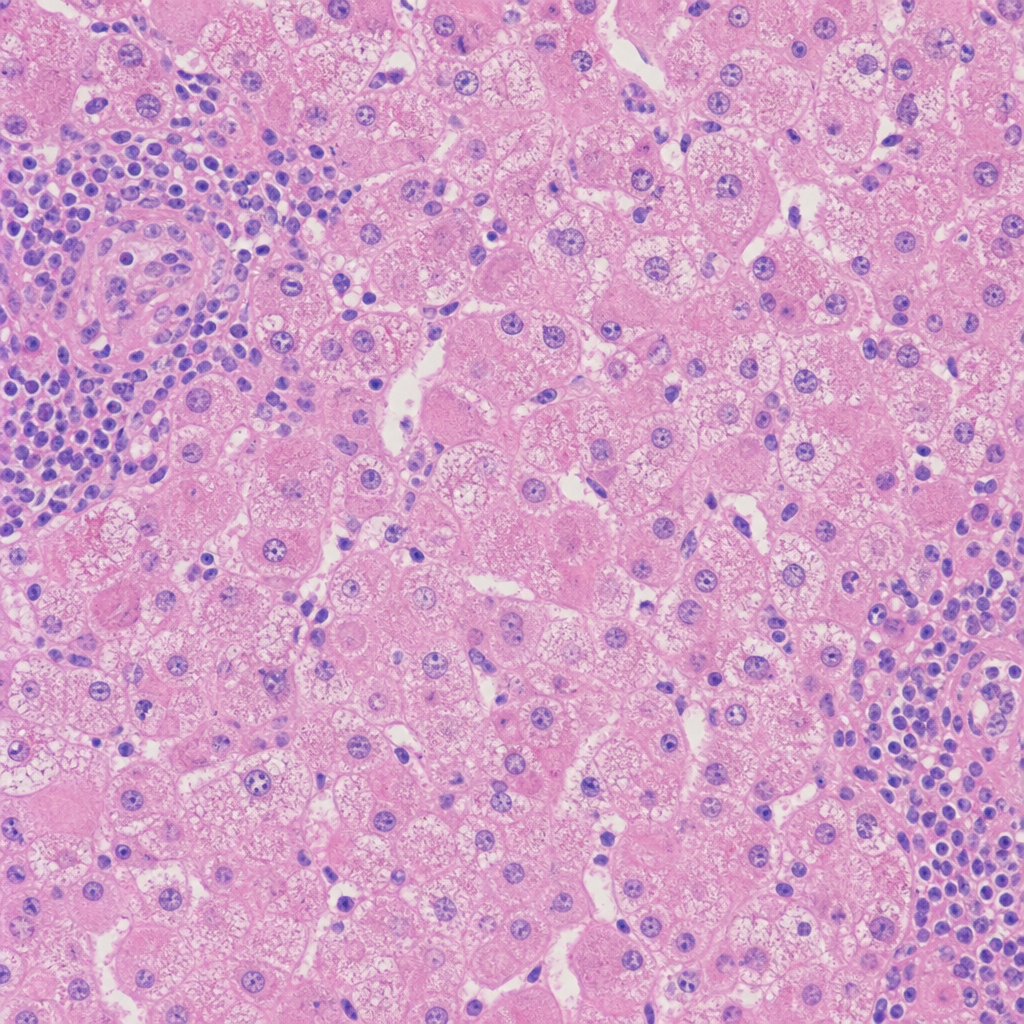
Which of the following is seen in obstructive jaundice?
Micronodular cirrhosis is seen in all of the following conditions EXCEPT?
What is the single most important indicator of the likelihood of rapid progression of hepatitis to liver cirrhosis?
Intrahepatic cholestasis is seen in which of the following conditions?
Which of the following conditions affect the intracanalicular hepatic apparatus?
Explanation: Alpha-fetoprotein (AFP) is a glycoprotein normally produced by the fetal yolk sac and liver. In adults, massive elevations (typically >400-500 ng/mL or >10 times the upper normal limit) are highly suggestive of specific malignancies, most notably **Hepatocellular Carcinoma (HCC)** and **Germ Cell Tumors (Yolk Sac Tumor)** [1]. **Why Option B is Correct:** While primary HCC is the classic cause of high AFP [1], **Metastatic carcinoma of the liver** (especially from the GI tract, pancreas, or lung) is a frequent clinical cause of significantly elevated AFP [2]. When the liver is extensively replaced by metastatic deposits, the surrounding regenerating hepatocytes or the tumor cells themselves (in certain foregut lineages) can lead to a marked rise in serum AFP levels, often exceeding 10 times the normal limit. **Analysis of Incorrect Options:** * **A. Seminoma:** This is a "pure" germ cell tumor. A key diagnostic feature is that **Seminomas do not produce AFP**. If AFP is elevated in a suspected seminoma, it indicates a mixed germ cell tumor component (specifically Yolk Sac elements). * **C. Cirrhosis of the liver:** Chronic liver inflammation and regeneration can cause AFP elevation, but it is usually **mild to moderate** (rarely exceeding 100-200 ng/mL) [1]. * **D. Oat cell tumor (Small Cell Lung Cancer):** This is a neuroendocrine tumor. While it may produce ectopic hormones (ACTH, ADH), it is not a recognized cause of significant AFP elevation. **High-Yield Pearls for NEET-PG:** * **AFP Cut-off:** In the context of a liver mass, an AFP >400 ng/mL is considered diagnostic for HCC in many clinical guidelines [1]. * **Yolk Sac Tumor (Endodermal Sinus Tumor):** AFP is the definitive marker; look for **Schiller-Duval bodies** on histology. * **Neural Tube Defects:** Elevated AFP in maternal screening suggests spina bifida or anencephaly, while **low AFP** is associated with Down Syndrome (Trisomy 21). **References:** [1] Cross SS. Underwood's Pathology: A Clinical Approach. 6th ed. Common Clinical Problems From Liver And Biliary System Disease, pp. 399-400. [2] Cross SS. Underwood's Pathology: A Clinical Approach. 6th ed. Common Clinical Problems From Liver And Biliary System Disease, pp. 398-399.
Explanation: **Explanation:** **Congestive splenomegaly** occurs due to chronic venous congestion, which leads to the pooling of blood within the splenic sinusoids [2]. **1. Why Cirrhosis is the correct answer:** Cirrhosis is the most common cause of **portal hypertension**, which is the primary driver of congestive splenomegaly [1]. In cirrhosis, the architectural distortion of the liver increases resistance to portal blood flow [1]. This backward pressure is transmitted to the splenic vein, causing chronic engorgement of the spleen. Over time, this leads to physical enlargement (splenomegaly) and histological changes such as fibrosis of the red pulp and the formation of **Gandy-Gamna bodies** (siderofibrotic nodules) [2]. **2. Analysis of Incorrect Options:** * **Chronic congestive cardiac failure (A):** While this causes systemic venous congestion and "nutmeg liver," the pressure is often dissipated through the systemic circulation. It causes splenic congestion, but it is a much less common cause of significant splenomegaly compared to portal hypertension [1]. * **Hepatic vein occlusion (C):** Also known as Budd-Chiari syndrome. While it causes portal hypertension and splenomegaly, it is a rare clinical entity compared to the high prevalence of cirrhosis [1]. * **Stenosis of the splenic vein (D):** This causes "left-sided" or "extrahepatic" portal hypertension [1]. While it leads to localized splenomegaly, it is an uncommon cause [2]. **High-Yield Pearls for NEET-PG:** * **Gandy-Gamna bodies:** These are small, brown nodules in the spleen containing iron and calcium deposits, characteristic of chronic congestive splenomegaly. * **Hypersplenism:** Enlargement often leads to increased sequestration of blood cells, resulting in anemia, leukopenia, or thrombocytopenia [2]. * **Weight:** A normal spleen weighs ~150g; in massive congestive splenomegaly (e.g., due to cirrhosis), it can exceed 1000g [2]. **References:** [1] Kumar V, Abbas AK, et al.. Robbins and Cotran Pathologic Basis of Disease. 9th ed. Liver and Gallbladder, pp. 834-835. [2] Kumar V, Abbas AK, et al.. Robbins and Cotran Pathologic Basis of Disease. 9th ed. Diseases of White Blood Cells, Lymph Nodes, Spleen, and Thymus, pp. 632-634.
Explanation: **Explanation:** The correct answer is **Hepatitis D**. **Anti-LKM (Liver-Kidney Microsomal) antibodies** are a group of autoantibodies directed against cytochrome P450 enzymes. While they are classic markers for Autoimmune Hepatitis (AIH), different subtypes are associated with specific viral triggers: 1. **Anti-LKM3:** These antibodies are directed against **UDP-glucuronosyltransferase (UGT1)**. They are highly specific for patients with **Chronic Hepatitis D (Delta)**, occurring in approximately 10-15% of cases. While they can rarely appear in Type 2 AIH, their presence in a clinical vignette involving viral co-infection points toward Hepatitis D. 2. **Anti-LKM1:** Directed against **CYP2D6** [1]. This is the hallmark of **Autoimmune Hepatitis Type 2** (typically seen in children/young females) and is also found in some cases of Hepatitis C [1]. 3. **Anti-LKM2:** Directed against **CYP2C9**. These were historically associated with **Ticrynafen-induced hepatitis** (drug-induced). **Why other options are incorrect:** * **Hepatitis B:** Does not typically induce LKM antibodies; diagnosis relies on HBsAg and HBV DNA. * **Hepatitis C:** Frequently associated with **Anti-LKM1**, not LKM3 [1]. * **Autoimmune Hepatitis:** AIH Type 2 is defined by **Anti-LKM1** [1]. While LKM3 can rarely be present, it is the classic "high-yield" association for Hepatitis D in competitive exams. **High-Yield Clinical Pearls for NEET-PG:** * **AIH Type 1:** Most common; positive for **ANA** and/or **Anti-Smooth Muscle Antibody (ASMA)** [1]. * **AIH Type 2:** Positive for **Anti-LKM1** and **Anti-LC1** (Liver Cytosol antigen) [1]. * **Soluble Liver Antigen (SLA/LP):** The most specific antibody for Autoimmune Hepatitis [1]. * **Hepatitis D requirement:** Remember that HDV is a defective RNA virus that requires the HBsAg coat from HBV for transmission. **References:** [1] Kumar V, Abbas AK, et al.. Robbins and Cotran Pathologic Basis of Disease. 9th ed. Liver and Gallbladder, pp. 845-846.
Explanation: **Explanation:** The question asks to identify the condition **not** primarily associated with abnormal copper metabolism. **1. Why Cardiomyopathy is the Correct Answer:** Cardiomyopathy is not a classic manifestation of copper metabolism disorders [3]. While severe copper deficiency (rare) can theoretically affect cardiac enzymes, it is not a recognized pathogenic feature of the major copper-related diseases like Wilson’s or Menkes. In Wilson disease, copper accumulates primarily in the **liver, brain (basal ganglia), and cornea (Kayser-Fleischer rings)**, but clinically significant cardiomyopathy is not a standard diagnostic or pathological feature [1], [2]. **2. Analysis of Incorrect Options:** * **Wilson Disease (Hepatolenticular Degeneration):** Caused by a mutation in the **ATP7B gene** on chromosome 13 [2]. This leads to impaired biliary excretion of copper and failure to incorporate copper into ceruloplasmin, resulting in toxic copper accumulation in the liver and brain [1]. * **Menkes Disease (Kinky Hair Syndrome):** Caused by a mutation in the **ATP7A gene**. This results in impaired intestinal absorption of copper, leading to severe systemic **copper deficiency**. Clinical features include "steely" or "kinky" hair, growth failure, and neurological degeneration. * **Indian Childhood Cirrhosis (ICC):** A progressive form of liver cirrhosis in children historically linked to high dietary copper intake (e.g., milk boiled in copper or brass vessels). It is characterized by massive copper deposition in hepatocytes and prominent Mallory-Denk bodies. **High-Yield Clinical Pearls for NEET-PG:** * **ATP7B = Wilson** (B for Biliary excretion/Basal ganglia). * **ATP7A = Menkes** (A for Absorption/Absence of copper). * **Diagnostic Triad for Wilson:** Low serum ceruloplasmin, increased urinary copper excretion, and Kayser-Fleischer (KF) rings on slit-lamp exam [1]. * **Stains for Copper:** Rhodanine stain (most specific), Orcein stain (stains copper-binding protein), and Timm’s silver sulfide stain. **References:** [1] Cross SS. Underwood's Pathology: A Clinical Approach. 6th ed. Common Clinical Problems From Liver And Biliary System Disease, pp. 394-395. [2] Kumar V, Abbas AK, et al.. Robbins and Cotran Pathologic Basis of Disease. 9th ed. Liver and Gallbladder, pp. 855-856. [3] Kumar V, Abbas AK, et al.. Robbins and Cotran Pathologic Basis of Disease. 9th ed. The Heart, pp. 572-576.
Explanation: **Explanation:** **Alpha-fetoprotein (AFP)** is a glycoprotein normally synthesized by the fetal liver and yolk sac. In adult pathology, it serves as a crucial tumor marker for Hepatocellular Carcinoma (HCC) and certain Germ Cell Tumors (Yolk Sac Tumor). [1] **Why Option B is correct:** Hepatoblastoma is the most common primary liver tumor in children. [2] AFP is an exceptionally sensitive marker for this condition, being elevated in approximately **90% of hepatoblastoma cases**. It is used not only for diagnosis but also for monitoring treatment response and detecting recurrence. **Analysis of Incorrect Options:** * **Options A & C (100%):** In clinical medicine, tumor markers are rarely 100% sensitivity. A small subset of tumors (especially poorly differentiated ones or specific variants) may not secrete the marker. [1] * **Option D (90% of HCC):** While AFP is the classic marker for HCC, its sensitivity is lower than in hepatoblastoma. AFP is elevated in roughly **60-70% of HCC cases**. Notably, the **Fibrolamellar variant of HCC** is characteristically associated with **normal AFP levels**, a high-yield distinction for exams. **High-Yield Clinical Pearls for NEET-PG:** * **AFP Cut-off:** In the context of a liver mass, an AFP level **>400 ng/mL** is highly suggestive of HCC. * **Other causes of elevated AFP:** Cirrhosis, chronic hepatitis (usually lower levels), and non-seminomatous germ cell tumors (Yolk Sac Tumor). * **Hepatoblastoma Association:** Often associated with **FAP (Familial Adenomatous Polyposis)** and Beckwith-Wiedemann syndrome. [2] * **Diagnosis of HCC:** Unlike most cancers, HCC can often be diagnosed based on imaging (triphasic CT/MRI showing arterial enhancement and venous washout) and elevated AFP without a mandatory biopsy. [1] **References:** [1] Cross SS. Underwood's Pathology: A Clinical Approach. 6th ed. Common Clinical Problems From Liver And Biliary System Disease, pp. 399-400. [2] Kumar V, Abbas AK, et al.. Robbins and Cotran Pathologic Basis of Disease. 9th ed. Liver and Gallbladder, pp. 875-876.
Explanation: In obstructive jaundice (post-hepatic jaundice), the physical blockage of the biliary tree (e.g., gallstones, carcinoma of the pancreas head) prevents conjugated bilirubin and bile salts from entering the duodenum [1]. **1. Why "Excess of bile salts in the urine" is correct:** When bile flow is obstructed, bile salts and conjugated bilirubin regurgitate from the hepatocytes into the systemic circulation [1]. Since bile salts are water-soluble, they are filtered by the kidneys and excreted in the urine. This is clinically detected using **Hay’s Test**. The presence of bile salts in urine is a hallmark of obstructive jaundice. **2. Why the other options are incorrect:** * **Option A (Urobilinogen):** Urobilinogen is formed by the action of intestinal bacteria on bilirubin. In complete obstruction, no bilirubin reaches the gut; therefore, no urobilinogen is produced [1]. Consequently, urobilinogen will be **absent** in the urine, and stools will appear "clay-colored" [1]. * **Option B (Unconjugated bilirubin):** Obstructive jaundice primarily causes an elevation of **conjugated (direct) bilirubin**, as the liver can still conjugate bilirubin but cannot excrete it [1]. Excess unconjugated bilirubin is characteristic of hemolytic (pre-hepatic) jaundice [1]. **NEET-PG High-Yield Pearls:** * **Van den Bergh Reaction:** Obstructive jaundice gives a **Direct Positive** reaction. * **Enzymatic Marker:** **Alkaline Phosphatase (ALP)** and GGT are significantly elevated in obstructive patterns, whereas ALT/AST are markers for hepatocellular injury [2]. * **Pruritus:** The accumulation of bile salts in the skin is the primary cause of intense itching in these patients [2]. * **Vitamin Deficiency:** Lack of bile salts in the gut leads to malabsorption of fat-soluble vitamins (A, D, E, **K**), often resulting in a prolonged Prothrombin Time (PT). **References:** [1] Cross SS. Underwood's Pathology: A Clinical Approach. 6th ed. Common Clinical Problems From Liver And Biliary System Disease, pp. 380-386. [2] Cross SS. Underwood's Pathology: A Clinical Approach. 6th ed. Common Clinical Problems From Liver And Biliary System Disease, pp. 392-393.
Explanation: **Explanation:** Cirrhosis is classified morphologically based on the size of the regenerating nodules: **Micronodular** (<3 mm) and **Macronodular** (>3 mm) [1]. **Why Wilson’s Disease is the Correct Answer:** In **Wilson’s disease**, the initial presentation may be micronodular; however, as the disease progresses and chronic inflammation persists, it characteristically evolves into **Macronodular cirrhosis** [3]. For NEET-PG purposes, Wilson’s disease, Chronic Hepatitis (B or C), and Alpha-1 antitrypsin deficiency are the classic prototypes for macronodular patterns. **Analysis of Incorrect Options:** * **Alcoholic Cirrhosis:** This is the classic prototype of **Micronodular cirrhosis** (Laennec’s cirrhosis) [1]. Chronic alcohol intake causes uniform nutritional insult, leading to small, regular nodules. (Note: It may convert to macronodular if the patient stops drinking [2]). * **Budd-Chiari Syndrome:** Chronic venous outflow obstruction leads to congestive hepatopathy. The resulting "cardiac cirrhosis" or "congestive cirrhosis" typically presents with a micronodular pattern due to the diffuse nature of the centrilobular necrosis. * **Indian Childhood Cirrhosis (ICC):** Characterized by excessive copper deposition and marked "creeping" fibrosis, ICC classically presents with a micronodular pattern and is often associated with the absence of distinct regenerative nodules in early stages. **High-Yield Clinical Pearls for NEET-PG:** * **Micronodular (<3mm):** Alcohol (most common), Malnutrition, Hemochromatosis (early), Biliary cirrhosis, Budd-Chiari. * **Macronodular (>3mm):** Post-necrotic (Viral hepatitis B/C), Wilson’s disease, Alpha-1 antitrypsin deficiency. * **Mixed Cirrhosis:** Often seen in long-standing cases where micronodules coalesce into larger ones. * **Key Histology:** Regardless of nodule size, the hallmark of cirrhosis is the presence of **regenerating nodules** completely surrounded by **fibrous bands** (Type I and III collagen) [1]. **References:** [1] Cross SS. Underwood's Pathology: A Clinical Approach. 6th ed. Common Clinical Problems From Liver And Biliary System Disease, pp. 395-396. [2] Kumar V, Abbas AK, et al.. Robbins and Cotran Pathologic Basis of Disease. 9th ed. Liver and Gallbladder, p. 834. [3] Cross SS. Underwood's Pathology: A Clinical Approach. 6th ed. Common Clinical Problems From Liver And Biliary System Disease, pp. 394-395.
Explanation: **Explanation:** The progression of chronic hepatitis to cirrhosis is not uniform; it is primarily determined by the **underlying etiology (Option B)** [2]. While histological features describe the current state of damage, the cause of the hepatitis dictates the natural history, the speed of fibrogenesis, and the response to treatment [2], [3]. For example, Hepatitis C (HCV) typically progresses slowly over decades, whereas Hepatitis B (HBV) with Delta virus co-infection or certain autoimmune hepatitides can progress much more rapidly to cirrhosis [2]. **Analysis of Incorrect Options:** * **Option A (Serological findings):** These are useful for diagnosis (e.g., HBsAg, Anti-HCV) and monitoring viral load, but they do not independently predict the rate of fibrosis as accurately as the specific disease entity itself [3]. * **Option C (Bridging necrosis):** This is a histological marker of **severity** and activity (grade) [3]. While bridging necrosis (portal-portal or portal-central) indicates a higher risk of progression compared to simple interface hepatitis, it is a snapshot in time rather than the primary driver of the disease's trajectory [4]. * **Option D (Mallory hyaline):** These are eosinophilic cytoplasmic inclusions (cytokeratin intermediate filaments) most commonly associated with Alcoholic Liver Disease [1]. While they indicate hepatocyte injury, they are not a prognostic indicator for the speed of progression to cirrhosis. **NEET-PG High-Yield Pearls:** * **Definition of Cirrhosis:** Characterized by three features: Bridging fibrosis, parenchymal nodules (regenerative), and disruption of the entire liver architecture. * **Ground Glass Hepatocytes:** Characteristic of Chronic Hepatitis B (due to HBsAg accumulation in the ER) [3], [5]. * **Lymphoid Aggregates:** Often seen in the portal tracts of Hepatitis C [3], [5]. * **Steatosis:** A common feature of Hepatitis C (Genotype 3) and Non-Alcoholic Fatty Liver Disease (NAFLD) [1], [3]. **References:** [1] Cross SS. Underwood's Pathology: A Clinical Approach. 6th ed. Common Clinical Problems From Liver And Biliary System Disease, pp. 389-390. [2] Cross SS. Underwood's Pathology: A Clinical Approach. 6th ed. Common Clinical Problems From Liver And Biliary System Disease, pp. 391-392. [3] Kumar V, Abbas AK, et al.. Robbins and Cotran Pathologic Basis of Disease. 9th ed. Liver and Gallbladder, pp. 843-844. [4] Kumar V, Abbas AK, et al.. Robbins and Cotran Pathologic Basis of Disease. 9th ed. Liver and Gallbladder, p. 844. [5] Kumar V, Abbas AK, et al.. Robbins and Cotran Pathologic Basis of Disease. 9th ed. Liver and Gallbladder, pp. 844-845.
Explanation: **Explanation:** **1. Why Galactosemia is Correct:** Galactosemia (specifically classic galactosemia due to GALT deficiency) is a metabolic disorder where the accumulation of galactose-1-phosphate leads to direct hepatotoxicity. In the liver, this manifests as early-onset **intrahepatic cholestasis**, severe steatosis, and bile ductular proliferation. The toxic metabolites cause damage to the hepatocytes and canalicular membranes, leading to the accumulation of bile within the liver parenchyma (cholestasis) and eventually progressing to cirrhosis if untreated. **2. Analysis of Incorrect Options:** * **Hypercalcemia:** While hypercalcemia can cause systemic issues like renal stones or pancreatitis, it does not directly cause intrahepatic cholestasis. * **Hemochromatosis:** This is a disorder of iron overload. While it leads to micronodular cirrhosis and significantly increases the risk of Hepatocellular Carcinoma (HCC), the primary pathology is the deposition of hemosiderin in hepatocytes, not cholestasis. * **Cystic Fibrosis:** While CF can cause liver disease, it typically results in **biliary cirrhosis** [1] due to inspissated (thickened) secretions within the *extrahepatic* or large intrahepatic bile ducts, rather than primary intrahepatic canalicular cholestasis. **3. NEET-PG High-Yield Pearls:** * **Histology of Galactosemia:** Look for "diffuse fatty change" and "pseudo-acinar" transformation of hepatocytes. * **Clinical Presentation:** An infant presenting with jaundice, hepatomegaly, and **cataracts** after starting milk feeds. * **Diagnostic Clue:** Presence of non-glucose reducing substances in the urine. * **Other causes of Intrahepatic Cholestasis:** Viral hepatitis, Alcoholic liver disease, Primary Biliary Cholangitis (PBC), and drugs (e.g., Anabolic steroids, OCPs) [2]. **References:** [1] Kumar V, Abbas AK, et al.. Robbins and Cotran Pathologic Basis of Disease. 9th ed. Liver and Gallbladder, p. 862. [2] Kumar V, Abbas AK, et al.. Robbins and Cotran Pathologic Basis of Disease. 9th ed. Liver and Gallbladder, pp. 860-862.
Explanation: The liver's biliary system is divided into **intrahepatic** (intracanalicular/ductular) and **extrahepatic** components [1]. This question tests the anatomical localization of biliary pathologies. ### **Explanation of the Correct Answer** **A. Alagille Syndrome:** This is an autosomal dominant disorder (typically involving the *JAG1* gene) characterized by **ductal paucity**. It specifically affects the **intracanalicular hepatic apparatus** (the small intrahepatic bile ducts). Histologically, there is a marked decrease in the ratio of bile ducts to portal tracts. Clinical features include cholestasis, butterfly vertebrae, and peripheral pulmonary artery stenosis. ### **Explanation of Incorrect Options** * **B. Cystic Fibrosis:** While it can cause biliary cirrhosis, the primary pathology involves the plugging of **medium-sized intrahepatic bile ducts** with inspissated secretions, rather than a primary defect of the intracanalicular apparatus. * **C. Congenital Hepatic Fibrosis:** This belongs to the spectrum of **fibropolycystic diseases** (ductal plate malformations). It involves the persistence of embryonic biliary structures and the formation of broad bands of fibrous tissue in the portal tracts, rather than the canalicular system. * **D. Caroli Disease:** This is characterized by segmental **cystic dilatation of the larger intrahepatic bile ducts**. ### **High-Yield NEET-PG Pearls** * **Ductal Paucity:** Defined as a bile duct-to-portal tract ratio of **<0.4** (Normal is 0.9–1.8). * **Alagille Syndrome Triad:** Cholestasis + Butterfly vertebrae + Posterior embryotoxon (eye finding). * **Fibropolycystic Diseases:** Often associated with **PKHD1** gene mutations and Polycystic Kidney Disease (ARPKD). * **Caroli Syndrome:** Caroli disease + Congenital hepatic fibrosis. **References:** [1] Kumar V, Abbas AK, et al.. Robbins and Cotran Pathologic Basis of Disease. 9th ed. Liver and Gallbladder, pp. 860-862.
Jaundice and Cholestasis
Practice Questions
Viral Hepatitis
Practice Questions
Alcoholic and Non-alcoholic Fatty Liver Disease
Practice Questions
Drug and Toxin Induced Liver Injury
Practice Questions
Cirrhosis and Its Complications
Practice Questions
Metabolic Liver Diseases
Practice Questions
Liver Tumors
Practice Questions
Gallbladder and Biliary Tract Diseases
Practice Questions
Congenital Liver Diseases
Practice Questions
Liver Transplantation Pathology
Practice Questions
Get full access to all questions, explanations, and performance tracking.
Start For Free