
Liver and Biliary Pathology — MCQs

On this page
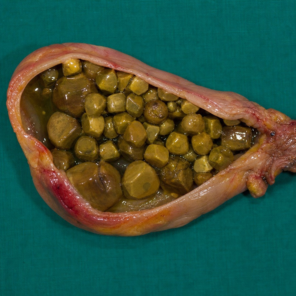
The image shows stones found in the gall bladder. Which of the following conditions is associated with their formation?
OVAL cells are seen in the stem cells of which organ?
A 42-year-old man experiences malaise and increasing icterus for 2 weeks. Physical examination shows jaundice, but there are no other significant findings. Serologic test results are positive for IgM anti-HAV and negative for anti-HCV, HBsAg, and IgM anti-HBc. Which of the following outcomes is most likely to occur in this man?
Bile infarct is related to which of the following conditions?
Ductopenia is a characteristic finding in which of the following conditions?
A 36-year-old woman has become increasingly icteric for 1 month. She has had several bouts of colicky, midabdominal pain for 3 years. On physical examination, she has generalized jaundice with scleral icterus. Her BMI is 32. There is tenderness in the right upper quadrant, and the liver span is normal. A liver biopsy is obtained, and microscopic examination shows bile duct proliferation and intracanalicular bile stasis, but no inflammation or hepatocyte necrosis. The level of which of the following is most likely to be increased in the patient's serum?
What is the most common paraneoplastic syndrome associated with hepatocellular carcinoma?
Mallory denk bodies are seen in all of the following conditions except?
Granulomatous hepatitis is not caused by which of the following?
"Onion-skin" fibrosis of bile duct is seen in?
Practice by Chapter
Jaundice and Cholestasis
Practice Questions
Viral Hepatitis
Practice Questions
Alcoholic and Non-alcoholic Fatty Liver Disease
Practice Questions
Drug and Toxin Induced Liver Injury
Practice Questions
Cirrhosis and Its Complications
Practice Questions
Metabolic Liver Diseases
Practice Questions
Liver Tumors
Practice Questions
Gallbladder and Biliary Tract Diseases
Practice Questions
Congenital Liver Diseases
Practice Questions
Liver Transplantation Pathology
Practice Questions
Want unlimited practice?
Get full access to all questions, explanations, and performance tracking.
Scan to download app