Which mediator of acute inflammation is not derived from cells?
A 35-year-old man has had increasing dyspnea for the past 24 hours. A chest radiograph shows large, bilateral pleural effusions. Thoracentesis yields 500 mL of slightly cloudy yellow fluid from the right pleural cavity. Cytologic examination of the fluid shows many neutrophils, but no lymphocytes or RBCs. Which of the following mechanisms contributes most to the pleural fluid accumulation?
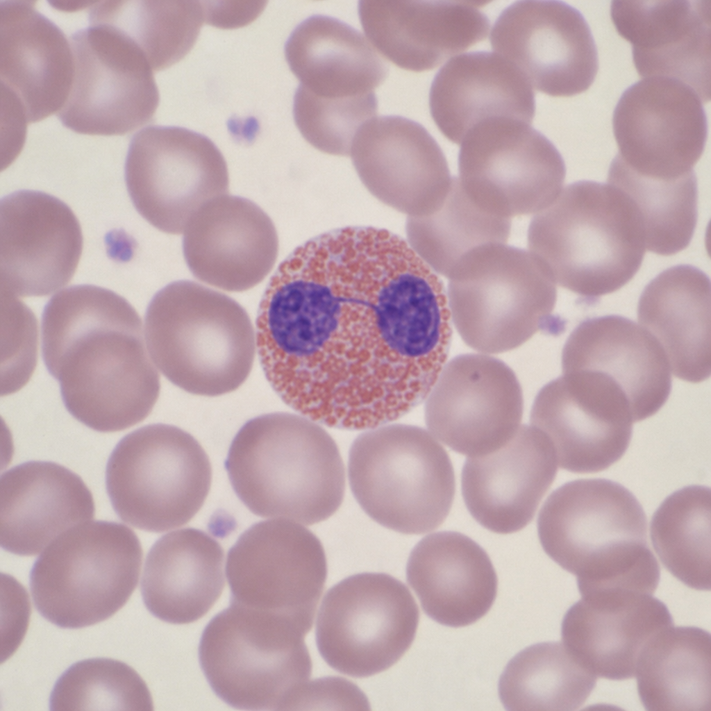
Which of the following has the least ability to regenerate?
Epithelioid granuloma consists mainly of which type of cells?
Which of the following substances most likely has the greatest affinity for neutrophils?
Which of the following acts as an acute phase reactant?
A 25-year-old machinist is injured by a metal sliver in his left hand. Over the next few days, the wounded area becomes reddened, tender, swollen, and feels warm to the touch. Redness at the site of injury in this patient is caused primarily by which of the following mechanisms?
Lipoxins synthesized from arachidonic acid act by?
What is the most pathognomonic sign of irreversible cell injury?
Leukotrienes cause all of the following effects, EXCEPT:
Explanation: **Explanation:** The mediators of inflammation are broadly classified into two categories based on their source: **Cell-derived** and **Plasma-derived** [3]. **1. Why Kinins is the correct answer:** Kinins (such as Bradykinin) are **plasma-derived mediators** [1]. They are produced primarily in the liver and circulate in the blood as inactive precursors (kininogens). They are activated through a proteolytic cascade, specifically triggered by the activation of **Hageman Factor (Factor XII)** [1]. Bradykinin is responsible for increasing vascular permeability, smooth muscle contraction, and is a potent mediator of **pain** [2]. **2. Why the other options are incorrect:** * **Histamine (Option A):** This is a preformed cell-derived mediator stored in the granules of **mast cells**, basophils, and platelets [3][4]. It is one of the first mediators released during the immediate transient response of inflammation. * **Leukotrienes (Option B):** These are newly synthesized cell-derived mediators produced from **arachidonic acid** via the lipoxygenase pathway in leukocytes [4]. * **Cytokines (Option D):** These are proteins produced mainly by activated **macrophages, lymphocytes, and endothelial cells** (e.g., TNF, IL-1) that modulate the functions of other cell types [5]. **High-Yield Clinical Pearls for NEET-PG:** * **Plasma-derived mediators** include the Complement system, the Kinin system, and the Coagulation/Fibrinolytic system [1]. All are synthesized in the **liver**. * **Factor XII (Hageman Factor)** is the "master switch" that links the kinin system, the clotting cascade, and the fibrinolytic system [1]. * **Pain mediators:** The two primary mediators responsible for pain in acute inflammation are **Bradykinin** and **Prostaglandins (PGE2)** [2]. * **Triple Response of Lewis:** Mediated primarily by Histamine. **References:** [1] Cross SS. Underwood's Pathology: A Clinical Approach. 6th ed. (Basic Pathology) introduces the student to key general principles of pathology, both as a medical science and as a clinical activity with a vital role in patient care. Part 2 (Disease Mechanisms) provides fundamental knowledge about the cellular and molecular processes involved in diseases, providing the rationale for their treatment. Part 3 (Systematic Pathology) deals in detail with specific diseases, with emphasis on the clinically important aspects., pp. 189-190. [2] Kumar V, Abbas AK, et al.. Robbins and Cotran Pathologic Basis of Disease. 9th ed. Inflammation and Repair, pp. 100-101. [3] Kumar V, Abbas AK, et al.. Robbins and Cotran Pathologic Basis of Disease. 9th ed. Inflammation and Repair, pp. 93-94. [4] Kumar V, Abbas AK, et al.. Robbins and Cotran Pathologic Basis of Disease. 9th ed. Inflammation and Repair, pp. 94-95. [5] Kumar V, Abbas AK, et al.. Robbins and Cotran Pathologic Basis of Disease. 9th ed. Inflammation and Repair, p. 97.
Explanation: ### Explanation The clinical presentation of acute dyspnea, bilateral pleural effusions, and a fluid analysis showing **many neutrophils** indicates an **acute inflammatory exudate** [1], [3]. **1. Why Endothelial Contraction is Correct:** In acute inflammation, the primary mechanism for increased vascular permeability (leading to exudate formation) is the formation of **endothelial gaps** in post-capillary venules [1], [3]. This is most commonly caused by **endothelial cell contraction** [1]. * **Mechanism:** Chemical mediators like histamine, bradykinin, and leukotrienes bind to specific receptors, triggering an intracellular signaling cascade that leads to the contraction of endothelial cells [1], [4]. * **Result:** This creates interendothelial gaps, allowing protein-rich fluid and leukocytes (neutrophils) to escape into the extravascular space (pleural cavity) [1], [2]. This is an immediate transient response (lasting 15–30 minutes) [1]. **2. Why Other Options are Incorrect:** * **A. Arteriolar vasoconstriction:** This is a transient, neurogenic reflex occurring immediately after injury, but it is followed by **vasodilation** [3]. Vasodilation increases hydrostatic pressure, contributing to transudate, but it does not create the gaps necessary for a neutrophil-rich exudate. * **C. Inhibition of platelet adherence:** Platelet adherence is crucial for hemostasis. Inhibiting it would lead to bleeding (hemorrhagic effusion), not a neutrophilic exudate. * **D. Lymphatic obstruction:** This typically results in **Chylous effusion** (milky fluid high in triglycerides) or a protein-poor transudate, not an acute inflammatory collection of neutrophils. **3. NEET-PG High-Yield Pearls:** * **Most common mechanism** of vascular leakage in acute inflammation: Endothelial cell contraction [1]. * **Site of leakage:** Exclusively in the **post-capillary venules** (not capillaries or arterioles) [1], [2]. * **Direct Endothelial Injury:** Occurs in severe injuries (burns, bacterial toxins) and affects all vessel levels (venules, capillaries, and arterioles) [1]. It results in "immediate sustained leakage." * **Transudate vs. Exudate:** Exudates (seen here) have high protein content (>3g/dL) and high LDH, usually due to increased permeability [3]. Transudates are due to increased hydrostatic or decreased oncotic pressure [3]. **References:** [1] Cross SS. Underwood's Pathology: A Clinical Approach. 6th ed. (Basic Pathology) introduces the student to key general principles of pathology, both as a medical science and as a clinical activity with a vital role in patient care. Part 2 (Disease Mechanisms) provides fundamental knowledge about the cellular and molecular processes involved in diseases, providing the rationale for their treatment. Part 3 (Systematic Pathology) deals in detail with specific diseases, with emphasis on the clinically important aspects., pp. 187-188. [2] Cross SS. Underwood's Pathology: A Clinical Approach. 6th ed. (Basic Pathology) introduces the student to key general principles of pathology, both as a medical science and as a clinical activity with a vital role in patient care. Part 2 (Disease Mechanisms) provides fundamental knowledge about the cellular and molecular processes involved in diseases, providing the rationale for their treatment. Part 3 (Systematic Pathology) deals in detail with specific diseases, with emphasis on the clinically important aspects., pp. 188-189. [3] Kumar V, Abbas AK, et al.. Robbins and Cotran Pathologic Basis of Disease. 9th ed. Inflammation and Repair, pp. 84-85. [4] Kumar V, Abbas AK, et al.. Robbins and Cotran Pathologic Basis of Disease. 9th ed. Inflammation and Repair, p. 101.
Explanation: ### Explanation The ability of a tissue to regenerate depends on its classification based on the cell cycle: **Labile** (continuously dividing), **Stable** (quiescent but can divide), or **Permanent** (non-dividing). **1. Why Striated Muscle is Correct:** Striated muscles (Skeletal and Cardiac) are classified as **Permanent tissues**. These cells have left the cell cycle and cannot undergo mitotic division in postnatal life. When skeletal muscle is damaged, it has very limited regenerative capacity via satellite cells; however, extensive injury typically results in **fibrosis (scarring)** rather than functional regeneration [1]. Cardiac muscle has virtually no regenerative capacity, making striated muscle the least able to regenerate among the choices. **2. Why the Other Options are Incorrect:** * **Liver (Stable Tissue):** The liver has a massive regenerative capacity. Hepatocytes are normally quiescent (G0 phase) but can re-enter the cell cycle rapidly following injury or partial hepatectomy. * **Bone (Stable/Labile properties):** Bone possesses excellent regenerative potential. Through the coordinated action of osteoblasts and progenitor cells, bone can heal via the formation of a callus, restoring original structural integrity. * **Collagen (Repair Component):** Collagen is a structural protein produced by fibroblasts. In the context of "regeneration," fibroblasts are highly active during wound healing and can proliferate extensively to fill defects. **3. NEET-PG High-Yield Pearls:** * **Permanent Cells:** Neurons, Cardiac myocytes, and Skeletal muscle. (Mnemonic: **N**ever **C**an **S**plit) [2]. * **Stable Cells:** Liver, Kidney, Pancreas, Vascular Endothelium, and Smooth Muscle. * **Labile Cells:** Surface epithelia (Skin, GI tract), Bone marrow, and Splenic lymphoid tissue. * **Key Concept:** If the **extracellular matrix (ECM)** is destroyed, even stable tissues cannot regenerate perfectly and will heal by scarring [1]. **References:** [1] Cross SS. Underwood's Pathology: A Clinical Approach. 6th ed. (Basic Pathology) introduces the student to key general principles of pathology, both as a medical science and as a clinical activity with a vital role in patient care. Part 2 (Disease Mechanisms) provides fundamental knowledge about the cellular and molecular processes involved in diseases, providing the rationale for their treatment. Part 3 (Systematic Pathology) deals in detail with specific diseases, with emphasis on the clinically important aspects., pp. 107-108. [2] Cross SS. Underwood's Pathology: A Clinical Approach. 6th ed. (Basic Pathology) introduces the student to key general principles of pathology, both as a medical science and as a clinical activity with a vital role in patient care. Part 2 (Disease Mechanisms) provides fundamental knowledge about the cellular and molecular processes involved in diseases, providing the rationale for their treatment. Part 3 (Systematic Pathology) deals in detail with specific diseases, with emphasis on the clinically important aspects., pp. 109-110.
Explanation: **Explanation:** The hallmark of granulomatous inflammation is the **epithelioid granuloma**. The correct answer is **Macrophages** because epithelioid cells are actually activated, modified macrophages that have undergone specific morphological changes [1]. **Why Macrophages are the correct answer:** Under the influence of cytokines (primarily **IFN-γ** secreted by Th1 cells), macrophages transform into epithelioid cells [1]. These cells are characterized by abundant, pale pink granular cytoplasm, indistinct cell boundaries (making them look like epithelial cells), and elongated, slipper-shaped nuclei [1]. Their primary function shifts from phagocytosis to **secretion**, playing a key role in sequestering indigestible antigens [2]. **Analysis of Incorrect Options:** * **A & B (B cells and T cells):** While lymphocytes (especially T cells) are present in the "lymphocytic rim" surrounding a granuloma, they are not the primary constituent of the epithelioid core itself [1]. * **C (Monocytes):** Monocytes are the precursors found in the blood. Once they migrate into the tissue and become activated in a chronic inflammatory setting, they differentiate into macrophages and then epithelioid cells [2]. **NEET-PG High-Yield Pearls:** * **Definition:** A granuloma is a focal collection of activated macrophages (epithelioid cells), surrounded by a rim of lymphocytes and often containing multinucleated giant cells [1], [2]. * **Giant Cells:** Formed by the fusion of epithelioid cells (e.g., **Langhans giant cells** in Tuberculosis, **Foreign body giant cells**) [1]. * **Key Cytokine:** **IFN-γ** is the most important cytokine for granuloma formation [1]. * **Caseating vs. Non-caseating:** Caseous necrosis (cheese-like) is characteristic of Tuberculosis, whereas non-caseating granulomas are seen in Sarcoidosis, Crohn’s disease, and Leprosy [2]. **References:** [1] Kumar V, Abbas AK, et al.. Robbins and Cotran Pathologic Basis of Disease. 9th ed. Inflammation and Repair, p. 109. [2] Cross SS. Underwood's Pathology: A Clinical Approach. 6th ed. (Basic Pathology) introduces the student to key general principles of pathology, both as a medical science and as a clinical activity with a vital role in patient care. Part 2 (Disease Mechanisms) provides fundamental knowledge about the cellular and molecular processes involved in diseases, providing the rationale for their treatment. Part 3 (Systematic Pathology) deals in detail with specific diseases, with emphasis on the clinically important aspects., pp. 198-200.
Explanation: **Explanation:** The question tests the concept of **Chemotaxis**, the process by which leukocytes are attracted to a site of injury by a chemical gradient. [1] **1. Why C5a is correct:** **C5a** is one of the most potent **chemotactic factors** (chemoattractants) for neutrophils. [1] It is a byproduct of the complement cascade (alternative, classical, or lectin pathways). C5a binds to specific G-protein coupled receptors on the neutrophil surface, triggering actin polymerization and directional movement toward the highest concentration of the substance. [2] Other major chemoattractants include Bacterial products (N-formylmethionine), Cytokines (IL-8), and Leukotriene B4 (LTB4). **2. Why the other options are incorrect:** * **Fucosyl transferase:** This is an enzyme required for the synthesis of Sialyl-Lewis X (the ligand on neutrophils). It is not a chemoattractant. * **P-selectin:** This is an adhesion molecule expressed on the surface of **endothelial cells** (stored in Weibel-Palade bodies). It mediates the "Rolling" phase of leukocyte extravasation, not chemotaxis. [2] * **TNF-α:** While TNF-α is a major pro-inflammatory cytokine that activates endothelial cells to express adhesion molecules (E-selectin, ICAM-1), it does not act as a direct chemoattractant with high affinity for neutrophils compared to C5a. **High-Yield Clinical Pearls for NEET-PG:** * **Mnemonic for Chemoattractants:** "B-C-I-L" (**B**acterial products, **C**5a, **I**L-8, **L**TB4). * **C5a** is also known as an **Anaphylatoxin** because it induces mast cell degranulation, increasing vascular permeability. [1] * **Deficiency of Fucosyl transferase** leads to **Leukocyte Adhesion Deficiency Type 2 (LAD-2)**, characterized by the absence of Sialyl-Lewis X and impaired rolling. [3] **References:** [1] Cross SS. Underwood's Pathology: A Clinical Approach. 6th ed. (Basic Pathology) introduces the student to key general principles of pathology, both as a medical science and as a clinical activity with a vital role in patient care. Part 2 (Disease Mechanisms) provides fundamental knowledge about the cellular and molecular processes involved in diseases, providing the rationale for their treatment. Part 3 (Systematic Pathology) deals in detail with specific diseases, with emphasis on the clinically important aspects., pp. 163-164. [2] Kumar V, Abbas AK, et al.. Robbins and Cotran Pathologic Basis of Disease. 9th ed. Migration in the tissues toward a chemotactic stimulus, pp. 86-87. [3] Kumar V, Abbas AK, et al.. Robbins and Cotran Pathologic Basis of Disease. 9th ed. Inflammation and Repair, pp. 87-89.
Explanation: **Explanation:** **Acute Phase Reactants (APRs)** are proteins whose plasma concentrations increase (positive APRs) or decrease (negative APRs) by at least 25% in response to inflammation, infection, or tissue injury. This process is primarily mediated by cytokines like **IL-6, IL-1, and TNF-α** [1]. **Why Serum Ferritin is Correct:** Serum ferritin is a **positive acute phase reactant**. During inflammation, the body sequesters iron to withhold it from invading pathogens (which require iron for metabolism) [1]. Ferritin levels rise to store this iron safely within cells and the reticuloendothelial system [1]. Consequently, in inflammatory states, ferritin levels can be falsely elevated, masking a true iron deficiency. **Analysis of Incorrect Options:** * **Serum Iron:** This is a **negative acute phase reactant**. During inflammation, levels decrease because iron is moved into storage (ferritin) and its absorption is blocked by increased **hepcidin** [1]. * **Total Iron-Binding Capacity (TIBC):** TIBC is an indirect measure of **Transferrin**. Transferrin is a **negative acute phase reactant**; its synthesis in the liver decreases during inflammation, leading to a low TIBC [1]. * **Percentage of Saturation:** Since both serum iron and TIBC decrease during inflammation (though disproportionately), the saturation percentage is not considered a primary acute phase reactant. **High-Yield Clinical Pearls for NEET-PG:** * **Positive APRs:** Ferritin, C-Reactive Protein (CRP), Fibrinogen, Haptoglobin, Hepcidin, and Serum Amyloid A (SAA). * **Negative APRs:** Albumin, Transferrin, and Transthyretin (Pre-albumin). * **ESR vs. CRP:** CRP is a more sensitive and rapid indicator of acute inflammation than ESR. * **Anemia of Chronic Disease:** Characterized by **High Ferritin** and **Low TIBC**, distinguishing it from Iron Deficiency Anemia (Low Ferritin, High TIBC) [1]. **References:** [1] Kumar V, Abbas AK, et al.. Robbins and Cotran Pathologic Basis of Disease. 9th ed. Red Blood Cell and Bleeding Disorders, pp. 657-662.
Explanation: ### Explanation The clinical presentation describes the classic **cardinal signs of acute inflammation** (*rubor, calor, tumor, dolor*) [1]. The redness (**rubor**) and warmth (**calor**) observed in this patient are primarily due to **vasodilation** of the precapillary arterioles [3]. **1. Why Vasodilation is Correct:** Following a brief, transient period of vasoconstriction, chemical mediators such as **histamine**, nitric oxide, and prostaglandins (PGI2) act on vascular smooth muscle [2]. This leads to vasodilation, which increases blood flow to the injured area (active hyperemia) [1]. This increased volume of packed red blood cells in the local microvasculature manifests clinically as redness and increased local temperature [3]. **2. Why the Other Options are Incorrect:** * **Hemorrhage:** This refers to the escape of blood from ruptured vessels into the extravascular space (e.g., a bruise or hematoma). While trauma occurred, the diffuse redness of inflammation is due to intravascular congestion, not active bleeding. * **Hemostasis:** This is the process of blood clotting to stop bleeding. It involves vasoconstriction and platelet plug formation, which would not cause the spreading redness and warmth characteristic of inflammation. * **Neutrophil Margination:** This is a cellular event where leukocytes move to the periphery of the vessel wall due to slowed blood flow (stasis). While it is a crucial step in acute inflammation, it does not contribute to the macroscopic appearance of redness. **3. NEET-PG High-Yield Pearls:** * **Cardinal Signs:** Rubor (Redness), Calor (Heat), Tumor (Swelling), Dolor (Pain), and *Functio Laesa* (Loss of function) [1]. * **Sequence of Vascular Events:** Transient vasoconstriction → Vasodilation (Rubor/Calor) → Increased permeability (Tumor) → Stasis → Leukocyte emigration [3]. * **Mediator of Vasodilation:** Histamine is the primary mediator responsible for the initial phase of vasodilation and increased vascular permeability [2]. * **Starling’s Law:** Swelling (edema) in inflammation is caused by increased **hydrostatic pressure** (due to vasodilation) and increased **interstitial osmotic pressure** (due to protein-rich exudate leaking out) [3]. **References:** [1] Cross SS. Underwood's Pathology: A Clinical Approach. 6th ed. (Basic Pathology) introduces the student to key general principles of pathology, both as a medical science and as a clinical activity with a vital role in patient care. Part 2 (Disease Mechanisms) provides fundamental knowledge about the cellular and molecular processes involved in diseases, providing the rationale for their treatment. Part 3 (Systematic Pathology) deals in detail with specific diseases, with emphasis on the clinically important aspects., pp. 185-186. [2] Kumar V, Abbas AK, et al.. Robbins and Cotran Pathologic Basis of Disease. 9th ed. Inflammation and Repair, p. 101. [3] Kumar V, Abbas AK, et al.. Robbins and Cotran Pathologic Basis of Disease. 9th ed. Inflammation and Repair, pp. 84-85.
Explanation: ### Explanation **Concept Overview:** Lipoxins (LXA4 and LXB4) are endogenous, anti-inflammatory lipid mediators derived from the **Arachidonic Acid (AA)** pathway via the **12-lipoxygenase** enzyme [1]. Unlike most AA metabolites (like leukotrienes and prostaglandins) which promote inflammation, lipoxins serve as "stop signals" that facilitate the **resolution phase** of inflammation. **Why Option A is Correct:** Lipoxins act as potent inhibitors of inflammation. Their primary mechanism involves: * **Inhibiting Neutrophil Recruitment:** They decrease the chemotaxis and adhesion of neutrophils to the endothelium. * **Promoting Resolution:** While they inhibit neutrophils, they actually *stimulate* the recruitment of non-phlogistic macrophages to clear apoptotic debris (efferocytosis). * **Antagonizing Leukotrienes:** They compete for receptors with Leukotriene B4 (LTB4), thereby halting the pro-inflammatory cycle. **Why Other Options are Incorrect:** * **Option B:** This describes the action of **Leukotriene B4 (LTB4)** and **C5a**, which are potent chemoattractants that recruit leukocytes to the site of injury [2]. * **Option C:** Lipoxins generally promote **vasodilation** (often by stimulating NO release). Vasoconstriction is a hallmark of **Thromboxane A2 (TXA2)** and **Leukotrienes C4, D4, and E4** [1]. * **Option D:** Increased vascular permeability is mediated by **Histamine**, **Bradykinin**, and **Leukotrienes (LTC4, LTD4, LTE4)** [2]. Lipoxins help stabilize the vascular barrier during the resolution phase. **NEET-PG High-Yield Pearls:** * **Dual Cell Requirement:** Lipoxin synthesis often requires two cell types (e.g., Neutrophils produce intermediates which are converted to Lipoxins by Platelets)—a process known as **transcellular biosynthesis**. * **Aspirin-Triggered Lipoxins (ATLs):** Aspirin acetylates COX-2, diverting the pathway to produce "epi-lipoxins," which contribute to aspirin’s anti-inflammatory profile. * **Resolution Mediators:** Along with Lipoxins, keep an eye on **Resolvins, Protectins, and Maresins** (derived from Omega-3 fatty acids), which also promote the resolution of inflammation. **References:** [1] Kumar V, Abbas AK, et al.. Robbins and Cotran Pathologic Basis of Disease. 9th ed. Inflammation and Repair, pp. 95-96. [2] Kumar V, Abbas AK, et al.. Robbins and Cotran Pathologic Basis of Disease. 9th ed. Inflammation and Repair, p. 101.
Explanation: ### Explanation The hallmark of **irreversible cell injury** is the inability to reverse mitochondrial dysfunction and profound disturbances in membrane function [1]. **Why Option A is Correct:** **Amorphous densities (flocculent densities)** in the mitochondrial matrix are the most pathognomonic sign of irreversible injury [1]. These densities represent large aggregates of denatured proteins and lipids. While mitochondrial swelling can occur in reversible injury, the presence of these large, opaque, flocculent densities indicates that the damage has crossed the "point of no return," leading to a permanent loss of ATP production and subsequent cell death (necrosis) [1], [3]. **Why Other Options are Incorrect:** * **B. Swelling of the cell membrane:** This is one of the earliest signs of **reversible injury**. It occurs due to the failure of energy-dependent ion pumps (like the Na+/K+ ATPase), leading to an influx of water (hydropic change) [1], [2]. * **C. Ribosomes detached from ER:** This occurs during **reversible injury** when the cell swells and the endoplasmic reticulum dilates [1]. It leads to a decrease in protein synthesis but can be corrected if oxygenation is restored. * **D. Clumping of nuclear chromatin:** This is an early change seen in **reversible injury**, primarily caused by a decrease in intracellular pH (lactic acidosis) due to anaerobic glycolysis [1], [4]. **NEET-PG High-Yield Pearls:** * **Irreversible Injury Landmarks:** 1. Severe mitochondrial damage (Amorphous densities); 2. Extensive plasma membrane damage; 3. Lysosomal rupture leading to autolysis [1], [2]. * **Nuclear Changes in Necrosis:** Pyknosis (shrinkage/condensation) $\rightarrow$ Karyorrhexis (fragmentation) $\rightarrow$ Karyolysis (dissolution) [4]. * **First sign of cell injury (Light Microscopy):** Cellular swelling (Cloudy swelling) [4]. * **First sign of cell injury (Electron Microscopy):** Mitochondrial swelling and ER dilation [1]. **References:** [1] Kumar V, Abbas AK, et al.. Robbins and Cotran Pathologic Basis of Disease. 9th ed. Cellular Responses to Stress and Toxic Insults: Adaptation, Injury, and Death, pp. 53-55. [2] Kumar V, Abbas AK, et al.. Robbins and Cotran Pathologic Basis of Disease. 9th ed. Cellular Responses to Stress and Toxic Insults: Adaptation, Injury, and Death, pp. 60-61. [3] Cross SS. Underwood's Pathology: A Clinical Approach. 6th ed. (Basic Pathology) introduces the student to key general principles of pathology, both as a medical science and as a clinical activity with a vital role in patient care. Part 2 (Disease Mechanisms) provides fundamental knowledge about the cellular and molecular processes involved in diseases, providing the rationale for their treatment. Part 3 (Systematic Pathology) deals in detail with specific diseases, with emphasis on the clinically important aspects., pp. 102-103. [4] Kumar V, Abbas AK, et al.. Robbins and Cotran Pathologic Basis of Disease. 9th ed. Cellular Responses to Stress and Toxic Insults: Adaptation, Injury, and Death, p. 53.
Explanation: ### Explanation The correct answer is **A. Vasodilation**. **1. Why Vasodilation is the correct answer:** Leukotrienes (LTs) are inflammatory mediators derived from arachidonic acid via the **lipoxygenase pathway**. While they are potent mediators of inflammation, they do **not** cause vasodilation [1]. In fact, the cysteinyl-containing leukotrienes (LTC4, LTD4, and LTE4) are potent **vasoconstrictors**. Vasodilation in the arachidonic acid cascade is primarily mediated by **Prostaglandins** (specifically PGI2, PGE1, PGE2, and PGD2) [1]. **2. Analysis of other options:** * **B. Vasoconstriction:** This is a classic effect of LTC4, LTD4, and LTE4 [1]. They cause intense contraction of smooth muscles, leading to both vasoconstriction and bronchoconstriction. * **C. Increased vascular permeability:** LTC4, LTD4, and LTE4 significantly increase vascular permeability by causing endothelial cell contraction, leading to the formation of intercellular gaps in post-capillary venules [1]. They are much more potent than histamine in this regard. **3. NEET-PG High-Yield Pearls:** * **LTB4:** A major chemotactic agent [1]. Remember the mnemonic: *"LTB4 helps Neutrophils reach the floor"* (Chemotaxis and Adhesion). * **LTC4, LTD4, LTE4:** Known as the "Slow Reacting Substance of Anaphylaxis" (SRS-A). They cause bronchospasm and increased vascular permeability. * **Lipoxins (LXA4, LXB4):** Unlike leukotrienes, these are **anti-inflammatory** and inhibit neutrophil chemotaxis and adhesion. * **Aspirin-induced Asthma:** Caused by the inhibition of the cyclooxygenase (COX) pathway, which shunts arachidonic acid metabolism toward the lipoxygenase pathway, leading to an overproduction of bronchoconstricting leukotrienes [2]. **References:** [1] Kumar V, Abbas AK, et al.. Robbins and Cotran Pathologic Basis of Disease. 9th ed. Inflammation and Repair, pp. 95-96. [2] Kumar V, Abbas AK, et al.. Robbins and Cotran Pathologic Basis of Disease. 9th ed. Inflammation and Repair, pp. 96-97.
Acute Inflammation: Vascular Events
Practice Questions
Acute Inflammation: Cellular Events
Practice Questions
Chemical Mediators of Inflammation
Practice Questions
Chronic Inflammation
Practice Questions
Granulomatous Inflammation
Practice Questions
Systemic Effects of Inflammation
Practice Questions
Wound Healing
Practice Questions
Tissue Regeneration
Practice Questions
Fibrosis and Repair
Practice Questions
Resolution of Inflammation
Practice Questions
Get full access to all questions, explanations, and performance tracking.
Start For Free