A 45-year old patient presented with fever, night sweats and weight loss. On X-ray, a mass was seen in apical lobe. On histopathology, caseous necrosis was present. What is the name of underlying process?
Which of the following is characteristically associated with sarcoidosis?
Granuloma is a pathological feature of all, except which of the following?
Which is NOT a feature of chronic inflammation?
The principal cell in a granuloma is
What is the primary reason for the development of granuloma formation in tertiary syphilis?
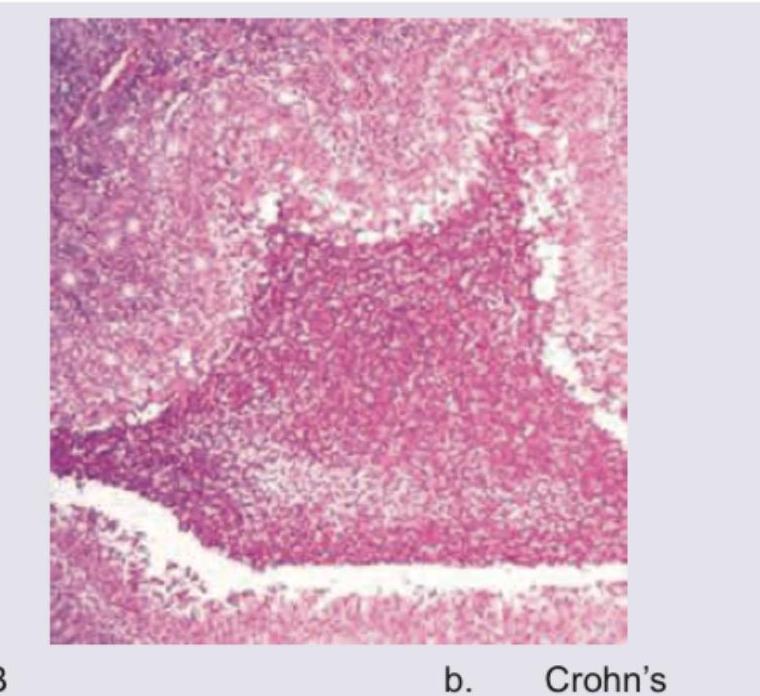
The image shows a histological section of intestinal tissue with a granuloma. What is the most likely diagnosis?

Which of the following does not cause granulomatous inflammation?
Which statement about macrophages is incorrect?
Most important for diapedesis is
Explanation: ***Granulomatous inflammation (Type IV hypersensitivity)*** - The presence of **caseous necrosis** on histopathology, combined with symptoms like fever, night sweats, and weight loss, and an apical lung mass, is highly characteristic of **tuberculosis** [1]. - Tuberculosis is a classic example of a **type IV hypersensitivity reaction** involving **epithelioid macrophages** (modified macrophages), **lymphocytes**, and **Langhans giant cells** forming **granulomas** with central **caseous necrosis** [2], [3]. - This represents **granulomatous inflammation**, which is the hallmark histopathological finding in tuberculosis [1]. *Decreased supply of growth factor* - This typically leads to **atrophy** or **apoptosis**, which are distinct from the inflammatory and necrotic process described. - It does not explain the characteristic histological finding of **caseous necrosis** or the systemic symptoms. *Acute decrease in blood supply* - An acute decrease in blood supply (ischemia or infarction) would lead to **coagulative necrosis** in most tissues, not the **caseous necrosis** seen in this presentation. - While infarction can cause tissue death, the specific histological and clinical picture points away from simple ischemia. *Enzymatic degeneration* - **Enzymatic degeneration**, particularly from pancreatic enzymes, is typical of **fat necrosis** (e.g., in acute pancreatitis), where fat cells are broken down. - This process does not produce the characteristic **caseous necrosis** and granulomatous inflammation seen in the patient's lung. **References:** [1] Kumar V, Abbas AK, et al.. Robbins and Cotran Pathologic Basis of Disease. 9th ed. Infectious Diseases, pp. 383-384. [2] Kumar V, Abbas AK, et al.. Robbins and Cotran Pathologic Basis of Disease. 9th ed. Inflammation and Repair, p. 109. [3] Kumar V, Abbas AK, et al.. Robbins and Cotran Pathologic Basis of Disease. 9th ed. Infectious Diseases, p. 360.
Explanation: ***Grossly bullous appearance*** - This feature is **not typically seen** in sarcoidosis; instead, sarcoidosis usually presents with various tissue infiltrations [1]. - Characteristically, sarcoidosis features **granulomas**, not a bullous skin manifestation [1]. *Asteroid bodies* - Asteroid bodies are **star-shaped inclusions** found within the non-caseating granulomas seen in sarcoidosis. - Their presence supports the diagnosis, indicating **typical histological findings** of the disease. *Non caseating granulomas* - Sarcoidosis is definitively characterized by the presence of **non-caseating granulomas**, which are key diagnostic features [1]. - These granulomas differentiate sarcoidosis from other granulomatous diseases, like tuberculosis [2]. *Schaumann bodies* - Schaumann bodies are **calcium-laden** structures found within the granulomas in sarcoidosis, further supporting the diagnosis. - The presence of these bodies alongside non-caseating granulomas is a classic histopathological finding in sarcoidosis. **References:** [1] Kumar V, Abbas AK, et al.. Robbins and Cotran Pathologic Basis of Disease. 9th ed. The Lung, pp. 700-701. [2] Kumar V, Abbas AK, et al.. Robbins and Cotran Pathologic Basis of Disease. 9th ed. Inflammation and Repair, p. 109.
Explanation: ***Microscopic polyangitis*** - This condition is associated with **necrotizing vasculitis** without significant **granulomatous inflammation** [1]. - Primarily affects small vessels and typically features **pauci-immune** glomerulonephritis [1]. *Giant cell arteritis* - Characterized by **granulomatous inflammation** in the temporal arteries, leading to headaches and vision loss [2]. - It often shows **multinucleated giant cells** in biopsy specimens, confirming the diagnosis [2]. *Churg strauss disease* - Also known as **Eosinophilic Granulomatosis with Polyangiitis**, it features **granulomas** and affects small to medium vessels. - Typically presents with asthma, nasal polyps, and significant **eosinophilia**. *Wegner's granulomatosis* - Now referred to as **Granulomatosis with Polyangiitis**, it prominently features **necrotizing granulomas** in the respiratory tract and kidneys [3]. - Associated with **c-ANCA** (anti-neutrophil cytoplasmic antibodies), confirming its granulomatous nature [3]. **References:** [1] Kumar V, Abbas AK, et al.. Robbins and Cotran Pathologic Basis of Disease. 9th ed. Diseases of Infancy and Childhood, pp. 518-519. [2] Kumar V, Abbas AK, et al.. Robbins and Cotran Pathologic Basis of Disease. 9th ed. Diseases of Infancy and Childhood, pp. 516-517. [3] Kumar V, Abbas AK, et al.. Robbins and Cotran Pathologic Basis of Disease. 9th ed. Diseases of Infancy and Childhood, pp. 519-520.
Explanation: ***Neutrophil predominance*** - **Neutrophil predominance** is characteristic of **acute inflammation**, where these cells are among the first responders to injury or infection [1]. - In chronic inflammation, neutrophils are typically present in much smaller numbers compared to mononuclear cells, or their presence indicates an acute exacerbation [3]. *Mononuclear cells* - **Mononuclear cells**, such as **macrophages**, **lymphocytes**, and **plasma cells**, are the hallmark cellular infiltrates of chronic inflammation [1]. - These cells are responsible for sustained immune responses, tissue destruction, and repair processes [2]. *Fibrosis* - **Fibrosis**, or the deposition of **collagen** by fibroblasts, is a common outcome of chronic inflammation as the body attempts to repair ongoing tissue damage [3]. - It leads to **scarring** and functional impairment of affected organs [4]. *Granulation tissue* - **Granulation tissue** is an early phase of **tissue repair** during chronic inflammation, characterized by the proliferation of **fibroblasts** and new **blood vessels (angiogenesis)** [5]. - It represents the body's effort to fill tissue defects and prepare for eventual fibrous scar formation [5]. **References:** [1] Cross SS. Underwood's Pathology: A Clinical Approach. 6th ed. (Basic Pathology) introduces the student to key general principles of pathology, both as a medical science and as a clinical activity with a vital role in patient care. Part 2 (Disease Mechanisms) provides fundamental knowledge about the cellular and molecular processes involved in diseases, providing the rationale for their treatment. Part 3 (Systematic Pathology) deals in detail with specific diseases, with emphasis on the clinically important aspects., pp. 195-196. [2] Kumar V, Abbas AK, et al.. Robbins and Cotran Pathologic Basis of Disease. 9th ed. Inflammation and Repair, pp. 107-109. [3] Cross SS. Underwood's Pathology: A Clinical Approach. 6th ed. (Basic Pathology) introduces the student to key general principles of pathology, both as a medical science and as a clinical activity with a vital role in patient care. Part 2 (Disease Mechanisms) provides fundamental knowledge about the cellular and molecular processes involved in diseases, providing the rationale for their treatment. Part 3 (Systematic Pathology) deals in detail with specific diseases, with emphasis on the clinically important aspects., pp. 196-197. [4] Cross SS. Underwood's Pathology: A Clinical Approach. 6th ed. (Basic Pathology) introduces the student to key general principles of pathology, both as a medical science and as a clinical activity with a vital role in patient care. Part 2 (Disease Mechanisms) provides fundamental knowledge about the cellular and molecular processes involved in diseases, providing the rationale for their treatment. Part 3 (Systematic Pathology) deals in detail with specific diseases, with emphasis on the clinically important aspects., pp. 200-202. [5] Cross SS. Underwood's Pathology: A Clinical Approach. 6th ed. (Basic Pathology) introduces the student to key general principles of pathology, both as a medical science and as a clinical activity with a vital role in patient care. Part 2 (Disease Mechanisms) provides fundamental knowledge about the cellular and molecular processes involved in diseases, providing the rationale for their treatment. Part 3 (Systematic Pathology) deals in detail with specific diseases, with emphasis on the clinically important aspects., pp. 194-195.
Explanation: ***Histiocyte*** - Histiocytes are the principal **macrophages** in a granuloma, playing a key role in the **immune response** [1][2]. - They are responsible for phagocytosis and the formation of **epithelioid cells**, which are typical of granulomatous inflammation [1]. *Giant cell* - While giant cells are present in granulomas, they are formed from the **fusion of macrophages** and are not the principal cell type [1]. - Their presence signifies a chronic inflammatory response but does not define the granuloma itself. *Langhans* - Langhans giant cells are a specific type of giant cell that may be found in granulomas, particularly in conditions like **tuberculosis** [1]. - They are characterized by a **multi-nucleated** appearance but are not the principal cell in granuloma formation [1]. *Lymphocyte* - Lymphocytes are part of the **adaptive immune response** and may be present in granulomas, but they are not the main cell type [1][3]. - Their role is more of a supportive function, typically acting later in the inflammatory response rather than in granuloma formation. **References:** [1] Kumar V, Abbas AK, et al.. Robbins and Cotran Pathologic Basis of Disease. 9th ed. Inflammation and Repair, p. 109. [2] Cross SS. Underwood's Pathology: A Clinical Approach. 6th ed. (Basic Pathology) introduces the student to key general principles of pathology, both as a medical science and as a clinical activity with a vital role in patient care. Part 2 (Disease Mechanisms) provides fundamental knowledge about the cellular and molecular processes involved in diseases, providing the rationale for their treatment. Part 3 (Systematic Pathology) deals in detail with specific diseases, with emphasis on the clinically important aspects., pp. 198-200. [3] Kumar V, Abbas AK, et al.. Robbins and Cotran Pathologic Basis of Disease. 9th ed. Infectious Diseases, pp. 360-362.
Explanation: ***Delayed hypersensitivity reaction to treponemal antigens*** - **Granuloma formation** in tertiary syphilis (gummas) is primarily an immune-mediated response [1]. - It represents a **Type IV delayed hypersensitivity reaction** to persistent **treponemal antigens**, leading to chronic inflammation and tissue destruction [2]. *Secondary bacterial infection of syphilitic lesions* - While secondary infections can occur in various skin lesions, they are **not the primary mechanism** for granuloma formation in tertiary syphilis. - Granulomas are a specific inflammatory response to the *Treponema pallidum* organism itself, not secondary bacterial pathogens. *Direct tissue invasion by spirochetes* - Although *Treponema pallidum* directly invades tissues during all stages of syphilis, the **sheer presence of spirochetes** is not the sole cause of the granulomatous reaction in tertiary syphilis [3]. - The few spirochetes present in tertiary lesions are insufficient to directly cause such extensive lesions; rather, the host's immune response to these persistent organisms is crucial. *Vasculitis leading to tissue ischemia* - **Obliterative endarteritis** (vasculitis of small vessels) is a common pathological feature of syphilis, contributing to tissue damage and necrosis. - However, it is an **associated pathological process**, not the primary immune mechanism driving the characteristic granuloma formation itself. **References:** [1] Cross SS. Underwood's Pathology: A Clinical Approach. 6th ed. (Basic Pathology) introduces the student to key general principles of pathology, both as a medical science and as a clinical activity with a vital role in patient care. Part 2 (Disease Mechanisms) provides fundamental knowledge about the cellular and molecular processes involved in diseases, providing the rationale for their treatment. Part 3 (Systematic Pathology) deals in detail with specific diseases, with emphasis on the clinically important aspects., pp. 173-174. [2] Kumar V, Abbas AK, et al.. Robbins and Cotran Pathologic Basis of Disease. 9th ed. Diseases of the Immune System, p. 218. [3] Kumar V, Abbas AK, et al.. Robbins and Cotran Pathologic Basis of Disease. 9th ed. Infectious Diseases, pp. 385-386.
Explanation: ***Crohn's*** - Crohn's disease is characterized by **non-caseating granulomas** in the intestinal wall, which are a key diagnostic feature [1][3]. - These granulomas are typically **non-necrotizing** and can be found in any part of the gastrointestinal tract from mouth to anus [1][2]. *TB* - Tuberculosis (TB) typically forms **caseating granulomas** with central necrosis, which is distinct from the non-caseating granulomas of Crohn's. - TB granulomas often contain **Langhans giant cells** [4] and are associated with a specific infectious etiology. *Cat scratch disease* - Cat scratch disease, caused by *Bartonella henselae*, presents with **stellate or suppurative granulomas** in lymph nodes. - These granulomas are characterized by a central area of **necrosis and neutrophils**, surrounded by epithelioid cells, differing from Crohn's. *Syphilis* - Syphilis, particularly in its tertiary stage, can form **gummas**, which are granulomatous lesions. - Gummas are characterized by **coagulative necrosis** and a chronic inflammatory infiltrate, but they are not typically found in the intestinal wall as a primary feature like in Crohn's. **References:** [1] Kumar V, Abbas AK, et al.. Robbins and Cotran Pathologic Basis of Disease. 9th ed. The Gastrointestinal Tract, pp. 806-807. [2] Cross SS. Underwood's Pathology: A Clinical Approach. 6th ed. Common Clinical Problems From Alimentary System Disease, pp. 365-366. [3] Cross SS. Underwood's Pathology: A Clinical Approach. 6th ed. Common Clinical Problems From Alimentary System Disease, pp. 366-367. [4] Kumar V, Abbas AK, et al.. Robbins and Cotran Pathologic Basis of Disease. 9th ed. Inflammation and Repair, p. 109.
Explanation: ***Pneumonia*** - **Pneumonia**, in its typical bacterial form, usually causes an **acute inflammatory response** with neutrophil infiltration in the alveoli [1], [2]. - While some atypical pneumonias can have chronic or granulomatous features, the term "pneumonia" alone generally refers to acute inflammation without granulomas. *Sarcoidosis* - **Sarcoidosis** is characterized by distinctive **non-caseating granulomas** in multiple organs, most commonly the lungs, lymph nodes, and skin [3], [4]. - The formation of these granulomas is a hallmark of the disease and is crucial for diagnosis [3]. *Tuberculosis* - **Tuberculosis** is classically characterized by the formation of **caseating granulomas** (tubercles) with central necrosis, surrounded by epithelioid macrophages and giant cells [2]. - The host immune response to *Mycobacterium tuberculosis* is primarily granulomatous, aiming to contain the infection. *Histoplasmosis* - **Histoplasmosis**, a fungal infection caused by *Histoplasma capsulatum*, often leads to the formation of **granulomas**, both caseating and non-caseating [2]. - The granulomatous response is an essential part of the host's defense mechanism against this intracellular pathogen. **References:** [1] Cross SS. Underwood's Pathology: A Clinical Approach. 6th ed. Common Clinical Problems From Respiratory Tract Disease, pp. 317-318. [2] Kumar V, Abbas AK, et al.. Robbins and Cotran Pathologic Basis of Disease. 9th ed. Infectious Diseases, p. 360. [3] Kumar V, Abbas AK, et al.. Robbins and Cotran Pathologic Basis of Disease. 9th ed. The Lung, pp. 700-701. [4] Cross SS. Underwood's Pathology: A Clinical Approach. 6th ed. (Basic Pathology) introduces the student to key general principles of pathology, both as a medical science and as a clinical activity with a vital role in patient care. Part 2 (Disease Mechanisms) provides fundamental knowledge about the cellular and molecular processes involved in diseases, providing the rationale for their treatment. Part 3 (Systematic Pathology) deals in detail with specific diseases, with emphasis on the clinically important aspects., pp. 198-200.
Explanation: ***M2 type involved in inflammation*** - The M2 macrophages are primarily associated with **anti-inflammatory responses** and tissue repair, not inflammation [1][2]. - They play a role in **wound healing** and **resolution of inflammation**, contrasting with the inflammatory role attributed in the statement [2][3]. *Phagocytic cells* - **Macrophages are indeed phagocytic**, meaning they ingest and eliminate pathogens and debris [1]. - This is a fundamental characteristic of macrophages, playing a crucial role in the **immune response**. *Activation by IFN-y* - **Interferon-gamma (IFN-y)** is known to activate macrophages, particularly enhancing their ability to kill intracellular pathogens [1][2]. - This activation is vital for macrophage's role in **cell-mediated immunity** [1]. *Major cells in chronic inflammation* - Macrophages are significant players in **chronic inflammation**, contributing to tissue remodeling and the persistent inflammatory state [1]. - They secrete various cytokines that maintain the pathological state seen in chronic inflammatory diseases. **References:** [1] Kumar V, Abbas AK, et al.. Robbins and Cotran Pathologic Basis of Disease. 9th ed. Inflammation and Repair, pp. 105-106. [2] Kumar V, Abbas AK, et al.. Robbins and Cotran Pathologic Basis of Disease. 9th ed. Inflammation and Repair, pp. 106-107. [3] Kumar V, Abbas AK, et al.. Robbins and Cotran Pathologic Basis of Disease. 9th ed. Inflammation and Repair, p. 115.
Explanation: ***Platelet Endothelial Cell Adhesion Molecule (PECAM)*** - PECAM plays a crucial role in the process of **diapedesis**, allowing white blood cells to pass through the endothelial barrier [1]. - It is specifically involved in **intercellular junctions**, facilitating the migration of leukocytes during **inflammation** [1]. *Selectins* - Selectins are important for **rolling** of leukocytes on the endothelium but do not directly mediate **diapedesis**. - They are crucial for initial attachment but do not promote the passage through the endothelial cell junctions. *Mucin like glycoprotein* - While mucin like glycoproteins can facilitate **cell adhesion**, they primarily contribute to the **rolling phase** rather than diapedesis itself. - They are not as directly involved in the **transmigration** across the endothelium as PECAM. *Integrins* - Integrins are involved in **firm adhesion** of leukocytes, but do not directly enable **diapedesis** across the endothelium. - They support the binding to the endothelium but play a lesser role compared to PECAM in the actual process of migration. **References:** [1] Kumar V, Abbas AK, et al.. Robbins and Cotran Pathologic Basis of Disease. 9th ed. Inflammation and Repair, pp. 87-89.
Get full access to all questions, explanations, and performance tracking.
Start For Free