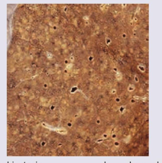
A 40-year-old man underwent lung transplantation. The resected lung specimen is shown below. Comment on the diagnosis.

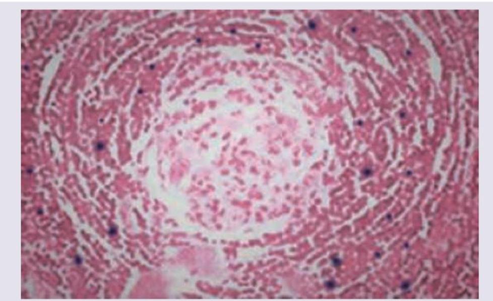
A patient presents with intermittent fever, no weight loss and enlarged retroperitoneal lymph nodes. Peripheral smear is normal. Gross sample and its histopathology slide is shown below. Comment on the diagnosis.

Which of the following tubes contain sodium fluoride as an anti-coagulant?
The earliest specific cystoscopic appearance of Bilharzial cystitis is :
"Collar-stud" abscess is seen in :
What is the primary reason for the development of granuloma formation in tertiary syphilis?
A 42 year-old female patient presents with cough, low-grade fever, and hemoptysis. Investigations reveal a cavitary lesion on her right lung apex, which on biopsy reveals caseous necrosis. The underlying pathophysiology is:
What is the mechanism by which Chlamydia trachomatis causes cellular damage in urethritis?
Cellulitis is characterized as:
Which of the following infectious diseases is the most likely cause of granuloma formation?
Explanation: ***Miliary TB*** - The image likely shows numerous small, uniform granulomas scattered throughout the lung parenchyma, characteristic of **miliary tuberculosis** [1]. - This widespread dissemination occurs when **Mycobacterium tuberculosis** spreads hematogenously, leading to a "millet seed" appearance on gross and microscopic examination [1]. *Bronchiectasis* - Bronchiectasis is characterized by **permanent dilation of bronchi** and bronchioles due to destruction of the muscle and elastic tissue [1]. - Grossly, it would show dilated, thickened airways often filled with mucus or pus, not diffuse small nodules. *Lung abscess* - A lung abscess is a **localized collection of pus** within the lung parenchyma, typically appearing as a single or a few large cavitary lesions [2]. - It would present as a well-defined cavity with necrotic debris, not numerous small, widely distributed lesions [2]. *Carcinoma lung* - Carcinoma of the lung typically presents as a **mass lesion**, nodule, or infiltrative growth, often with irregular borders [3]. - While multiple metastases can occur, the uniform, small, and widely distributed pattern seen in miliary TB is not typical for primary lung carcinoma [4]. **References:** [1] Cross SS. Underwood's Pathology: A Clinical Approach. 6th ed. Common Clinical Problems From Respiratory Tract Disease, pp. 320-321. [2] Kumar V, Abbas AK, et al.. Robbins and Cotran Pathologic Basis of Disease. 9th ed. The Lung, pp. 715-716. [3] Cross SS. Underwood's Pathology: A Clinical Approach. 6th ed. Common Clinical Problems From Respiratory Tract Disease, pp. 336-337. [4] Kumar V, Abbas AK, et al.. Robbins and Cotran Pathologic Basis of Disease. 9th ed. The Lung, pp. 723-724.
Explanation: ***Castleman disease*** - The image shows **"lollipop lesions"** or **"onion-skinning"** of follicles with **regressed germinal centers** and prominent mantle zones, characteristic of the hyaline-vascular variant of Castleman disease. - Clinical features like **intermittent fever** and **enlarged retroperitoneal lymph nodes** with **no weight loss** can be seen in Castleman disease, particularly the unicentric type. *Non-Hodgkin's lymphoma* - This would typically show effacement of the normal lymph node architecture by a **monotonous proliferation of atypical lymphocytes**. - While it can cause lymphadenopathy and fever, the specific histological features presented do not align with typical non-Hodgkin's lymphoma. *Angiolymphoid hyperplasia* - Characterized by **multiple vascular channels** lined by plump endothelial cells with **lymphoid infiltrates**. - It often presents as subcutaneous nodules, usually in the head and neck region, and lacks the follicular changes seen here. *IgG4 related disease* - Histologically, this disease is characterized by a **dense lymphoplasmacytic infiltrate rich in IgG4-positive plasma cells**, storiform fibrosis, and obliterative phlebitis. - While it can cause lymph node enlargement, the specific follicular changes in the image are not typical of IgG4-related disease.
Explanation: ***a. Gray-top tube*** - The **gray-top tube** contains **sodium fluoride** and **potassium oxalate** as anticoagulants. - Sodium fluoride acts as both an **anticoagulant** and an **antiglycolytic agent**, preserving glucose concentration by inhibiting glycolysis. - This tube is the **standard choice for glucose testing** in clinical laboratories. *b. Green-top tube* - The **green-top tubes** contain **heparin** (either sodium, lithium, or ammonium heparin) as an anticoagulant. - These tubes are used for various tests including **plasma chemistry** and some hormone assays, but not for glucose preservation with sodium fluoride. *c. Pink-top tube* - The **pink-top tube** generally contains **EDTA** (ethylenediaminetetraacetic acid) and is specifically designed for **blood bank** tests due to its special labeling and anticoagulant properties for cross-matching. - While it contains an anticoagulant, it is not sodium fluoride and is not used for glucose measurement. *d. Blue-top tube with yellow checkerboard pattern* - While some manufacturers may use different labeling systems, the **standard tube for sodium fluoride** is the gray-top tube, not the blue-top with checkerboard pattern. - Blue-top tubes typically contain **sodium citrate** and are used for coagulation studies (PT, PTT, INR).
Explanation: ***Sandy patches*** - **Sandy patches** are the **earliest and most characteristic** specific cystoscopic finding in Bilharzial cystitis (urinary schistosomiasis). [1] - They appear as fine, yellow-golden granules resembling grains of sand, visible through the bladder mucosa, representing **calcified *Schistosoma haematobium* eggs** deposited in the submucosa. - This pathognomonic finding typically appears in the trigone and posterior bladder wall and is the hallmark early sign during cystoscopy. *Pseudo tubercles* - **Pseudo tubercles (bilharzial tubercles)** represent a **later stage** of the disease, occurring after sandy patches. - They are organized granulomatous reactions (granulomas) that form around egg deposits within the bladder wall, appearing as small, whitish-yellow elevated lesions. [1] - While specific for schistosomiasis, they develop after the initial egg deposition phase marked by sandy patches. *Nodules* - **Nodules** represent more advanced inflammatory changes or can be associated with chronic schistosomiasis and potential neoplastic transformation. - They are non-specific and can occur in various bladder pathologies, not characteristic of early disease. *Ulcers* - **Ulcers** develop in advanced stages of Bilharzial cystitis due to chronic inflammation, tissue necrosis, or secondary bacterial infection. - They indicate significant mucosal damage and are not early manifestations of the disease. **References:** [1] Kumar V, Abbas AK, et al.. Robbins and Cotran Pathologic Basis of Disease. 9th ed. Infectious Diseases, pp. 405-406.
Explanation: ***Tuberculosis*** - A "**collar-stud**" abscess is a classic presentation of **tuberculous lymphadenitis**, particularly in the neck. - This type of abscess forms when pus from an infected deep lymph node erodes through the deep fascia but is contained by the superficial fascia, creating a dumbbell or "collar-stud" shape. *Lymphomatous degeneration* - **Lymphomatous degeneration** refers to the transformation of a benign lymphoid process into lymphoma. - While lymph nodes are involved, it typically presents as **lymphadenopathy** (enlargement of lymph nodes) and does not characteristically form an abscess with this specific morphology. *Pseudomonas infection* - **Pseudomonas infections** can cause abscesses, especially in immunocompromised individuals or associated with contaminated wounds or medical devices. - However, they do not specifically form a "**collar-stud**" abscess, which is a hallmark of tuberculous infection of lymph nodes. *Streptococcal infection* - **Streptococcal infections** frequently cause cellulitis, erysipelas, and various forms of abscesses, such as peritonsillar or skin abscesses. - While streptococci can cause **suppurative lymphadenitis**, they do not typically produce the distinctive "**collar-stud**" morphology seen in tuberculosis.
Explanation: ***Delayed hypersensitivity reaction to treponemal antigens*** - **Granuloma formation** in tertiary syphilis (gummas) is primarily an immune-mediated response [1]. - It represents a **Type IV delayed hypersensitivity reaction** to persistent **treponemal antigens**, leading to chronic inflammation and tissue destruction [2]. *Secondary bacterial infection of syphilitic lesions* - While secondary infections can occur in various skin lesions, they are **not the primary mechanism** for granuloma formation in tertiary syphilis. - Granulomas are a specific inflammatory response to the *Treponema pallidum* organism itself, not secondary bacterial pathogens. *Direct tissue invasion by spirochetes* - Although *Treponema pallidum* directly invades tissues during all stages of syphilis, the **sheer presence of spirochetes** is not the sole cause of the granulomatous reaction in tertiary syphilis [3]. - The few spirochetes present in tertiary lesions are insufficient to directly cause such extensive lesions; rather, the host's immune response to these persistent organisms is crucial. *Vasculitis leading to tissue ischemia* - **Obliterative endarteritis** (vasculitis of small vessels) is a common pathological feature of syphilis, contributing to tissue damage and necrosis. - However, it is an **associated pathological process**, not the primary immune mechanism driving the characteristic granuloma formation itself. **References:** [1] Cross SS. Underwood's Pathology: A Clinical Approach. 6th ed. (Basic Pathology) introduces the student to key general principles of pathology, both as a medical science and as a clinical activity with a vital role in patient care. Part 2 (Disease Mechanisms) provides fundamental knowledge about the cellular and molecular processes involved in diseases, providing the rationale for their treatment. Part 3 (Systematic Pathology) deals in detail with specific diseases, with emphasis on the clinically important aspects., pp. 173-174. [2] Kumar V, Abbas AK, et al.. Robbins and Cotran Pathologic Basis of Disease. 9th ed. Diseases of the Immune System, p. 218. [3] Kumar V, Abbas AK, et al.. Robbins and Cotran Pathologic Basis of Disease. 9th ed. Infectious Diseases, pp. 385-386.
Explanation: ***Type 4 hypersensitivity reaction*** - **Caseous necrosis**, characteristic of **tuberculosis**, is mediated by a **Type 4 hypersensitivity reaction** to the mycobacterial antigens [1], [3]. - This delayed-type hypersensitivity involves activated **macrophages** and **T-lymphocytes** forming **granulomas** with central caseous necrosis [2], [4]. *Sudden cut off of blood supply* - This mechanism typically leads to **coagulative necrosis** (e.g., in myocardial infarction), where the tissue architecture is preserved for some time. - **Infarction** due to loss of blood supply generally does not result in the distinct cheesy, crumbly appearance of caseous necrosis. *Enzyme degradation* - This mechanism describes **liquefactive necrosis**, where dead cells are digested by hydrolytic enzymes, resulting in a viscous fluid. - Liquefactive necrosis is common in bacterial infections and central nervous system infarcts, which is not consistent with the morphology of caseous necrosis. *Fibrinoid deposition* - **Fibrinoid necrosis** involves immune complex deposition in arterial walls, leading to leakage of plasma proteins and fibrin. - This type of necrosis is characteristic of **vasculitis** and immunologic reactions in vessels, not the widespread tissue destruction seen in caseous necrosis. **References:** [1] Cross SS. Underwood's Pathology: A Clinical Approach. 6th ed. (Basic Pathology) introduces the student to key general principles of pathology, both as a medical science and as a clinical activity with a vital role in patient care. Part 2 (Disease Mechanisms) provides fundamental knowledge about the cellular and molecular processes involved in diseases, providing the rationale for their treatment. Part 3 (Systematic Pathology) deals in detail with specific diseases, with emphasis on the clinically important aspects., pp. 173-174. [2] Kumar V, Abbas AK, et al.. Robbins and Cotran Pathologic Basis of Disease. 9th ed. Inflammation and Repair, p. 109. [3] Kumar V, Abbas AK, et al.. Robbins and Cotran Pathologic Basis of Disease. 9th ed. Infectious Diseases, p. 380. [4] Kumar V, Abbas AK, et al.. Robbins and Cotran Pathologic Basis of Disease. 9th ed. Infectious Diseases, pp. 383-384.
Explanation: ***Through intracellular replication leading to cell lysis*** - **Chlamydia trachomatis** is an **obligate intracellular bacterium** that replicates within the host cell's cytoplasm, forming **inclusion bodies** [1]. - As the bacteria multiply, they eventually overwhelm and lyse the host cell to release new elementary bodies, causing tissue damage and inflammation in the urethra [1]. *Through direct cytolysis* - This mechanism implies the bacterium directly ruptures the host cell without prior replication or involvement of a complex lifecycle. - **Chlamydia** does not directly lyse cells upon entry; instead, it has an **intracellular developmental cycle** that culminates in cell lysis [1]. *By toxin production* - While some bacteria cause damage through toxins, **Chlamydia trachomatis** is not known to produce potent exotoxins or endotoxins that are the primary drivers of cellular damage in urethritis. - Its pathogenicity is principally due to its **intracellular lifestyle** and subsequent cell destruction. *Through elementary body replication* - **Elementary bodies (EBs)** are the **infectious, metabolically inert** form of Chlamydia that attach to and enter host cells [1]. - EBs do not replicate; once inside the cell, they differentiate into **reticulate bodies (RBs)**, which are the metabolically active, replicative form [1]. **References:** [1] Cross SS. Underwood's Pathology: A Clinical Approach. 6th ed. Common Clinical Problems From Diseases Of The Urinary And Male Genital Tracts, pp. 507-508.
Explanation: ***Nonsuppurative and invasive*** - Cellulitis is considered **nonsuppurative** as it typically lacks macroscopic pus formation, distinguishing it from abscesses. - It is **invasive** because it involves the dermal and subcutaneous tissues, spreading through fascial planes. *Suppurative and invasive* - This description is more indicative of conditions like an **abscess**, which involves localized collections of pus. - While abscesses are invasive, cellulitis characteristically lacks the discrete pus collection. *Nonsuppurative and non-invasive* - Conditions that are nonsuppurative and non-invasive might include self-limiting skin rashes or superficial inflammatory processes. - Cellulitis involves deeper tissue infection, which inherently makes it invasive. *Suppurative and non-invasive* - A condition that is suppurative but non-invasive would be rare and contradictory, as pus formation often indicates a tissue response that is at least locally invasive. - Superficial pustules might be considered suppurative and relatively non-invasive, but cellulitis clearly extends beyond such superficial lesions.
Explanation: ***Leprosy*** - Leprosy is a classic example of a chronic infection that leads to **granuloma formation**, particularly in its tuberculoid and borderline tuberculoid forms [1]. - The immune response to *Mycobacterium leprae* involves the formation of **macrophage-rich granulomas** to contain the infection, often affecting the skin and nerves [2]. *Syphilis* - While syphilis can cause **gummas**, which are granulomatous lesions, these are typically seen in tertiary syphilis and are less characteristic of granuloma formation across all stages compared to leprosy [3]. - Gummas are often **necrotic** and can be widespread, but the primary pathology of syphilis involves vasculitis and inflammation rather than classic granulomatous tissue reaction. *Cat scratch disease* - Caused by *Bartonella henselae*, this infection typically leads to **lymphadenopathy** with **stellate microabscesses** and sometimes epithelioid granulomas, but the granulomas are usually less prominent and distinct than those seen in leprosy. - The histological features are dominated by **suppurative necrosis** within lymphoid tissue rather than well-formed, non-caseating granulomas. *Trench fever* - Trench fever, caused by *Bartonella quintana*, primarily presents with **fever**, **bone pain**, and a **maculopapular rash**. - It does not typically cause **granuloma formation**; the pathology is more related to bacteremia and inflammation of vascular endothelium. **References:** [1] Kumar V, Abbas AK, et al.. Robbins and Cotran Pathologic Basis of Disease. 9th ed. Infectious Diseases, pp. 385-386. [2] Cross SS. Underwood's Pathology: A Clinical Approach. 6th ed. Disorders Involving Inflammatory And Haemopoietic Cells, pp. 638-639. [3] Kumar V, Abbas AK, et al.. Robbins and Cotran Pathologic Basis of Disease. 9th ed. Infectious Diseases, p. 360.
Host-Pathogen Interactions
Practice Questions
Bacterial Infections
Practice Questions
Viral Infections
Practice Questions
Fungal Infections
Practice Questions
Parasitic Diseases
Practice Questions
Emerging Infections
Practice Questions
Healthcare-Associated Infections
Practice Questions
Infectious Disease Pathology in Immunocompromised Hosts
Practice Questions
Laboratory Diagnosis of Infections
Practice Questions
Antimicrobial Resistance
Practice Questions
Get full access to all questions, explanations, and performance tracking.
Start For Free