All the following malignancies are associated with HIV, except?
Which of the following is a difference between herpangina and primary herpetic stomatitis?
What is the best laboratory test for diagnosing Lupus vulgaris in the oral cavity?
What are the clinical features of infectious mononucleosis?
What is a tuberculoma?
A 25-year-old presents with painful vesicular lesions on the lips. A Tzanck smear from the lesion base shows multinucleated giant cells. What is the most likely causative agent?
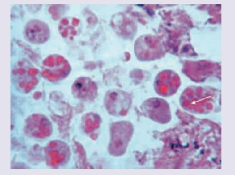
The given inclusion bodies are characteristic of which of the following organisms? 
A male patient presents with fever, cough, and hemoptysis. Bronchoalveolar lavage (BAL) fluid examination shows septate hyphae with acute angle (dichotomous) branching under microscopy. What is the most likely diagnosis?
Identify the Anopheles mosquito larva from the image shown below:

A 23-year-old male presented with abdominal pain and bloody diarrhea of one week duration. The following colonoscopic biopsy is diagnostic of infection with:

Explanation: **Explanation:** The association between HIV and malignancy is primarily driven by profound immunosuppression (low CD4+ counts) and the oncogenic potential of co-infecting viruses [1]. **Why Astrocytoma is the correct answer:** Astrocytomas are primary glial tumors of the CNS. Unlike Primary CNS Lymphoma, **Astrocytomas have no established epidemiological or pathogenetic link to HIV infection.** Their incidence in HIV-positive patients is similar to that of the general population. **Analysis of other options:** * **Kaposi’s Sarcoma (KS):** This is an **AIDS-defining illness** caused by Human Herpesvirus 8 (HHV-8) [1]. It is the most common neoplasm in HIV patients, characterized by vascular proliferations. * **Non-Hodgkin’s Lymphoma (NHL):** HIV patients have a significantly higher risk of aggressive B-cell lymphomas (e.g., Diffuse Large B-cell Lymphoma or Burkitt Lymphoma), often associated with **Epstein-Barr Virus (EBV)** [2]. * **Gastric Adenocarcinoma:** While less common than KS or Lymphoma, HIV patients have an increased risk of various non-AIDS-defining cancers, including gastrointestinal malignancies. This is attributed to chronic inflammation, lifestyle factors, and potential co-infection with *H. pylori*. **High-Yield Clinical Pearls for NEET-PG:** 1. **AIDS-Defining Malignancies:** Kaposi’s Sarcoma (HHV-8), Non-Hodgkin’s Lymphoma (EBV), and Invasive Cervical Carcinoma (HPV) [1]. 2. **Primary CNS Lymphoma:** This is the most common CNS tumor in HIV patients (strongly linked to EBV) and must be differentiated from Toxoplasmosis on imaging [3]. 3. **Anal Cancer:** There is a markedly increased incidence of squamous cell carcinoma of the anus in HIV-positive MSM (Men who have Sex with Men) due to HPV co-infection [1]. 4. **Trend:** Since the advent of HAART, the incidence of AIDS-defining cancers has decreased, while non-AIDS-defining cancers (Lung, Liver, Hodgkin’s) are increasing as patients live longer. **References:** [1] Kumar V, Abbas AK, et al.. Robbins and Cotran Pathologic Basis of Disease. 9th ed. Diseases of the Immune System, pp. 261-262. [2] Kumar V, Abbas AK, et al.. Robbins and Cotran Pathologic Basis of Disease. 9th ed. Diseases of the Immune System, pp. 262-263. [3] Kumar V, Abbas AK, et al.. Robbins and Cotran Pathologic Basis of Disease. 9th ed. The Central Nervous System, pp. 1315-1316.
Explanation: This question tests the ability to clinically differentiate between two common viral oropharyngeal infections: **Herpangina** (caused by Coxsackievirus A) and **Primary Herpetic Gingivostomatitis** (caused by HSV-1). [1] ### **Explanation of the Correct Answer** The primary distinguishing factor between these two conditions is the **anatomical distribution** of the lesions: * **Herpangina:** Characteristically involves the **posterior** oropharynx. Lesions (vesicles that progress to ulcers) are typically found on the **anterior faucial pillars**, soft palate, uvula, and tonsils. * **Primary Herpetic Stomatitis:** Characteristically involves the **anterior** oral cavity. It affects the gingiva (causing diffuse marginal gingivitis), labial mucosa, buccal mucosa, and the tongue. [1] ### **Analysis of Incorrect Options** * **Option A (Prodromal symptoms):** Both conditions are preceded by systemic prodromal symptoms, including high fever, malaise, and sore throat. Therefore, this is a similarity, not a difference. * **Option B (Unilateral nature):** Both conditions typically present with **bilateral** and diffuse involvement of the oral mucosa. [1] Unilateral lesions are more characteristic of Herpes Zoster (shingles). [2] * **Option D (Viral etiology):** Both are viral infections. Herpangina is an **Enterovirus** (Coxsackie A), while Herpetic Stomatitis is a **Herpesvirus** (HSV-1). [1] ### **High-Yield Clinical Pearls for NEET-PG** * **Herpangina:** Look for "Posterior" (P for Pillars, P for Posterior). It is usually seasonal (summer/autumn). * **Hand-Foot-Mouth Disease:** Also caused by Coxsackie A16; similar to herpangina but includes a maculopapular rash on the palms and soles. * **Herpetic Gingivostomatitis:** Look for **"Gingival involvement"** (punched-out erosions on gums) and Tzanck smear showing multinucleated giant cells with Cowdry Type A bodies. [1] * **Treatment:** Both are generally self-limiting; however, Acyclovir is effective for HSV if started early, whereas treatment for Herpangina is purely supportive. **References:** [1] Kumar V, Abbas AK, et al.. Robbins and Cotran Pathologic Basis of Disease. 9th ed. Infectious Diseases, p. 366. [2] Kumar V, Abbas AK, et al.. Robbins and Cotran Pathologic Basis of Disease. 9th ed. Infectious Diseases, pp. 366-367.
Explanation: **Explanation:** **Lupus vulgaris** is a chronic, progressive form of cutaneous tuberculosis occurring in individuals with a high degree of immunity against *Mycobacterium tuberculosis*. In the oral cavity, it typically presents as "apple-jelly" nodules that may ulcerate or cause significant tissue destruction. 1. **Why Biopsy is the Correct Answer:** The gold standard for diagnosing Lupus vulgaris is a **Biopsy** followed by histopathological examination. The characteristic finding is the presence of **tuberculoid granulomas** (epithelioid cells, Langhans giant cells, and lymphocytes) with minimal or absent caseous necrosis [1]. Because the bacterial load is extremely low in these lesions (paucibacillary), tissue architecture is the most reliable diagnostic feature [1]. 2. **Why Other Options are Incorrect:** * **Bacterial Smear:** Since Lupus vulgaris is a paucibacillary form of TB, Acid-Fast Bacilli (AFB) are rarely seen on a direct smear [1]. This leads to a very high false-negative rate. * **Blood Studies (CBC/ESR):** While the ESR may be elevated, these tests are non-specific and cannot differentiate TB from other inflammatory or infectious conditions. * **Blood Chemistry:** Tests like LFTs or RFTs provide information on organ function but have no diagnostic value for cutaneous or oral tuberculosis. **High-Yield Clinical Pearls for NEET-PG:** * **Clinical Sign:** The "Apple-jelly" appearance on **diascopy** (blanching with a glass slide) is a classic clinical clue. * **Pathology:** It is a **paucibacillary** condition; therefore, Culture and PCR are more sensitive than smears, but Biopsy remains the primary diagnostic tool [1]. * **Differential Diagnosis:** Must be differentiated from Sarcoidosis and Leprosy (both also show granulomas). * **Treatment:** Standard anti-tubercular therapy (ATT) for 6 months. **References:** [1] Kumar V, Abbas AK, et al.. Robbins and Cotran Pathologic Basis of Disease. 9th ed. Infectious Diseases, pp. 381-385.
Explanation: **Explanation:** Infectious Mononucleosis (IM), also known as "Glandular Fever," is a clinical syndrome most commonly caused by the **Epstein-Barr Virus (EBV)**. It primarily affects adolescents and young adults and is characterized by a classic triad of fever, pharyngitis, and lymphadenopathy. **Why "All of the above" is correct:** * **Glandular involvement (Option A):** This is a hallmark of the disease [3]. Patients typically present with symmetric **posterior cervical lymphadenopathy** [1]. Additionally, splenomegaly is seen in about 50% of cases due to lymphoid proliferation. * **Febrile (Option B):** Fever is one of the most consistent clinical findings, often accompanied by malaise, fatigue, and chills [2]. * **Palatine petechiae (Option C):** This is a high-yield physical finding. Small red spots (petechiae) at the junction of the soft and hard palate are seen in approximately 25–60% of patients and are highly suggestive of IM. **High-Yield Clinical Pearls for NEET-PG:** * **Hematology:** The characteristic laboratory finding is **atypical lymphocytosis** (Downey cells), which are activated T-cells (CD8+) reacting against EBV-infected B-cells [1]. * **Diagnosis:** The **Monospot test** (detecting heterophile antibodies) is the screening test of choice. * **Complication:** Avoid prescribing Ampicillin or Amoxicillin if IM is suspected, as it can trigger a characteristic **maculopapular rash**. * **Management:** Patients must avoid contact sports for 3–4 weeks to prevent **splenic rupture**, a rare but life-threatening complication. **References:** [1] Kumar V, Abbas AK, et al.. Robbins and Cotran Pathologic Basis of Disease. 9th ed. Infectious Diseases, pp. 368-370. [2] Kumar V, Abbas AK, et al.. Robbins and Cotran Pathologic Basis of Disease. 9th ed. Inflammation and Repair, pp. 110-111. [3] Cross SS. Underwood's Pathology: A Clinical Approach. 6th ed. Common Clinical Problems From Diseases Of The Urinary And Male Genital Tracts, pp. 549-551.
Explanation: **Explanation:** The term **tuberculoma** refers to a localized, tumor-like mass of inflammatory tissue caused by *Mycobacterium tuberculosis*. While the term is most commonly associated with the Central Nervous System (CNS) in clinical practice [4], in the context of specific dental and oral pathology, it refers to a **tuberculous periapical granuloma**. 1. **Why Option B is Correct:** In oral pathology, a tuberculoma is defined as a chronic tuberculous infection at the apex of a tooth. It occurs when tubercle bacilli reach the periapical area (usually via the bloodstream or through an open pulp canal), leading to the formation of a granuloma characterized by central caseous necrosis surrounded by epithelioid cells and Langhans giant cells [1][3]. 2. **Why Options A and C are Incorrect:** * **Option A:** A granuloma in the lungs is typically referred to as a **Ghon focus** (if primary) or simply a pulmonary granuloma. While a large mass-like lesion in the lung can be called a tuberculoma, it is not the specific definition used in this context. * **Option C:** Tuberculous involvement of the lymph nodes is termed **tuberculous lymphadenitis** (or **Scrofula** when involving cervical nodes). 3. **NEET-PG High-Yield Pearls:** * **CNS Tuberculoma:** This is the most common "tumor" in the brain in developing countries. On MRI, it often shows "ring enhancement" with a target sign [4]. * **Histology:** The hallmark of any tuberculoma is **caseating granulomatous inflammation** [1]. * **Differential Diagnosis:** In the brain, it must be differentiated from Neurocysticercosis (NCC) and metastasis [4]. In the periapical region, it must be differentiated from a standard periapical cyst or granuloma [2]. **References:** [1] Kumar V, Abbas AK, et al.. Robbins and Cotran Pathologic Basis of Disease. 9th ed. Infectious Diseases, pp. 383-384. [2] Kumar V, Abbas AK, et al.. Robbins and Cotran Pathologic Basis of Disease. 9th ed. Head and Neck, pp. 741-742. [3] Kumar V, Abbas AK, et al.. Robbins and Cotran Pathologic Basis of Disease. 9th ed. Inflammation and Repair, p. 109. [4] Cross SS. Underwood's Pathology: A Clinical Approach. 6th ed. Common Clinical Manifestations Of Central And Peripheral Nervous System Disease, pp. 709-710.
Explanation: ***Herpes simplex virus*** - The presence of **multinucleated giant cells** on a **Tzanck smear** is a characteristic finding of **herpesvirus infections** (HSV and VZV) [3]. - In the context of **painful vesicular lesions on the lips** in a young adult, **HSV-1** is the most likely causative agent, causing **herpes labialis** (cold sores) [1]. - The Tzanck smear is a rapid, inexpensive diagnostic method that detects the cytopathic effect of herpesviruses (cell fusion creating multinucleated giant cells) [3]. - **HSV-1** is the predominant cause of orolabial herpes, while HSV-2 more commonly causes genital herpes. *Varicella-zoster virus* - **VZV** also produces **multinucleated giant cells** on Tzanck smear (indistinguishable from HSV cytologically). - However, VZV typically presents as **chickenpox** (generalized vesicular rash) in primary infection or **shingles** (dermatomal distribution) in reactivation, not isolated lip lesions [4]. - The clinical presentation of localized lip vesicles in a young adult makes HSV far more likely than VZV. *Human papillomavirus* - HPV infection is characterized by **koilocytes** (cells with perinuclear clearing and nuclear atypia), not multinucleated giant cells. - HPV causes **warts** and mucosal papillomas, not vesicular lesions [2]. *Coxsackievirus A16* - This virus causes **Hand, Foot, and Mouth Disease** with vesicles in characteristic distribution (hands, feet, oral mucosa). - Coxsackievirus does **not** produce multinucleated giant cells on cytology. - Diagnosis relies on clinical presentation or PCR, not Tzanck smear. **References:** [1] Kumar V, Abbas AK, et al.. Robbins and Cotran Pathologic Basis of Disease. 9th ed. Infectious Diseases, p. 366. [2] Cross SS. Underwood's Pathology: A Clinical Approach. 6th ed. Common Clinical Problems From Diseases Of The Urinary And Male Genital Tracts, pp. 503-504. [3] Kumar V, Abbas AK, et al.. Robbins and Cotran Pathologic Basis of Disease. 9th ed. Infectious Diseases, pp. 365-366. [4] Kumar V, Abbas AK, et al.. Robbins and Cotran Pathologic Basis of Disease. 9th ed. Infectious Diseases, pp. 366-367.
Explanation: ***Cytomegalovirus*** - The image displays cells with marked enlargement (**cytomegaly**) and large, basophilic intranuclear inclusion bodies surrounded by a clear halo, which is the classic **"owl's eye"** appearance pathognomonic for Cytomegalovirus (CMV) infection [1]. - These inclusions are composed of viral particles and are typically seen in various tissues, such as the lungs, kidneys, and gastrointestinal tract, especially in **immunocompromised** patients [1]. *Human papillomavirus* - HPV infection is histologically characterized by **koilocytes**, which are squamous epithelial cells with a non-staining perinuclear halo and a wrinkled, hyperchromatic nucleus. - These changes are typically seen in cervical smears (Pap smears) or skin warts and are distinct from the large intranuclear inclusions of CMV. *Epstein-Barr virus* - EBV infection, particularly in infectious mononucleosis, is identified by the presence of **atypical lymphocytes** (Downey cells) in the peripheral blood, which have abundant cytoplasm and indented nuclei. - EBV does not produce the characteristic "owl's eye" intranuclear inclusions seen in the provided image. *Herpes simplex virus* - HSV infection is characterized by **multinucleated giant cells** with molded nuclei and eosinophilic intranuclear inclusions known as **Cowdry type A bodies**. - While both are herpesviruses, the inclusions in HSV typically give a **"ground-glass"** appearance to the nucleus, which is different from the distinct, haloed "owl's eye" inclusion of CMV. **References:** [1] Kumar V, Abbas AK, et al.. Robbins and Cotran Pathologic Basis of Disease. 9th ed. Infectious Diseases, pp. 367-368.
Explanation: ***Aspergillosis*** - The characteristic finding of **septate hyphae** displaying uniform **acute angle (dichotomous) branching** (typically 45°) in the **Bronchoalveolar lavage (BAL)** fluid is the defining microscopic feature of *Aspergillus* infection [1], [2]. - The clinical picture of fever, cough, and **hemoptysis** suggests an invasive pulmonary fungal infection, which *Aspergillus* commonly causes, especially in immunocompromised hosts [1], [2]. *Mucormycosis* - This diagnosis is characterized by **broad, non-septate (aseptate) hyphae** that exhibit irregular branching, typically at a **wide angle (90°)**. - The absence of septae and the differing angle of branching rule out mucormycosis based on the microscopic findings. *Histoplasmosis* - *Histoplasma capsulatum* appears in tissue and BAL primarily as **small, oval, budding yeast forms** (2–4 µm) that are often **intracellular** within macrophages. - It is not a hyphal infection in tissue form and therefore does not show septate hyphae with dichotomous branching. *Candidiasis* - *Candida* is identified by the presence of both **budding yeast cells** and **pseudohyphae** (links of elongated yeast cells) [2]. - Although true septate hyphae can occasionally be seen, it lacks the highly characteristic, uniform **acute-angle dichotomous branching** that is specific to *Aspergillus*. **References:** [1] Kumar V, Abbas AK, et al.. Robbins and Cotran Pathologic Basis of Disease. 9th ed. Infectious Diseases, pp. 396-397. [2] Cross SS. Underwood's Pathology: A Clinical Approach. 6th ed. Common Clinical Problems From Respiratory Tract Disease, pp. 318-319.
Explanation: ***Anopheles*** - The first image, showing a larva resting **parallel to the water surface** with a **rudimentary breathing tube**, is characteristic of *Anopheles* mosquito larvae. - Unlike *Culex* or *Aedes* larvae, *Anopheles* lack a prominent siphon, allowing them to lie flat against the water for respiration. *Option B* - This option likely refers to the second or third image, which depicts larvae resting at an angle to the water surface with distinct breathing tubes (siphons). These characteristics are typical of *Culex* or *Aedes* larvae, not *Anopheles*. - The presence of a short or long air tube (siphon) differentiates these from the *Anopheles* larva's rudimentary breathing mechanism. *Option C* - Similar to Option B, this refers to larvae with prominent breathing tubes and an angular resting position. These are features of *Culex* or *Aedes* species. - The specific description of the air tube (short and stout or long and slender with hair tufts) helps further distinguish between *Culex* and *Aedes*, neither of which matches the *Anopheles* morphology shown in the first panel. *Option D* - This option also describes larval types with clear breathing tubes and an angled resting posture, which are not characteristics of *Anopheles* species. - The distinct morphology of the first larva, particularly its parallel resting position and rudimentary breathing tube, is unique to *Anopheles*.
Explanation: ***Amoebiasis*** - **Amoebiasis** caused by *Entamoeba histolytica* is characterized by **bloody diarrhea** and **abdominal pain**, which are key clinical features in this case [1] [2]. - Colonoscopic biopsy in amoebiasis often shows **flask-shaped ulcers** and trophozoites of *Entamoeba histolytica* invading the colonic mucosa [1]. *Giardiasis* - **Giardiasis** typically presents with **non-bloody, watery diarrhea**, malabsorption, and flatulence, not bloody diarrhea. - It primarily affects the **small intestine** and is diagnosed by finding cysts or trophozoites in stool, not typically via colonoscopic biopsy for bloody diarrhea. *Enterobius* - **Enterobius vermicularis** (pinworm) infection primarily causes **perianal itching**, especially at night. - It does not typically cause **bloody diarrhea** or significant colonic inflammation visible on colonoscopic biopsy. *Severe bacterial infection* - While severe bacterial infections can cause **bloody diarrhea** (e.g., *Shigella*, *E. coli* O157:H7), the question implies a specific diagnostic finding from the colonoscopic biopsy that points to a parasitic infection. - The term "severe bacterial infection" is broad, and without specific bacterial findings or a characteristic pattern, it is less precise than a specific parasitic diagnosis suggested by the biopsy. **References:** [1] Cross SS. Underwood's Pathology: A Clinical Approach. 6th ed. Common Clinical Problems From Alimentary System Disease, pp. 364-365. [2] Kumar V, Abbas AK, et al.. Robbins and Cotran Pathologic Basis of Disease. 9th ed. The Gastrointestinal Tract, pp. 801-802.
Host-Pathogen Interactions
Practice Questions
Bacterial Infections
Practice Questions
Viral Infections
Practice Questions
Fungal Infections
Practice Questions
Parasitic Diseases
Practice Questions
Emerging Infections
Practice Questions
Healthcare-Associated Infections
Practice Questions
Infectious Disease Pathology in Immunocompromised Hosts
Practice Questions
Laboratory Diagnosis of Infections
Practice Questions
Antimicrobial Resistance
Practice Questions
Get full access to all questions, explanations, and performance tracking.
Start For Free