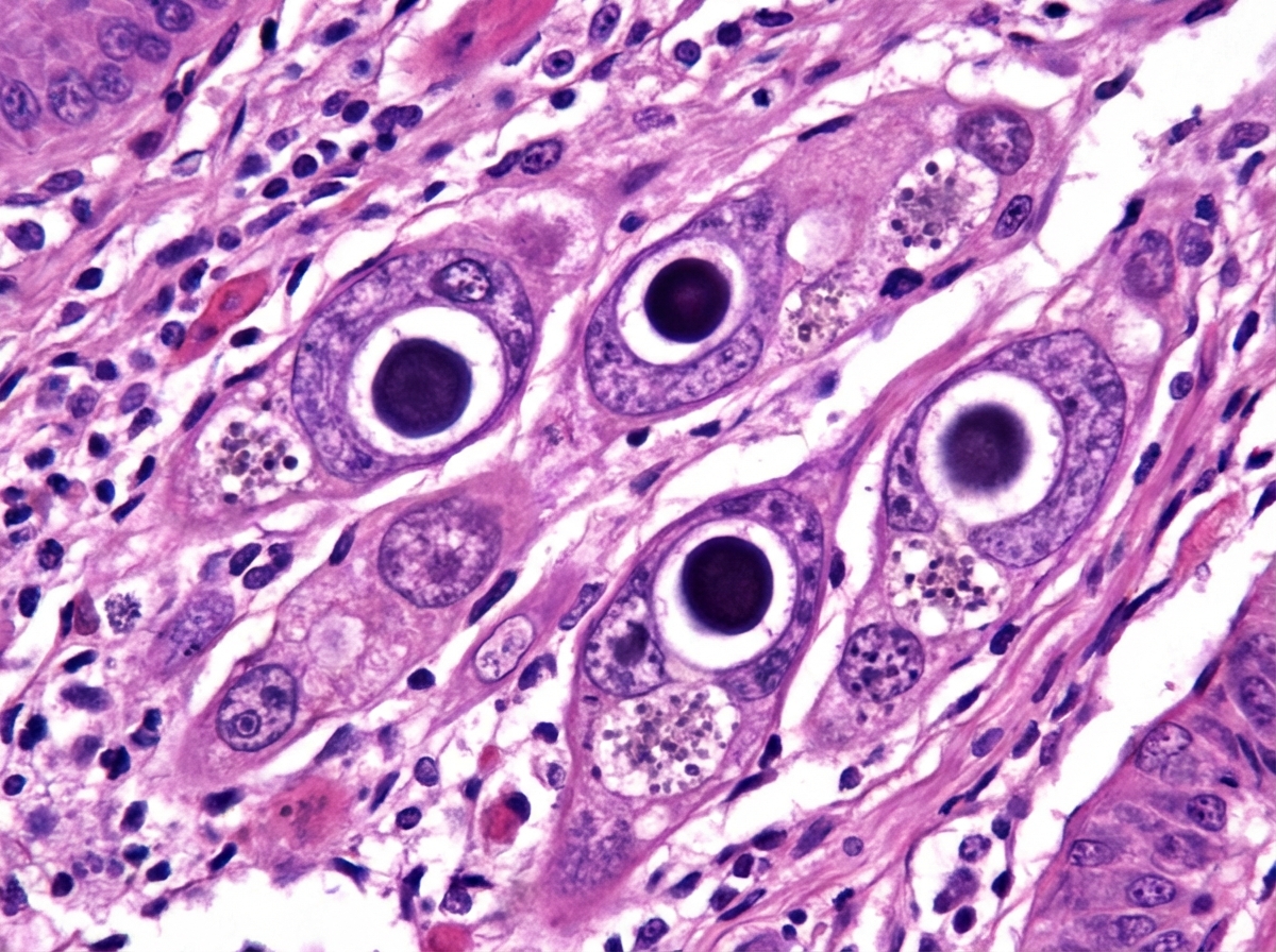
A 45-year-old HIV-positive male presented with dysphagia. Endoscopy and biopsy were performed. What is the most likely diagnosis based on the endoscopic and histological findings?

Which of the following is the brownish-colored substance seen in heart failure cells?
What is the multifocal tumor of vascular origin commonly seen in a patient with AIDS?
Hepatitis B virus (HBV) infection is associated with all of the following conditions except:
Erythrophagia and Mononuclear cell infiltration ulcers are seen in which condition?
In Tuberculosis, what type of cells primarily provide immunity?
A 37-year-old man presents with a productive cough, fever, and night sweats. A chest X-ray reveals an ill-defined area of consolidation at the periphery of the right middle lobe and mediastinal lymphadenopathy. Sputum culture is positive for acid-fast bacilli. What pathologic finding is most likely to be seen in a lymph node biopsy from this patient?
Which of the following statements is NOT TRUE regarding primary Herpes Simplex Virus (HSV) infections?
Which of the following is NOT a disease caused by coxsackievirus?
Which of the following statements regarding Viridans streptococci is TRUE?
Explanation: ***Cytomegalovirus (CMV) esophagitis*** - Characterized by **large linear ulcerations** on endoscopy, typically seen in severely immunocompromised HIV patients with **CD4+ count <50 cells/μL**. - Histologically shows pathognomonic **owl-eye nuclear inclusions** (large eosinophilic intranuclear inclusions with surrounding halo) in infected cells. *Herpes esophagitis* - Endoscopically presents with **small, punched-out ulcers** rather than large linear ulcerations seen in CMV. - Histologically shows **multinucleated giant cells** with **Cowdry A inclusions**, not the characteristic owl-eye inclusions of CMV. *Candida esophagitis* - Endoscopically appears as **white plaques** or **pseudomembranes** that can be wiped off, revealing erythematous mucosa underneath. - Histologically shows **pseudohyphae and budding yeasts** on PAS or GMS staining, without the viral inclusions described. *Pseudomonas esophagitis* - Extremely rare cause of esophagitis, typically occurring only in **severely neutropenic patients** or those with extensive burns. - Would show **gram-negative rod bacteria** on histology and **necrotizing inflammation**, not the viral cytopathic effects described.
Explanation: **Explanation:** **Heart failure cells** are alveolar macrophages containing a brownish-granular pigment called **Hemosiderin**. **Why Hemosiderin is correct:** In chronic left-sided heart failure, there is increased pressure in the pulmonary capillaries (pulmonary congestion). This causes red blood cells (RBCs) to leak into the alveolar spaces. Alveolar macrophages phagocytose these extravasated RBCs and break down the hemoglobin [2]. The iron released from the heme is stored as **hemosiderin**, giving the macrophages a characteristic golden-brown appearance [2]. These cells are a hallmark of **Chronic Passive Congestion (CPC) of the lung**. **Why other options are incorrect:** * **Lipofuscin:** Known as the "wear and tear" or "aging" pigment. It is a yellowish-brown, finely granular lipid-containing pigment found in the heart (brown atrophy) and liver of elderly or malnourished patients. * **Myoglobin:** An iron- and oxygen-binding protein found in muscle tissue. While it contains iron, it does not form the granular brown deposits seen in alveolar macrophages. * **Bilirubin:** A yellow breakdown product of normal heme catabolism [2]. While it can cause tissue discoloration (jaundice) [1], it is typically seen in the liver or skin and does not form the coarse brown granules characteristic of heart failure cells. **High-Yield Pearls for NEET-PG:** * **Stain:** Hemosiderin is best visualized using the **Prussian Blue (Perl’s) stain**, which stains the iron blue [3]. * **Clinical Significance:** Presence of these cells in sputum or lung biopsy indicates chronic pulmonary congestion, most commonly due to **Mitral Stenosis** or **Left Ventricular Failure**. * **Nutmeg Liver:** This is the corresponding gross appearance of the liver in chronic passive congestion (usually due to right-sided heart failure). **References:** [1] Kumar V, Abbas AK, et al.. Robbins and Cotran Pathologic Basis of Disease. 9th ed. Red Blood Cell and Bleeding Disorders, pp. 639-640. [2] Kumar V, Abbas AK, et al.. Robbins and Cotran Pathologic Basis of Disease. 9th ed. Cellular Responses to Stress and Toxic Insults: Adaptation, Injury, and Death, pp. 75-76. [3] Kumar V, Abbas AK, et al.. Robbins and Cotran Pathologic Basis of Disease. 9th ed. Liver and Gallbladder, pp. 854-855.
Explanation: **Kaposi Sarcoma (KS)** is the correct answer because it is a low-grade vascular tumor strongly associated with **Human Herpesvirus 8 (HHV-8)** and is the most common neoplasm seen in patients with AIDS [1]. It typically presents as multifocal, purplish-red cutaneous nodules or plaques but can also involve visceral organs like the lungs and GI tract [2]. Pathologically, it is characterized by the proliferation of spindle cells, slit-like vascular spaces, and extravasated red blood cells [2]. **Analysis of Incorrect Options:** * **Astrocytoma:** While brain tumors occur in immunocompromised states, astrocytomas are not specifically associated with AIDS or vascular origin. * **Gastric Carcinoma:** Though AIDS patients have a higher risk of certain GI malignancies, gastric adenocarcinoma is not a vascular tumor and is primarily linked to *H. pylori* or genetic factors. * **Primary CNS Lymphoma:** This is the second most common malignancy in AIDS patients (after KS). However, it is a **B-cell neoplasm** associated with **EBV**, not a tumor of vascular origin [1]. **High-Yield Clinical Pearls for NEET-PG:** * **Defining Characteristic:** KS is an **AIDS-defining illness**. * **Histology:** Look for "spindle cells" and "slit-like spaces" containing RBCs [2]. * **Four Clinical Variants:** Classic (European), Endemic (African), Transplant-associated (Immunosuppression), and Epidemic (AIDS-associated). * **Treatment:** Highly Active Antiretroviral Therapy (HAART) often leads to the regression of AIDS-related KS lesions [3]. **References:** [1] Kumar V, Abbas AK, et al.. Robbins and Cotran Pathologic Basis of Disease. 9th ed. Diseases of the Immune System, pp. 261-262. [2] Kumar V, Abbas AK, et al.. Robbins and Cotran Pathologic Basis of Disease. 9th ed. Diseases of Infancy and Childhood, pp. 526-527. [3] Kumar V, Abbas AK, et al.. Robbins and Cotran Pathologic Basis of Disease. 9th ed. Diseases of the Immune System, pp. 260-261.
Explanation: **Explanation:** The correct answer is **Hepatic adenoma**. Hepatic adenoma is a benign liver tumor primarily associated with **oral contraceptive pill (OCP) use**, anabolic steroid use, and glycogen storage diseases (Type I and III) [1]. It is not linked to viral hepatitis infections. **Why the other options are incorrect:** Hepatitis B Virus (HBV) is a DNA virus known for its potential to cause persistent infection and genomic integration, leading to: * **Chronic Hepatitis:** Approximately 5-10% of adults infected with HBV develop chronic hepatitis, characterized by the persistence of HBsAg for more than 6 months [3]. * **Cirrhosis:** Chronic inflammation and repeated cycles of hepatocyte death and regeneration lead to extensive fibrosis and nodule formation (cirrhosis) [2]. * **Hepatic Cancer (Hepatocellular Carcinoma - HCC):** HBV is a major risk factor for HCC [2]. It can cause cancer both indirectly (via cirrhosis) and directly (via the **HBx protein**, which disrupts cell cycle control and inhibits p53) [3]. Notably, HBV can cause HCC even in the absence of cirrhosis [2]. **High-Yield Clinical Pearls for NEET-PG:** * **HBV vs. HCV:** HBV is a DNA virus; HCV is an RNA virus. HBV has a higher risk of vertical transmission, while HCV has a higher risk of progressing to chronicity (~80%). * **Ground Glass Hepatocytes:** A classic histopathological finding in chronic HBV, representing the accumulation of HBsAg in the endoplasmic reticulum [4]. * **Tumor Marker:** Alpha-fetoprotein (AFP) is the screening marker for HCC in patients with HBV/cirrhosis. * **Hepatic Adenoma Risk:** The most significant risk is **rupture and intraperitoneal hemorrhage**, especially during pregnancy. **References:** [1] Kumar V, Abbas AK, et al.. Robbins and Cotran Pathologic Basis of Disease. 9th ed. Liver and Gallbladder, p. 874. [2] Kumar V, Abbas AK, et al.. Robbins and Cotran Pathologic Basis of Disease. 9th ed. Liver and Gallbladder, pp. 876-877. [3] Kumar V, Abbas AK, et al.. Robbins and Cotran Pathologic Basis of Disease. 9th ed. Liver and Gallbladder, pp. 838-840. [4] Kumar V, Abbas AK, et al.. Robbins and Cotran Pathologic Basis of Disease. 9th ed. Liver and Gallbladder, pp. 844-845.
Explanation: **Explanation:** The correct answer is **Typhoid ulcers (D)**. Typhoid fever, caused by *Salmonella typhi*, primarily affects the GALT (Gut-Associated Lymphoid Tissue), specifically the **Peyer’s patches** in the terminal ileum [1]. **Pathogenesis & Histology:** The hallmark of Typhoid is the proliferation of mononuclear cells (macrophages) known as **Typhoid cells**. These are activated macrophages that exhibit **erythrophagia**—the ingestion of red blood cells, lymphocytes, and necrotic debris. These cells infiltrate the Peyer’s patches, leading to necrosis of the overlying mucosa and the formation of characteristic **longitudinal ulcers** (oriented along the long axis of the bowel). **Why other options are incorrect:** * **Necrotising colitis:** Typically seen in neonates (NEC) or ischemic conditions; characterized by transmural necrosis and pneumatosis intestinalis, not specific erythrophagic mononuclear infiltration. * **Ulcerative Colitis:** A chronic inflammatory bowel disease characterized by **crypt abscesses**, pseudopolyps, and continuous mucosal inflammation starting from the rectum. * **Crohn’s Disease:** Characterized by transmural inflammation, **non-caseating granulomas**, and "skip lesions." Ulcers are typically aphthous or linear (cobblestone appearance), not longitudinal with erythrophagia. **High-Yield Clinical Pearls for NEET-PG:** * **Ulcer Orientation:** Typhoid ulcers are **longitudinal** (parallel to the long axis), whereas Tuberculous ulcers are **transverse** (circumferential). * **Widal Test:** Significant titers are usually seen in the 2nd week of infection. * **Complication:** The most serious complication of typhoid ulcers is intestinal perforation, usually occurring in the 3rd week. * **Mallory Bodies:** Do not confuse these with "Mallory's spots" (Typhoid nodules) found in the liver, which also contain Typhoid cells. **References:** [1] Cross SS. Underwood's Pathology: A Clinical Approach. 6th ed. Common Clinical Problems From Alimentary System Disease, pp. 362-363.
Explanation: **Explanation:** The immunity against *Mycobacterium tuberculosis* is primarily **cell-mediated immunity (CMI)**, as the pathogen is an intracellular bacterium that survives within macrophages [1]. **Why CD4+ cells are correct:** CD4+ T-helper cells (specifically **Th1 cells**) play the central role in the immune response to TB [3]. When macrophages ingest the bacilli, they present antigens via MHC Class II molecules to CD4+ T cells. These T cells then secrete **Interferon-gamma (IFN-γ)**, which is the critical cytokine required to activate macrophages [3]. Activated macrophages then increase their production of nitric oxide and reactive oxygen species to kill the intracellular bacteria and form the characteristic **granuloma** [1], [2]. **Why other options are incorrect:** * **CD8+ cells:** While CD8+ cytotoxic T cells do play a minor role by killing infected macrophages, they are not the primary mediators of protective immunity compared to the orchestrating role of CD4+ cells. * **IgG and IgM antibodies:** These represent **humoral immunity** [4]. Since *M. tuberculosis* resides inside cells, antibodies are largely ineffective at reaching or neutralizing the bacilli. Humoral immunity does not provide significant protection against TB. **High-Yield Clinical Pearls for NEET-PG:** * **Cytokine Profile:** The "protective" response is driven by **IL-12** (which stimulates Th1 differentiation) and **IFN-γ** (which activates macrophages) [3]. * **TNF-α Role:** TNF-α is essential for maintaining granuloma integrity. Patients on **Anti-TNF therapy** (e.g., Infliximab) are at high risk for reactivation of latent TB [1]. * **HIV Correlation:** The loss of CD4+ cells in HIV patients is the primary reason for their extreme susceptibility to disseminated and primary Tuberculosis [2]. **References:** [1] Kumar V, Abbas AK, et al.. Robbins and Cotran Pathologic Basis of Disease. 9th ed. Infectious Diseases, pp. 380-381. [2] Kumar V, Abbas AK, et al.. Robbins and Cotran Pathologic Basis of Disease. 9th ed. Infectious Diseases, pp. 383-384. [3] Kumar V, Abbas AK, et al.. Robbins and Cotran Pathologic Basis of Disease. 9th ed. Diseases of the Immune System, p. 206. [4] Kumar V, Abbas AK, et al.. Robbins and Cotran Pathologic Basis of Disease. 9th ed. Diseases of the Immune System, pp. 206-207.
Explanation: ### Explanation **Correct Option: A. Caseating granulomas** The clinical presentation (fever, night sweats, productive cough) and sputum culture positive for **acid-fast bacilli (AFB)** are pathognomonic for **Tuberculosis (TB)** [1]. The chest X-ray findings—a peripheral consolidation (Ghon focus) combined with mediastinal lymphadenopathy—describe the **Ghon complex**, which is the hallmark of primary tuberculosis. Pathologically, TB is characterized by **Type IV hypersensitivity**, leading to the formation of **granulomas** [2]. These consist of activated macrophages (epithelioid cells), Langhans giant cells, and a rim of lymphocytes. The defining feature of TB granulomas is **caseous necrosis** (cheese-like, acellular debris at the center), caused by the host's immune response to *Mycobacterium tuberculosis* [1]. **Why the other options are incorrect:** * **B. Follicular hyperplasia:** This is a non-specific reactive change in lymph nodes characterized by B-cell proliferation in germinal centers, typically seen in viral infections or chronic inflammatory states, not TB. * **C. Nodular amyloidosis:** This involves the extracellular deposition of misfolded proteins. While chronic TB can lead to *Secondary (AA) Amyloidosis*, it presents as systemic organ involvement rather than the primary diagnostic finding in an acute nodal biopsy. * **D. Noncaseating granulomas:** These lack central necrosis. While they can be seen in early TB, they are the classic hallmark of **Sarcoidosis**, Crohn’s disease, or Berylliosis [1]. In the presence of AFB, caseation is the expected finding. ### NEET-PG High-Yield Pearls * **Ghon Complex:** Ghon focus (parenchymal lesion) + Lymph nodal involvement. * **Ranke Complex:** A radiologically visible, calcified Ghon complex. * **Epithelioid cells:** These are the most essential components of a granuloma; they are modified macrophages transformed by **IFN-gamma** [2]. * **Stain:** Ziehl-Neelsen (ZN) stain is used to identify AFB; they appear as bright red, slightly curved rods [1]. **References:** [1] Kumar V, Abbas AK, et al.. Robbins and Cotran Pathologic Basis of Disease. 9th ed. Infectious Diseases, pp. 382-384. [2] Kumar V, Abbas AK, et al.. Robbins and Cotran Pathologic Basis of Disease. 9th ed. Infectious Diseases, pp. 380-381.
Explanation: **Explanation:** The correct answer is **C (Occurs epidemically)** because Herpes Simplex Virus type 1 (HSV-1) infections are **endemic**, not epidemic. HSV is ubiquitous worldwide; most individuals are infected in childhood through direct contact with infected secretions (like saliva). It does not occur in large-scale seasonal outbreaks or localized epidemics but rather persists in the population through continuous transmission and lifelong latency. **Analysis of other options:** * **Option A & B:** Primary HSV-1 infection in children most commonly manifests as **Acute Herpetic Gingivostomatitis** [1]. It characteristically involves the **anterior portion of the mouth**, including the gingiva (gums), tongue, and labial mucosa, presenting with painful vesicles that rupture into ulcers [1]. * **Option D:** Primary infections are frequently preceded by **prodromal symptoms** such as high-grade fever, malaise, irritability, and regional lymphadenopathy before the appearance of oral lesions [1]. **High-Yield Clinical Pearls for NEET-PG:** * **Latency:** HSV-1 remains latent in the **Trigeminal ganglion**, while HSV-2 typically resides in the **Sacral ganglia**. * **Diagnosis:** The gold standard for rapid bedside diagnosis is the **Tzanck Smear**, which shows **Multinucleated Giant Cells** with Cowdry Type A inclusion bodies [1]. * **Histopathology:** Look for "acantholysis," "ballooning degeneration" of keratinocytes, and "margination of chromatin" [1]. * **Encephalitis:** HSV-1 is the most common cause of sporadic fatal viral encephalitis, typically involving the **temporal lobes** [2]. **References:** [1] Kumar V, Abbas AK, et al.. Robbins and Cotran Pathologic Basis of Disease. 9th ed. Infectious Diseases, pp. 365-366. [2] Kumar V, Abbas AK, et al.. Robbins and Cotran Pathologic Basis of Disease. 9th ed. The Central Nervous System, p. 1278.
Explanation: **Explanation:** The correct answer is **D. Herpes**. This is because Herpes is caused by the **Herpes Simplex Virus (HSV)**, a DNA virus belonging to the *Herpesviridae* family [1]. In contrast, Coxsackievirus is a member of the *Picornaviridae* family (genus Enterovirus) and is a positive-sense RNA virus. **Analysis of Options:** * **Herpangina (Option A):** Characterized by fever, sore throat, and small vesicular/ulcerative lesions on the posterior oropharynx (tonsillar pillars, soft palate). It is primarily caused by **Coxsackievirus Group A**. * **Hand, Foot, and Mouth Disease (Option B):** A common childhood illness presenting with vesicular eruptions on the palms, soles, and oral mucosa. It is most commonly caused by **Coxsackievirus A16** (and Enterovirus 71). * **Acute Lymphonodular Pharyngitis (Option C):** A variant of pharyngitis where white-to-yellow nodules (packed lymphocytes) appear on the posterior pharynx. This is specifically associated with **Coxsackievirus A10**. **NEET-PG High-Yield Pearls:** * **Coxsackie Group A:** Primarily affects mucous membranes and skin (Herpangina, HFMD). * **Coxsackie Group B:** Primarily affects the heart and body walls. It is the most common cause of **viral myocarditis** and **pericarditis**, as well as **Bornholm disease** (epidemic pleurodynia/Devil’s grip). * **Aseptic Meningitis:** Both Group A and B can cause viral meningitis, but Group B is more frequently implicated. * **Type 1 Diabetes:** Coxsackie B4 has been epidemiologically linked to the triggering of islet cell antibodies. **References:** [1] Kumar V, Abbas AK, et al.. Robbins and Cotran Pathologic Basis of Disease. 9th ed. Infectious Diseases, p. 366.
Explanation: **Explanation** **Viridans group streptococci (VGS)** are a large, heterogeneous group of alpha-hemolytic streptococci that are normal commensals of the oral cavity, gastrointestinal tract, and genitourinary tract [1]. **Why the correct answer is right:** * **Option B:** The term "Viridans" is derived from the Latin word *viridis* (green). These bacteria **reliably produce alpha-hemolysis** (partial hemolysis) on blood agar, which creates a characteristic green discoloration around the colonies. This occurs due to the oxidation of hemoglobin to methemoglobin by hydrogen peroxide produced by the bacteria. **Why the other options are incorrect:** * **Option A:** While *S. mutans, S. mitis, S. sanguis,* and *S. salivarius* are indeed members of the Viridans group, this option is technically incomplete as the group contains over 50 species. In the context of multiple-choice questions, "Reliably produces hemolysis" is the defining microbiological characteristic. * **Option C:** This is incorrect because Viridans streptococci are the **most common cause of subacute bacterial endocarditis (SBE)**, typically affecting previously damaged or prosthetic heart valves following dental procedures [1]. * **Option D:** While *S. mutans* is the primary cariogenic (cavity-causing) organism, the statement "The main strains" is imprecise compared to the definitive microbiological property of hemolysis [2]. **High-Yield NEET-PG Pearls:** * **Differentiation:** Unlike *S. pneumoniae*, Viridans streptococci are **Optochin resistant** and **Bile insoluble**. * **S. sanguis:** Specifically associated with endocarditis; it produces extracellular polysaccharides (dextrans) that allow it to adhere to fibrin-platelet aggregates on damaged heart valves. * **S. bovis (Group D):** If isolated in blood cultures, it is a high-yield marker for underlying **colonic carcinoma**. * **Prophylaxis:** Patients with prosthetic valves undergoing dental work often require antibiotic prophylaxis to prevent VGS bacteremia. **References:** [1] Kumar V, Abbas AK, et al.. Robbins and Cotran Pathologic Basis of Disease. 9th ed. The Heart, pp. 567-568. [2] Kumar V, Abbas AK, et al.. Robbins and Cotran Pathologic Basis of Disease. 9th ed. Infectious Diseases, pp. 372-374.
Host-Pathogen Interactions
Practice Questions
Bacterial Infections
Practice Questions
Viral Infections
Practice Questions
Fungal Infections
Practice Questions
Parasitic Diseases
Practice Questions
Emerging Infections
Practice Questions
Healthcare-Associated Infections
Practice Questions
Infectious Disease Pathology in Immunocompromised Hosts
Practice Questions
Laboratory Diagnosis of Infections
Practice Questions
Antimicrobial Resistance
Practice Questions
Get full access to all questions, explanations, and performance tracking.
Start For Free