
Immunopathology — MCQs

On this page
MALT is most commonly present in which part of the small intestine?
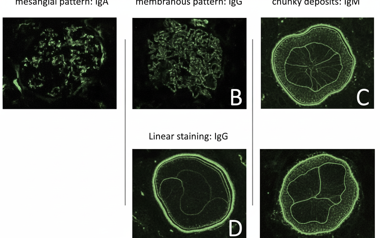
Which of the following types of hypersensitivity reactions is associated with the given condition?
Which of the following organs is NOT typically affected in Graft-Versus-Host disease?
The interaction of antigens with antibodies on the surface of a mast cell leads to degranulation and anaphylaxis. The mast cell granules produce anaphylaxis because they contain:
T cells recognize which antigen during graft rejection?
Which of the following interleukins is characteristically produced in a TH1 response?
What is a common consequence of hypogammaglobulinemia?
Allograft rejection is an example of which type of immune response?
All are true about severe combined immunodeficiency except?
A 63-year-old man presents with a 15-year history of chronic arthritis. Physical examination reveals ulnar deviation, bony ankylosis, and swan neck deformities of the fingers. Laboratory findings include 4.2 g of protein in a 24-hour urine collection, a serum creatinine of 3.1 mg/dL, and blood urea nitrogen of 30 mg/dL. The C-reactive protein level is markedly elevated. A rectal biopsy shows amorphous pink material deposited in the mucosa, which stains positive with Congo red on H&E staining. Which of the following proteins is the most likely precursor to this material in the mucosa?
Practice by Chapter
Cells and Tissues of the Immune System
Practice Questions
Innate Immunity
Practice Questions
Adaptive Immunity
Practice Questions
Hypersensitivity Reactions
Practice Questions
Autoimmune Diseases
Practice Questions
Immunodeficiency Disorders
Practice Questions
Transplantation Immunopathology
Practice Questions
Immune Response to Infections
Practice Questions
Immunologic Laboratory Techniques
Practice Questions
Tumor Immunology
Practice Questions
Want unlimited practice?
Get full access to all questions, explanations, and performance tracking.
Scan to download app