Which of the following features is shared in common between lymphocyte-rich and lymphocyte-predominant types of Hodgkin's lymphoma?
All of the following statements about the Philadelphia chromosome in CML are true, except:
Working formulation in staging of non-Hodgkin's lymphoma is based on which of the following?
What is true about follicular lymphoma?
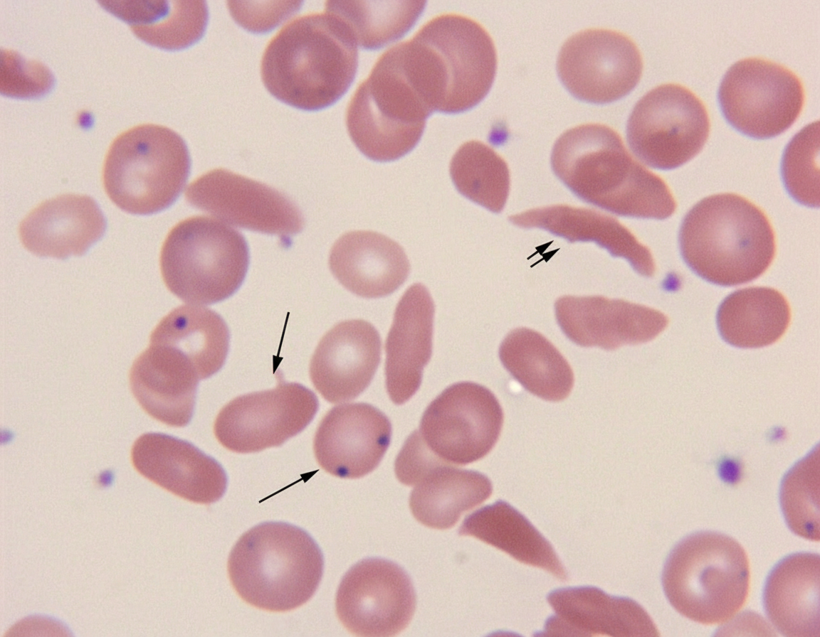
The above marked structures are seen in all except?

Non-specific esterase is positive in all categories of AML except?
Popcorn cells are characteristic of which of the following hematological malignancies?
Reticulocytes are stained by which method?
Radiological examination shows evidence of bone infarct in a child. Which of the following conditions may be responsible?
Which of the following is NOT typically seen in Disseminated Intravascular Coagulation (DIC)?
Explanation: **Explanation:** Both **Lymphocyte-Rich Hodgkin Lymphoma (LRHL)** and **Nodular Lymphocyte-Predominant Hodgkin Lymphoma (NLPHL)** are characterized by an excellent clinical outcome [1]. They typically present at early stages (Stage I or II) and have a very high cure rate, making **"Good prognosis"** the defining shared feature. **Analysis of Options:** * **A. Paucity of diagnostic Reed-Sternberg (RS) cells:** This is true for **LRHL**, where classic RS cells are rare against a background of small lymphocytes [2]. However, **NLPHL** does not contain "classic" RS cells at all; instead, it features **L&H (Lymphocytic and Histiocytic) cells**, also known as "Popcorn cells" [1]. * **B. EBV Association:** LRHL is associated with EBV in about 40% of cases. In contrast, **NLPHL is almost never associated with EBV** [1], making this a distinguishing factor rather than a shared one. * **C. CD20 Positivity:** This is the hallmark of **NLPHL** (Popcorn cells are CD20+, CD45+, but CD15- and CD30-) [1]. In **LRHL**, the RS cells follow the classic immunophenotype: **CD15+, CD30+, and CD20 negative**. **High-Yield Pearls for NEET-PG:** * **NLPHL** is now considered a distinct entity from Classic Hodgkin Lymphoma (CHL) due to its B-cell marker expression (CD20) [1]. * **LRHL** is a subtype of **Classic Hodgkin Lymphoma** but mimics NLPHL morphologically [2]. * **Popcorn cells (L&H cells)** = NLPHL [1]. * **Most common subtype** of HL = Nodular Sclerosis. * **Best prognosis** = NLPHL / LRHL; **Worst prognosis** = Lymphocyte Depleted. **References:** [1] Kumar V, Abbas AK, et al.. Robbins and Cotran Pathologic Basis of Disease. 9th ed. Diseases of White Blood Cells, Lymph Nodes, Spleen, and Thymus, pp. 616-618. [2] Cross SS. Underwood's Pathology: A Clinical Approach. 6th ed. Common Clinical Problems From Diseases Of The Urinary And Male Genital Tracts, pp. 559-560.
Explanation: ### Explanation The Philadelphia chromosome (Ph) is the hallmark of Chronic Myeloid Leukemia (CML) [1], [2]. It results from a **reciprocal translocation, t(9;22)(q34;q11)** [4]. **Why Option D is the correct answer (the False statement):** While the translocation involves both chromosomes 9 and 22, the resulting **Philadelphia chromosome is the derivative chromosome 22 (22q-)**. This shortened chromosome 22 harbors the chimeric **BCR-ABL1 fusion gene**, which encodes a constitutively active tyrosine kinase responsible for leukemogenesis [1]. The fusion gene is NOT located on chromosome 9. **Analysis of Incorrect Options (True statements):** * **Option A:** It is indeed a reciprocal translocation between the long arms (q) of chromosomes 9 and 22 [4]. * **Option B:** The *BCR* (Breakpoint Cluster Region) gene is located on chromosome 22 at band q11. * **Option C:** The *ABL* (Abelson murine leukemia viral oncogene homolog 1) proto-oncogene is located on chromosome 9 at band q34. **High-Yield Clinical Pearls for NEET-PG:** * **Fusion Protein:** The most common variant in CML is the **p210** protein. The **p190** variant is more commonly associated with Ph+ Acute Lymphoblastic Leukemia (ALL), while **p230** is linked to Chronic Neutrophilic Leukemia (CNL). * **Diagnostic Gold Standard:** While conventional karyotyping identifies the Ph chromosome, **FISH** or **RT-PCR** are used to detect the BCR-ABL1 rearrangement, especially in "Ph-negative" CML [2]. * **Treatment:** The discovery of this mutation led to the development of **Imatinib**, a selective tyrosine kinase inhibitor (TKI), which is the first-line therapy [3]. **References:** [1] Kumar V, Abbas AK, et al.. Robbins and Cotran Pathologic Basis of Disease. 9th ed. Diseases of White Blood Cells, Lymph Nodes, Spleen, and Thymus, p. 624. [2] Cross SS. Underwood's Pathology: A Clinical Approach. 6th ed. (Basic Pathology) introduces the student to key general principles of pathology, both as a medical science and as a clinical activity with a vital role in patient care. Part 2 (Disease Mechanisms) provides fundamental knowledge about the cellular and molecular processes involved in diseases, providing the rationale for their treatment. Part 3 (Systematic Pathology) deals in detail with specific diseases, with emphasis on the clinically important aspects., pp. 225-226. [3] Kumar V, Abbas AK, et al.. Robbins and Cotran Pathologic Basis of Disease. 9th ed. Neoplasia, pp. 295-296. [4] Cross SS. Underwood's Pathology: A Clinical Approach. 6th ed. Common Clinical Problems From Blood And Bone Marrow Disease, pp. 605-607.
Explanation: The **Working Formulation (WF)**, introduced in 1982, was a classification system designed to provide a uniform language for clinicians to categorize Non-Hodgkin Lymphomas (NHL) based on their clinical behavior. ### Why the Correct Answer is Right: The Working Formulation is strictly based on **morphology** (the architectural pattern and cytological features of the cells). It categorizes lymphomas into three prognostic groups (Low, Intermediate, and High grade) based on two main morphological criteria: 1. **Growth Pattern:** Whether the cells are arranged in a **Follicular** (nodular) or **Diffuse** pattern. 2. **Cell Type:** The size and shape of the cells (e.g., small cleaved, large cell, or immunoblastic). ### Why the Other Options are Wrong: * **B. Cell surface markers:** These are the basis for **Immunophenotyping** (CD markers). While crucial for the modern WHO classification [1], the Working Formulation was created before these markers were routinely used for classification. * **C. Survival characteristics:** While the WF *correlates* with survival (prognosis), the classification itself is built on morphology, not the actual survival data of the patient. * **D. Cellular genetics:** Cytogenetics (e.g., t(14;18) in Follicular Lymphoma) are central to the **REAL** and **WHO** classifications [1] but were not part of the Working Formulation. ### High-Yield Facts for NEET-PG: * **Evolution of Classifications:** Rappaport (Morphology) → Working Formulation (Morphology + Clinical Grade) → REAL Classification (Immunophenotype) → **WHO Classification** (Morphology + Immunophenotype + Genetics + Clinical features) [1]. * **The Gold Standard:** The current gold standard for NHL diagnosis is the **WHO Classification** [1]. * **WF Limitation:** The Working Formulation did not distinguish between B-cell and T-cell lineages, which is its primary clinical drawback. **References:** [1] Kumar V, Abbas AK, et al.. Robbins and Cotran Pathologic Basis of Disease. 9th ed. Diseases of White Blood Cells, Lymph Nodes, Spleen, and Thymus, pp. 596-598.
Explanation: **Follicular Lymphoma (FL)** is the most common indolent (slow-growing) Non-Hodgkin Lymphoma (NHL) [1]. Understanding its clinical and molecular profile is high-yield for NEET-PG. ### **Explanation of Options** * **Option A (Correct):** The most common clinical presentation of FL is **painless, generalized lymphadenopathy**. Patients often describe a "waxing and waning" course where lymph nodes fluctuate in size over several years before a diagnosis is made [2]. * **Option B (Technicality):** While FL is classically associated with **BCL-2 expression** (due to the **t(14;18)** translocation), this option is often considered "less correct" than the clinical presentation in standardized exams if the question asks for the "most true" clinical feature or if the examiner follows specific textbook hierarchies (like Robbins) [1]. *Note: In many contexts, BCL-2 positivity is a hallmark, but clinical presentation remains the primary diagnostic trigger.* * **Option C (Incorrect):** FL is a B-cell neoplasm that is **CD5 negative** and **CD10 positive** (germinal center origin) [1]. CD5 positivity is characteristic of Chronic Lymphocytic Leukemia (CLL/SLL) and Mantle Cell Lymphoma. * **Option D (Incorrect):** There is no significant female predilection; FL affects males and females almost **equally**, typically occurring in older adults (median age ~60 years). ### **High-Yield Clinical Pearls for NEET-PG** * **Cytogenetics:** **t(14;18)** translocation involving the *IGH* gene (Ch 14) and *BCL-2* (Ch 18), leading to overexpression of the anti-apoptotic BCL-2 protein [2]. * **Morphology:** Shows a nodular/follicular growth pattern. Unlike reactive follicles, these lack **tingible body macrophages** and show a loss of polarization [1]. * **Transformation:** In 30-50% of cases, FL can transform into a more aggressive **Diffuse Large B-Cell Lymphoma (DLBCL)** (Richter-like transformation). * **Immunophenotype:** CD19+, CD20+, CD10+, BCL-2+, **CD5-**, **CD23-** [1]. **References:** [1] Kumar V, Abbas AK, et al.. Robbins and Cotran Pathologic Basis of Disease. 9th ed. Diseases of White Blood Cells, Lymph Nodes, Spleen, and Thymus, pp. 602-604. [2] Cross SS. Underwood's Pathology: A Clinical Approach. 6th ed. Common Clinical Problems From Diseases Of The Urinary And Male Genital Tracts, pp. 561-562.
Explanation: ***Thalassemia*** - The spleen remains **functionally intact** in thalassemia and actively removes **Howell-Jolly bodies** from circulation. - Despite splenomegaly from hemolysis, splenic function is preserved, effectively culling nuclear remnants from RBCs. *Sickle cell anemia* - **Functional asplenia** develops due to repeated **sickling crises** causing splenic autoinfarction and fibrosis. - The damaged spleen cannot remove **Howell-Jolly bodies**, leading to their persistence in peripheral blood. *Liver cirrhosis* - Advanced cirrhosis causes **splenic dysfunction** and **hyposplenism** due to portal hypertension and compromised splenic blood flow. - The dysfunctional spleen fails to filter **Howell-Jolly bodies**, allowing them to remain in circulation. *Chronic myeloid leukemia (CML)* - **Leukemic cell infiltration** of the spleen impairs its normal filtering function and causes splenic dysfunction. - The compromised spleen cannot effectively remove **Howell-Jolly bodies** from the bloodstream.
Explanation: Explanation: The diagnosis of Acute Myeloid Leukemia (AML) subtypes often relies on cytochemical staining to identify the lineage of the blast cells. **Non-specific esterase (NSE)**, such as alpha-naphthyl acetate esterase, is a marker primarily used to identify **monocytic differentiation** [1]. 1. **Why M6 is the correct answer:** AML-M6 (Erythroleukemia) involves the proliferation of erythroid precursors. These cells are typically **NSE negative**. Instead, they characteristically show **PAS (Periodic Acid-Schiff) positivity** in a chunky or globular pattern. Since M6 lacks a monocytic component, it does not show NSE positivity. 2. **Analysis of incorrect options:** * **M4 (Acute Myelomonocytic Leukemia):** This subtype contains both granulocytic and monocytic lineages. Therefore, it is **positive** for both Myeloperoxidase (MPO) and NSE [1]. * **M5 (Acute Monocytic Leukemia):** This subtype is the "classic" positive for NSE [1]. It shows **strong, diffuse NSE positivity** which is characteristically inhibited by the addition of sodium fluoride. * **M3 (Acute Promyelocytic Leukemia):** While M3 is primarily known for intense MPO positivity, it can occasionally show weak or focal NSE positivity [1]. However, in the context of this question, M6 is the definitive "negative" because it belongs to an entirely different lineage (erythroid). **High-Yield Clinical Pearls for NEET-PG:** * **MPO:** Best marker for myeloid lineage (M1, M2, M3, M4). * **NSE:** Best marker for monocytic lineage (M4, M5). * **Sodium Fluoride Inhibition Test:** Used to confirm that NSE positivity is specifically from monocytes (M5). * **PAS Positive:** Characteristic of M6 (Erythroleukemia) and L1/L2 (ALL). * **M7 (Megakaryoblastic):** Positive for Platelet Peroxidase (PPO) and markers like CD41/CD61. **References:** [1] Kumar V, Abbas AK, et al.. Robbins and Cotran Pathologic Basis of Disease. 9th ed. Diseases of White Blood Cells, Lymph Nodes, Spleen, and Thymus, pp. 620-621.
Explanation: **Explanation:** **Popcorn cells** (also known as L&H cells—Lymphocytic and Histiocytic variants) are the hallmark diagnostic feature of **Nodular Lymphocyte Predominant Hodgkin Lymphoma (NLPHL)**, a specific subtype of Hodgkin’s Lymphoma [1]. These cells are characterized by large, multi-lobed nuclei resembling a kernel of popped corn, with inconspicuous nucleoli. Unlike the classic Reed-Sternberg (RS) cells found in other subtypes, popcorn cells are **CD20 positive** and CD15/CD30 negative [1]. **Analysis of Options:** * **Option A (Correct):** Popcorn cells are a variant of RS cells specific to NLPHL [1]. While classic Hodgkin Lymphoma features "Owl-eye" RS cells, NLPHL is unique for these "popcorn" variants. * **Option B:** Non-Hodgkin’s Lymphoma (NHL) consists of a diverse group of B-cell and T-cell malignancies (like Follicular or Diffuse Large B-cell Lymphoma) that do not typically feature popcorn cells. * **Option C:** Burkitt’s Lymphoma is characterized by a **"Starry-sky" appearance** on low power, consisting of medium-sized B-cells with high mitotic activity and tingible body macrophages. * **Option D:** Multiple Myeloma is a plasma cell dyscrasia characterized by "Clock-face" chromatin in plasma cells, Mott cells, and Flame cells, rather than popcorn cells. **High-Yield Clinical Pearls for NEET-PG:** * **Immunophenotype of Popcorn Cells:** CD20+, CD45+, but **CD15- and CD30-** (the opposite of classic Hodgkin Lymphoma). * **Classic RS Cell:** Binucleated with prominent eosinophilic nucleoli ("Owl-eye"). * **Lacunar Cells:** Characteristic of the **Nodular Sclerosis** subtype of Hodgkin Lymphoma. * **Mnemonic:** NLPHL = **N**egative for 15/30, **L**&H cells, **P**opcorn appearance. **References:** [1] Kumar V, Abbas AK, et al.. Robbins and Cotran Pathologic Basis of Disease. 9th ed. Diseases of White Blood Cells, Lymph Nodes, Spleen, and Thymus, pp. 614-618.
Explanation: **Explanation:** **1. Why Supravital Stain is Correct:** Reticulocytes are immature red blood cells that contain residual ribosomal RNA (rRNA). Because these cells are still living and do not have a nucleus, they cannot be identified on a routine peripheral smear. **Supravital staining** involves staining living cells in a test tube before a smear is made [1]. The dyes—most commonly **New Methylene Blue** or **Brilliant Cresyl Blue**—cause the residual RNA to precipitate into a visible blue, granular network or "reticulum," hence the name "reticulocyte." **2. Why Other Options are Incorrect:** * **Prussian Blue Stain:** Also known as Perls’ stain, this is used to detect **ferric iron**. It is the gold standard for identifying sideroblasts in bone marrow or Pappenheimer bodies in peripheral blood. * **Hematoxylin and Eosin (H&E) Stain:** This is the standard tissue stain. On H&E or Romanowsky stains (like Leishman), reticulocytes appear as slightly larger, bluish-grey cells known as **polychromatophilic cells**, but the specific reticulum is not visible. * **Reticulin Stain:** Despite the similar name, this uses **silver impregnation** to visualize **Type III collagen fibers** (reticulin fibers) in organs like the liver or bone marrow to assess fibrosis [2]. **3. High-Yield Clinical Pearls for NEET-PG:** * **Normal Reticulocyte Count:** 0.5% – 2.5% in adults. * **Corrected Reticulocyte Count (CRC):** Essential in anemia; $CRC = \text{Observed Retic \%} \times (\text{Patient Hct} / \text{Normal Hct})$. * **Reticulocyte Production Index (RPI):** An RPI > 3 indicates an adequate bone marrow response (e.g., hemolysis); RPI < 2 indicates an inadequate response (e.g., iron deficiency). * **Automated Counting:** Modern labs use flow cytometry with fluorescent dyes (like Thiazole Orange) for more accurate reticulocyte quantification. **References:** [1] Cross SS. Underwood's Pathology: A Clinical Approach. 6th ed. Common Clinical Problems From Blood And Bone Marrow Disease, pp. 578-579. [2] Cross SS. Underwood's Pathology: A Clinical Approach. 6th ed. (Basic Pathology) introduces the student to key general principles of pathology, both as a medical science and as a clinical activity with a vital role in patient care. Part 2 (Disease Mechanisms) provides fundamental knowledge about the cellular and molecular processes involved in diseases, providing the rationale for their treatment. Part 3 (Systematic Pathology) deals in detail with specific diseases, with emphasis on the clinically important aspects., pp. 257-258.
Explanation: ### Explanation **Correct Answer: C. Sickle cell disease** **Why it is correct:** The hallmark of **Sickle Cell Disease (SCD)** is the polymerization of deoxygenated Hemoglobin S (HbS), which causes red blood cells to assume a "sickle" shape [1]. These rigid cells lead to **Vaso-occlusive Crises (VOC)**. When sickled cells obstruct the microvasculature supplying the bone marrow and cortical bone, it results in ischemia and subsequent **bone infarction** [1]. In children, this often manifests as "Hand-Foot Syndrome" (dactylitis) [2], while in older patients, it commonly leads to avascular necrosis of the femoral or humeral heads. **Why the other options are incorrect:** * **A. Iron Deficiency Anemia:** This is a microcytic hypochromic anemia caused by lack of iron. It does not involve red cell rigidity or vascular occlusion; therefore, it does not cause bone infarcts. * **B. Thalassemia:** While Thalassemia involves ineffective erythropoiesis and massive bone marrow expansion (leading to "crew-cut" appearance on X-ray and "chipmunk facies"), it is not a primary vaso-occlusive disorder. Bone infarcts are not a standard feature. * **D. Hereditary Spherocytosis:** This is a membrane defect (spectrin/ankyrin deficiency) leading to extravascular hemolysis in the spleen. It does not cause microvascular occlusion or bone infarction. **High-Yield Clinical Pearls for NEET-PG:** * **Salmonella Osteomyelitis:** Patients with SCD and bone infarcts are uniquely predisposed to *Salmonella* osteomyelitis (though *S. aureus* remains a common cause). * **Radiology:** Chronic bone infarcts may appear as "H-shaped vertebrae" (Lincoln Log vertebrae) due to central endplate depression. * **Autosplenectomy:** Repeated splenic infarcts in SCD lead to a shrunken, fibrotic spleen by adulthood, often showing **Howell-Jolly bodies** on peripheral smear [2]. **References:** [1] Cross SS. Underwood's Pathology: A Clinical Approach. 6th ed. Common Clinical Problems From Blood And Bone Marrow Disease, pp. 598-599. [2] Kumar V, Abbas AK, et al.. Robbins and Cotran Pathologic Basis of Disease. 9th ed. Red Blood Cell and Bleeding Disorders, pp. 645-646.
Explanation: **Explanation:** Disseminated Intravascular Coagulation (DIC) is a complex thrombohemorrhagic disorder characterized by the systemic activation of the coagulation cascade, leading to the widespread formation of microthrombi and the subsequent consumption of clotting factors and platelets [1]. **Why Option C is the Correct Answer:** In DIC, there is massive consumption of factors in both the intrinsic and extrinsic pathways. Since **APTT** measures the integrity of the intrinsic and common pathways (Factors XII, XI, IX, VIII, X, V, II, and I), it will be **prolonged**, not normal [1]. A normal APTT is highly unlikely in an acute DIC scenario. **Analysis of Incorrect Options:** * **A. Decreased Fibrinogen:** Fibrinogen (Factor I) is consumed during the widespread conversion of fibrinogen to fibrin clots [2]. Low levels are a hallmark of acute DIC. * **B. Thrombocytopenia:** Platelets are trapped within the extensive microthrombi throughout the microvasculature, leading to a significant drop in the peripheral platelet count (consumption thrombocytopenia) [1]. * **D. Elevated Prothrombin Time (PT):** PT measures the extrinsic and common pathways. Because Factor VII and other common pathway factors are consumed, the PT is characteristically prolonged [1]. **High-Yield Clinical Pearls for NEET-PG:** * **Schistocytes:** Look for fragmented RBCs on a peripheral smear (Microangiopathic Hemolytic Anemia - MAHA) [1]. * **Best Screening Test:** Platelet count and PT/APTT. * **Most Specific Test:** Elevated **D-dimer** or Fibrin Degradation Products (FDPs), indicating active fibrinolysis [1]. * **Common Triggers:** Sepsis (most common), Obstetric complications (Abruptio placentae), and Malignancy (APML - M3 subtype). **References:** [1] Cross SS. Underwood's Pathology: A Clinical Approach. 6th ed. Common Clinical Problems From Blood And Bone Marrow Disease, pp. 625-626. [2] Cross SS. Underwood's Pathology: A Clinical Approach. 6th ed. (Basic Pathology) introduces the student to key general principles of pathology, both as a medical science and as a clinical activity with a vital role in patient care. Part 2 (Disease Mechanisms) provides fundamental knowledge about the cellular and molecular processes involved in diseases, providing the rationale for their treatment. Part 3 (Systematic Pathology) deals in detail with specific diseases, with emphasis on the clinically important aspects., pp. 151-152.
Anemias: Classification and Approach
Practice Questions
Hemolytic Anemias
Practice Questions
Myeloproliferative Neoplasms
Practice Questions
Myelodysplastic Syndromes
Practice Questions
Acute Leukemias
Practice Questions
Chronic Leukemias
Practice Questions
Lymphomas and Lymphoid Neoplasms
Practice Questions
Plasma Cell Disorders
Practice Questions
Bleeding Disorders
Practice Questions
Thrombotic Disorders
Practice Questions
Get full access to all questions, explanations, and performance tracking.
Start For Free