Which of the following statements about platelet function defects is true?
A 30-year-old male presents with cervical lymphadenopathy for 2 months. There is no past history of TB or TB contact. On examination, two lymph nodes measuring 1x1 cm are palpable, rubbery in consistency without matting. Histopathological biopsy findings were noted (image not provided, assume characteristic findings for the diagnosis). Based on the clinical presentation and likely histopathological findings, what is the most likely diagnosis among the subtypes of Hodgkin's lymphoma?
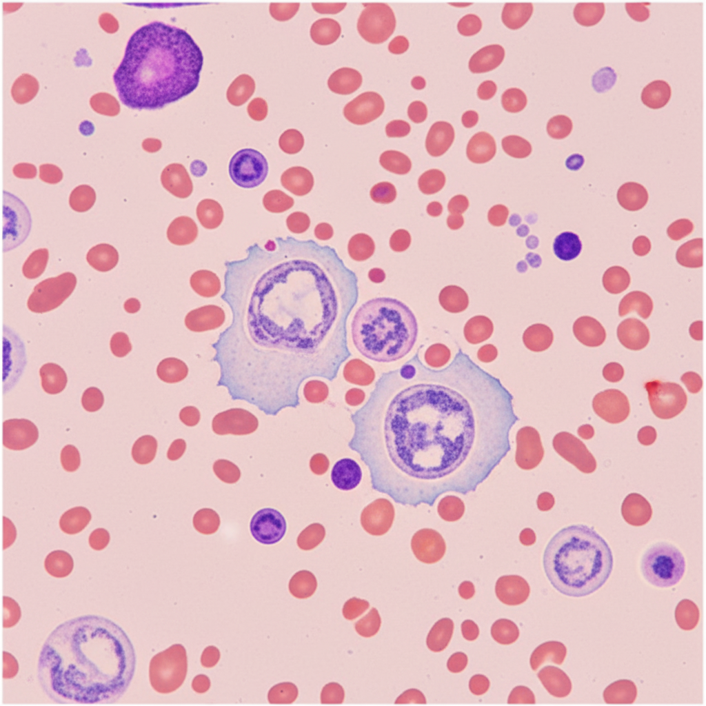
Gamma-Gandy bodies are seen in which organ?
Leucopenia is not seen in which of the following conditions?
Maximum magnitude of lymphocytosis is seen with which of the following conditions?
Sudden severe hemorrhage in a normal person causes all except:
All the following conditions cause thrombocytopenia except?
Abnormally high LAP score is seen in?
A leukemic patient develops disseminated intravascular coagulation. Examination of the marrow reveals hypergranular promyelocytes, some of which contain multiple Auer rods. The diagnosis of acute promyelocytic leukemia is made. Which of the following translocations is associated with the development of this disorder?
Crew haircut appearance on X-ray skull and Gandy gamma bodies are characteristic findings in which of the following conditions?
Explanation: **Explanation:** The core concept in hematopathology is distinguishing between **quantitative** and **qualitative** platelet disorders. **1. Why Option A is Correct:** Platelet function defects (e.g., Glanzmann Thrombasthenia, Bernard-Soulier Syndrome, or aspirin-induced dysfunction) are **qualitative** disorders [1]. In these conditions, the **number** of platelets is typically within the reference range (1.5–4.5 lakh/µL), but their ability to form a primary hemostatic plug is impaired [4]. Since **Bleeding Time (BT)** is the clinical marker for primary hemostasis (platelet-vessel wall interaction), it becomes **prolonged** despite a normal count. **2. Why Other Options are Incorrect:** * **Option B:** This describes **Thrombocytopenia** (e.g., ITP) [5]. While BT is prolonged here, it is due to a lack of numbers, not a functional defect. * **Option C:** Thrombocytosis (high count) usually presents with normal or decreased BT, though extreme thrombocytosis (Essential Thrombocythemia) can paradoxically cause bleeding due to acquired von Willebrand Syndrome. * **Option D:** This represents normal physiology or secondary hemostasis defects (e.g., Hemophilia), where platelets are functional, but clotting factors are deficient. **3. High-Yield Clinical Pearls for NEET-PG:** * **Bernard-Soulier Syndrome:** Deficiency of **GpIb-IX-V** (Adhesion defect) [2]. Characterized by **Giant Platelets** and mild thrombocytopenia (the only major exception where count may be low). * **Glanzmann Thrombasthenia:** Deficiency of **GpIIb/IIIa** (Aggregation defect) [2]. Platelet morphology is normal. * **Ristocetin Aggregation Test:** Absent in Bernard-Soulier and vWD; corrected by adding normal plasma only in vWD. * **Drug-induced:** Aspirin causes irreversible inhibition of **COX-1**, leading to a functional defect for the lifespan of the platelet (7–10 days). Other drugs like Clopidogrel inhibit ADP-mediated activation [3]. **References:** [1] Kumar V, Abbas AK, et al.. Robbins and Cotran Pathologic Basis of Disease. 9th ed. Red Blood Cell and Bleeding Disorders, pp. 668-669. [2] Kumar V, Abbas AK, et al.. Robbins and Cotran Pathologic Basis of Disease. 9th ed. Hemodynamic Disorders, Thromboembolic Disease, and Shock, p. 128. [3] Cross SS. Underwood's Pathology: A Clinical Approach. 6th ed. Common Clinical Problems From Blood And Bone Marrow Disease, pp. 621-622. [4] Cross SS. Underwood's Pathology: A Clinical Approach. 6th ed. Common Clinical Problems From Blood And Bone Marrow Disease, pp. 619-620. [5] Kumar V, Abbas AK, et al.. Robbins and Cotran Pathologic Basis of Disease. 9th ed. Red Blood Cell and Bleeding Disorders, pp. 666-667.
Explanation: ### Explanation **Correct Answer: D. Nodular Lymphocyte Predominant Hodgkin's Lymphoma (NLPHL)** **Why it is correct:** The clinical presentation of a young male with localized, slow-growing, asymptomatic (no "B" symptoms), and non-matted cervical lymphadenopathy is classic for NLPHL [1]. Unlike Classical Hodgkin Lymphoma (CHL), NLPHL often presents in younger males (3rd–4th decade) as localized Stage I or II disease [1]. Histologically, it is characterized by **"Popcorn cells"** (L&H cells—Lymphocytic and Histiocytic variants) which express B-cell markers like **CD20 and CD45**, while being negative for CD15 and CD30 [1]. **Why incorrect options are wrong:** * **A. Mixed Cellularity HL:** Typically presents with systemic "B" symptoms (fever, weight loss) and is strongly associated with EBV [1]. The background shows a pleomorphic infiltrate of eosinophils, plasma cells, and macrophages [1]. * **B. Lymphocyte Depleted HL:** The rarest and most aggressive subtype, usually seen in older or HIV-positive patients with advanced-stage disease and a poor prognosis [2]. * **C. Nodular Sclerosis HL:** While it is the most common subtype overall, it typically presents in young females with a **mediastinal mass** [1]. Histology shows lacunar cells and collagen bands encircling nodules [1]. **NEET-PG High-Yield Pearls:** * **NLPHL Marker Profile:** CD20+, CD45+, CD15–, CD30– (The "Non-Classical" profile) [1]. * **Classical HL Marker Profile:** CD15+, CD30+, CD20–, CD45–. * **Consistency:** "Rubbery" lymph nodes are a hallmark of lymphomas, whereas "matted" nodes suggest TB or metastatic carcinoma. * **Popcorn Cells:** These are the diagnostic Reed-Sternberg variants specific to NLPHL [1]. **References:** [1] Kumar V, Abbas AK, et al.. Robbins and Cotran Pathologic Basis of Disease. 9th ed. Diseases of White Blood Cells, Lymph Nodes, Spleen, and Thymus, pp. 616-618. [2] Cross SS. Underwood's Pathology: A Clinical Approach. 6th ed. Common Clinical Problems From Diseases Of The Urinary And Male Genital Tracts, pp. 559-560.
Explanation: **Explanation:** **Gamma-Gandy bodies** (also known as Siderofibrotic nodules) are small, firm, brownish-yellow nodules found in the **Spleen**. They represent organized areas of focal hemorrhage within the splenic parenchyma [1]. **Why Spleen is the correct answer:** The underlying mechanism is chronic venous congestion, most commonly due to **Portal Hypertension** (secondary to Cirrhosis) or Congestive Heart Failure [1]. The increased pressure leads to the rupture of small follicular arteries. As the blood extravasates, it undergoes organization, resulting in deposits of **hemosiderin** (iron), **calcium salts**, and **fibrous tissue** on the splenic connective tissue framework. On microscopic examination, these appear as characteristic "tobacco-leaf" or "bamboo-stick" shaped structures when stained with Perls’ Prussian Blue [1]. **Why other options are incorrect:** * **Liver:** While the liver is the primary site of pathology in portal hypertension (cirrhosis), Gamma-Gandy bodies are a sequela of the resulting splenic congestion, not a feature of the liver itself. * **Kidney & Lung:** These organs do not typically exhibit the specific fibro-siderotic organization seen in the spleen. While the lungs show "Heart Failure Cells" (hemosiderin-laden macrophages) in chronic congestion, they do not form organized Gamma-Gandy nodules. **High-Yield Clinical Pearls for NEET-PG:** * **Composition:** Iron (hemosiderin), Calcium, and Fibrous tissue. * **Stain:** Perls’ Prussian Blue (highlights the iron content). * **Classic Association:** Portal Hypertension and Sickle Cell Anemia (due to chronic congestion and infarcts) [1]. * **Imaging:** On MRI, they appear as "signal voids" (hypointense) due to the paramagnetic effect of iron. **References:** [1] Kumar V, Abbas AK, et al.. Robbins and Cotran Pathologic Basis of Disease. 9th ed. Diseases of White Blood Cells, Lymph Nodes, Spleen, and Thymus, pp. 631-634.
Explanation: **Explanation:** The correct answer is **Pyogenic bacterial infection**. **1. Why Pyogenic bacterial infection is the correct answer:** Pyogenic (pus-forming) bacterial infections, such as those caused by *Staphylococcus aureus* or *Streptococcus pneumoniae*, typically trigger a **Leucocytosis** (specifically neutrophilia) [1], [2]. This occurs due to the release of inflammatory cytokines (IL-1, TNF-α) that stimulate the bone marrow to release stored neutrophils and increase production [3]. In contrast, leucopenia refers to a decrease in the total white blood cell count (<4,000/mm³). **2. Analysis of Incorrect Options:** * **Viral infection:** Most viral infections (e.g., Influenza, Hepatitis, HIV) characteristically cause leucopenia and relative lymphocytosis due to the redistribution of lymphocytes and bone marrow suppression [3]. * **Malnutrition:** Severe nutritional deficiencies (e.g., Vitamin B12, Folic acid, or protein-energy malnutrition) lead to impaired DNA synthesis and marrow hypoplasia, resulting in leucopenia or even pancytopenia. * **Enteric fever:** Caused by *Salmonella typhi*, this is a classic exception to the rule of bacterial infections. It typically presents with **leucopenia and eosinopenia** due to the sequestration of cells in the spleen and bone marrow suppression by endotoxins [3]. **3. High-Yield Clinical Pearls for NEET-PG:** * **Left Shift:** The presence of immature neutrophils (band cells, metamyelocytes) in the peripheral blood during pyogenic infections. * **Leukemoid Reaction:** An extreme elevation of WBC count (>50,000/mm³) mimicking leukemia, often seen in severe pyogenic infections or acute hemolysis [2]. * **Exceptions:** While most bacterial infections cause leucocytosis, **Enteric fever, Brucellosis, and Miliary Tuberculosis** are notable bacterial causes of leucopenia [3]. * **Eosinopenia:** A highly specific finding in the early stages of Enteric fever. **References:** [1] Kumar V, Abbas AK, et al.. Robbins and Cotran Pathologic Basis of Disease. 9th ed. Diseases of White Blood Cells, Lymph Nodes, Spleen, and Thymus, p. 592. [2] Cross SS. Underwood's Pathology: A Clinical Approach. 6th ed. Common Clinical Problems From Blood And Bone Marrow Disease, pp. 580-581. [3] Kumar V, Abbas AK, et al.. Robbins and Cotran Pathologic Basis of Disease. 9th ed. Inflammation and Repair, pp. 110-111.
Explanation: **Explanation:** **Infectious Mononucleosis (IM)**, caused by the Epstein-Barr Virus (EBV), is characterized by a profound absolute lymphocytosis. [1] The hallmark of IM is the presence of **atypical lymphocytes (Downey cells)**, which are actually activated CD8+ T-killer cells responding to EBV-infected B-cells. [1] In IM, the total leukocyte count typically ranges from 12,000 to 18,000 cells/µL, with lymphocytes often exceeding 60% of the differential count. [1] This represents the highest magnitude of reactive lymphocytosis among the given options. **Analysis of Incorrect Options:** * **Pulmonary Tuberculosis:** While chronic infections like TB can cause lymphocytosis, it is usually mild to moderate. [3] The primary response is often granulomatous inflammation rather than a massive surge in peripheral blood lymphocytes. * **Typhoid Fever:** Classically presents with **leukopenia** and relative lymphocytosis, but the absolute magnitude does not reach the levels seen in viral infections like IM. [2] * **Bacterial Pneumonia:** Typically results in **neutrophilic leukocytosis** with a "left shift" (increased band cells). [3] Lymphocyte counts are usually normal or relatively decreased. **High-Yield Clinical Pearls for NEET-PG:** * **Atypical Lymphocytes:** Also seen in CMV, Toxoplasmosis, and Viral Hepatitis, but most characteristic of IM. * **Paul Bunnell Test / Monospot Test:** Detects heterophile antibodies produced during EBV infection. * **Triad of IM:** Fever, Pharyngitis, and Lymphadenopathy (often posterior cervical). [1] * **Warning:** Avoid Ampicillin/Amoxicillin in suspected IM as it can trigger a characteristic maculopapular rash. **References:** [1] Kumar V, Abbas AK, et al.. Robbins and Cotran Pathologic Basis of Disease. 9th ed. Infectious Diseases, pp. 368-370. [2] Kumar V, Abbas AK, et al.. Robbins and Cotran Pathologic Basis of Disease. 9th ed. Inflammation and Repair, pp. 110-111. [3] Kumar V, Abbas AK, et al.. Robbins and Cotran Pathologic Basis of Disease. 9th ed. Diseases of White Blood Cells, Lymph Nodes, Spleen, and Thymus, p. 592.
Explanation: **Explanation:** In the event of **sudden severe hemorrhage** (acute blood loss), the body loses whole blood—meaning both plasma and red blood cells are lost in equal proportions. **Why "Low MCV" is the correct answer:** MCV (Mean Corpuscular Volume) measures the average size of red blood cells. In acute hemorrhage, the remaining RBCs in circulation are those that were already present before the bleed; they are mature, normocytic (normal size), and normochromic [1]. Microcytosis (Low MCV) only occurs in **chronic** blood loss, where iron stores are gradually depleted, leading to iron deficiency anemia [2]. Therefore, acute hemorrhage results in a **normocytic normochromic anemia**, not a low MCV. **Analysis of Incorrect Options:** * **Low red cell mass:** This is a direct consequence of hemorrhage. While the initial hematocrit may appear normal due to proportional loss of plasma and cells, the absolute total red cell mass in the body is significantly reduced [3]. * **Reticulocytosis:** Within 5–7 days of a major bleed, the kidneys sense hypoxia and release erythropoietin. This stimulates the bone marrow to produce new RBCs, leading to an increase in immature red cells (reticulocytes) in the peripheral blood. * **Thrombocytosis:** Acute blood loss triggers a "stress response." The bone marrow increases production of all cell lines, and platelets are released rapidly from the splenic pool and marrow to aid in hemostasis [2]. **High-Yield Clinical Pearls for NEET-PG:** * **Immediate Phase:** Hematocrit/Hemoglobin may be normal immediately after a bleed because plasma and cells are lost together. * **24–72 Hours:** Hemodilution occurs as interstitial fluid shifts into the vascular space, causing the Hb/Hct to drop. * **Morphology:** Acute blood loss = Normocytic Normochromic; Chronic blood loss = Microcytic Hypochromic (Iron deficiency). **References:** [1] Kumar V, Abbas AK, et al.. Robbins and Cotran Pathologic Basis of Disease. 9th ed. Red Blood Cell and Bleeding Disorders, p. 638. [2] Cross SS. Underwood's Pathology: A Clinical Approach. 6th ed. Common Clinical Problems From Blood And Bone Marrow Disease, pp. 590-591. [3] Cross SS. Underwood's Pathology: A Clinical Approach. 6th ed. Common Clinical Problems From Blood And Bone Marrow Disease, pp. 587-588.
Explanation: **Explanation:** The correct answer is **Iron deficiency anemia (IDA)** because it is typically associated with **reactive thrombocytosis** (an increase in platelet count) rather than thrombocytopenia. This occurs because erythropoietin, which is elevated in IDA, has a structural similarity to thrombopoietin, leading to cross-stimulation of megakaryocytes. **Analysis of Options:** * **Iron deficiency anemia (Option D):** In most cases, IDA presents with a normal or elevated platelet count. Thrombocytopenia is extremely rare in IDA and only occurs in cases of severe, long-standing nutritional depletion. * **Giant hemangioma (Option A):** This refers to **Kasabach-Merritt Syndrome**, where platelets are sequestered and consumed within the vascular channels of a large hemangioma, leading to consumptive thrombocytopenia [2]. * **Infectious mononucleosis (Option B):** Caused by the Epstein-Barr virus (EBV), it can cause thrombocytopenia through immune-mediated destruction (anti-platelet antibodies) or transient bone marrow suppression. * **HIV infection (Option C):** Thrombocytopenia is a common hematological manifestation of HIV [1]. It occurs via multiple mechanisms: direct infection of megakaryocytes, immune-mediated destruction (ITP-like picture), and hypersplenism [1]. **NEET-PG High-Yield Pearls:** 1. **Kasabach-Merritt Syndrome:** Triad of Giant Hemangioma + Thrombocytopenia + Consumptive Coagulopathy. 2. **Reactive Thrombocytosis:** Common causes include IDA, acute hemorrhage, inflammation, and post-splenectomy. 3. **HIV & Platelets:** Thrombocytopenia can be the initial presenting sign of an undiagnosed HIV infection. **References:** [1] Kumar V, Abbas AK, et al.. Robbins and Cotran Pathologic Basis of Disease. 9th ed. Red Blood Cell and Bleeding Disorders, pp. 665-667. [2] Cross SS. Underwood's Pathology: A Clinical Approach. 6th ed. Common Clinical Problems From Blood And Bone Marrow Disease, pp. 619-621.
Explanation: ### Explanation **Leukocyte Alkaline Phosphatase (LAP)**, also known as the Neutrophil Alkaline Phosphatase (NAP) score, measures the activity of the enzyme alkaline phosphatase within the secondary granules of mature neutrophils. **1. Why Polycythemia Vera (PV) is correct:** Polycythemia Vera is a chronic myeloproliferative neoplasm (MPN) characterized by the autonomous overproduction of erythroid, myeloid, and megakaryocytic cells [1]. In PV, the neutrophils produced are functionally mature and possess increased enzyme activity, leading to an **elevated LAP score**. This helps distinguish PV from secondary polycythemia (where the score is usually normal) [3]. **2. Why the other options are incorrect:** * **Chronic Myeloid Leukemia (CML):** This is a classic high-yield distinction. In CML, despite a high white cell count, the LAP score is **characteristically low or zero**. This is because the neoplastic neutrophils are biochemically defective. (Note: The score may rise during a "blast crisis" or concurrent infection). * **Paroxysmal Nocturnal Hemoglobinuria (PNH):** This is an acquired stem cell disorder where there is a deficiency of GPI-anchored proteins on the cell membrane. Since LAP is a GPI-linked protein, its expression is **decreased** in PNH. **3. NEET-PG High-Yield Pearls:** * **High LAP Score:** Leukemoid reaction (most common cause), Polycythemia Vera, Essential Thrombocythemia, Myelofibrosis, Pregnancy, and use of Steroids [2]. * **Low LAP Score:** CML, PNH, Hypophosphatasia, and sometimes Aplastic Anemia or Pernicious Anemia. * **Normal Range:** 40–100. * **Key Differentiator:** The LAP score is the gold standard for differentiating a **Leukemoid Reaction (High)** from **CML (Low)**. **References:** [1] Kumar V, Abbas AK, et al.. Robbins and Cotran Pathologic Basis of Disease. 9th ed. Diseases of White Blood Cells, Lymph Nodes, Spleen, and Thymus, pp. 626-627. [2] Kumar V, Abbas AK, et al.. Robbins and Cotran Pathologic Basis of Disease. 9th ed. Diseases of White Blood Cells, Lymph Nodes, Spleen, and Thymus, p. 624. [3] Kumar V, Abbas AK, et al.. Robbins and Cotran Pathologic Basis of Disease. 9th ed. Red Blood Cell and Bleeding Disorders, pp. 663-664.
Explanation: **Explanation:** The clinical presentation of hypergranular promyelocytes, multiple Auer rods (faggot cells), and the life-threatening complication of Disseminated Intravascular Coagulation (DIC) is pathognomonic for **Acute Promyelocytic Leukemia (APL)**, classified as AML-M3 in the FAB system [1]. **1. Why t(15;17) is correct:** The molecular hallmark of APL is the reciprocal translocation **t(15;17)(q22;q12)**. This results in the fusion of the **PML** (Promyelocytic Leukemia) gene on chromosome 15 and the **RARA** (Retinoic Acid Receptor Alpha) gene on chromosome 17 [1]. The resulting PML-RARA fusion protein blocks myeloid differentiation at the promyelocytic stage. Treatment with All-Trans Retinoic Acid (ATRA) overcomes this block, inducing differentiation of the malignant cells. **2. Analysis of Incorrect Options:** * **t(4;11):** Associated with **MLL (KMT2A)** gene rearrangement, typically seen in infant ALL or therapy-related AML; it carries a poor prognosis. * **t(6;9):** Associated with **DEK-NUP214** fusion in AML; often presents with basophilia and multilineage dysplasia. * **t(8;14):** The classic translocation of **Burkitt Lymphoma**, involving the c-MYC oncogene and the Ig heavy chain locus [2]. **High-Yield Clinical Pearls for NEET-PG:** * **Faggot Cells:** Promyelocytes containing bundles of Auer rods (seen in APL) [1]. * **DIC Risk:** APL is a medical emergency because the primary granules contain procoagulants and fibrinolytic enzymes that trigger DIC upon release. * **Treatment:** ATRA + Arsenic Trioxide (ATO) is now the standard of care. * **Differentiation Syndrome:** A potential complication of ATRA therapy characterized by fever, dyspnea, and pulmonary infiltrates. **References:** [1] Kumar V, Abbas AK, et al.. Robbins and Cotran Pathologic Basis of Disease. 9th ed. Diseases of White Blood Cells, Lymph Nodes, Spleen, and Thymus, pp. 620-621. [2] Kumar V, Abbas AK, et al.. Robbins and Cotran Pathologic Basis of Disease. 9th ed. Neoplasia, pp. 324-325.
Explanation: ### Explanation **Correct Option: D. Sickle cell anemia** The "crew-cut" (or hair-on-end) appearance on a skull X-ray is a classic sign of **extramedullary hematopoiesis**. In chronic hemolytic anemias like Sickle Cell Anemia (SCA) and Thalassemia, the bone marrow undergoes massive expansion to compensate for the shortened lifespan of RBCs [1]. This expansion thins the outer table of the skull and causes new bone trabeculae to orient perpendicularly, creating the characteristic radiographic appearance [2]. **Gandy-Gamma bodies** (Siderofibrotic nodules) are small, firm, brown-yellow nodules found in the spleen. They consist of organized areas of hemorrhage containing deposits of iron (hemosiderin) and calcium salts on a fibrous stroma. In SCA, chronic congestion and repeated splenic infarcts lead to the formation of these bodies, eventually progressing to **autosplenectomy** [1]. --- ### Why other options are incorrect: * **A. G-6-PD deficiency:** This is an episodic hemolytic anemia triggered by oxidative stress. It does not typically cause the chronic, massive marrow expansion required for a crew-cut appearance or the chronic splenic congestion seen in SCA. * **B. Hodgkin's lymphoma:** This is a malignancy of the lymphoid tissue. While it can involve the spleen, it typically presents with "Reed-Sternberg cells" and "Mottled/Porphyry spleen" (Hard-bake spleen), not Gandy-Gamma bodies. * **C. Hereditary spherocytosis:** While it can cause a crew-cut appearance due to chronic hemolysis, the spleen in HS is characterized by **splenomegaly** (congestion of the Cords of Billroth) rather than the fibrotic, shrunken state associated with Gandy-Gamma bodies and autosplenectomy. --- ### High-Yield Pearls for NEET-PG: * **Crew-cut appearance:** Most common in **Thalassemia major**, followed by Sickle Cell Anemia [2]. * **Autosplenectomy:** A hallmark of Sickle Cell Anemia (usually complete by adulthood) [1]. * **Gandy-Gamma bodies:** Also seen in Portal Hypertension and Congestive Splenomegaly. * **Howell-Jolly bodies:** Nuclear remnants in RBCs seen on peripheral smears of patients with SCA due to functional asplenia [1]. **References:** [1] Kumar V, Abbas AK, et al.. Robbins and Cotran Pathologic Basis of Disease. 9th ed. Red Blood Cell and Bleeding Disorders, pp. 644-645. [2] Kumar V, Abbas AK, et al.. Robbins and Cotran Pathologic Basis of Disease. 9th ed. Red Blood Cell and Bleeding Disorders, pp. 648-649.
Anemias: Classification and Approach
Practice Questions
Hemolytic Anemias
Practice Questions
Myeloproliferative Neoplasms
Practice Questions
Myelodysplastic Syndromes
Practice Questions
Acute Leukemias
Practice Questions
Chronic Leukemias
Practice Questions
Lymphomas and Lymphoid Neoplasms
Practice Questions
Plasma Cell Disorders
Practice Questions
Bleeding Disorders
Practice Questions
Thrombotic Disorders
Practice Questions
Get full access to all questions, explanations, and performance tracking.
Start For Free