All of the following are features of reversible cell injury, except?
Which of the following statements regarding pyroptosis is not true?
Which of the following is NOT affected in Graft-Versus-Host disease?
All of the following are autosomal dominant disorders, except?
Which of the following is NOT a component of the Virchow triad?
Psammoma bodies are seen in which of the following conditions?
Aneuploidy is seen with all except:
What does 'adeno lymphoma' refer to?
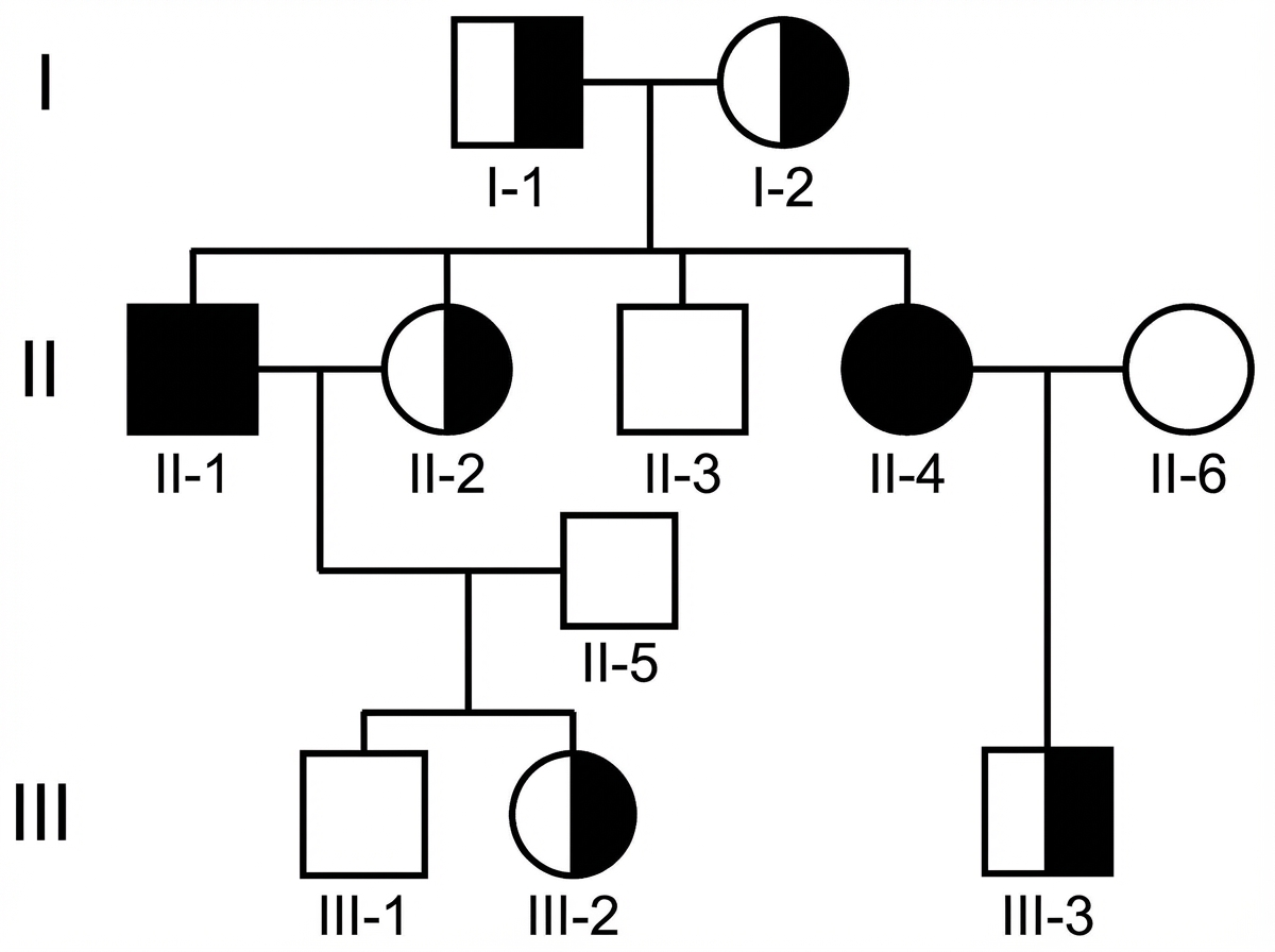
Examine the above pedigree chart. Which one of the following diseases is the most likely for this situation?

Trisomy 13 is identified as:
Explanation: ### Explanation The core concept in distinguishing reversible from irreversible cell injury lies in the **integrity of the nucleus and the cell membrane** [1]. **Why Karyorrhexis is the correct answer:** Karyorrhexis refers to the fragmentation of the pyknotic (condensed) nucleus. It is a hallmark of **irreversible cell injury** and cell death (necrosis) [4]. Once the nucleus undergoes significant structural changes—namely **Pyknosis** (clumping), **Karyorrhexis** (fragmentation), or **Karyolysis** (dissolution)—the cell has passed the "point of no return" and cannot recover. **Analysis of Incorrect Options (Features of Reversible Injury):** * **A. Endoplasmic reticulum (ER) swelling:** This occurs due to the failure of ATP-dependent ion pumps, leading to an influx of water (hydropic change) [1]. It is one of the earliest signs of reversible injury [2]. * **C. Plasma membrane blebbing:** While the membrane shows alterations like blebbing or loss of microvilli due to cytoskeletal damage, it remains **intact** [4]. If the membrane ruptures, the injury becomes irreversible [3]. * **D. Detachment of ribosomes:** Reduced ATP leads to the swelling of the ER, which causes ribosomes to detach from the Rough ER, resulting in decreased protein synthesis. This is reversible if oxygenation is restored. **NEET-PG High-Yield Pearls:** 1. **The "Point of No Return":** Irreversible injury is characterized by two main phenomena: the inability to reverse **mitochondrial dysfunction** and profound **disturbances in membrane function** (especially lysosomal and plasma membranes) [3]. 2. **Light Microscopy:** The earliest change visible under a light microscope in reversible injury is **cellular swelling** (cloudy swelling) [2]. 3. **Mitochondrial Changes:** Small, amorphous densities in mitochondria are seen in reversible injury, whereas **large, flocculent, electron-dense deposits** indicate irreversible injury [5]. 4. **Myocardial Infarction:** In cardiac muscle, irreversible injury typically occurs after 20–40 minutes of severe ischemia. **References:** [1] Kumar V, Abbas AK, et al.. Robbins and Cotran Pathologic Basis of Disease. 9th ed. Cellular Responses to Stress and Toxic Insults: Adaptation, Injury, and Death, pp. 49-50. [2] Kumar V, Abbas AK, et al.. Robbins and Cotran Pathologic Basis of Disease. 9th ed. Cellular Responses to Stress and Toxic Insults: Adaptation, Injury, and Death, pp. 51-53. [3] Kumar V, Abbas AK, et al.. Robbins and Cotran Pathologic Basis of Disease. 9th ed. Cellular Responses to Stress and Toxic Insults: Adaptation, Injury, and Death, pp. 60-61. [4] Kumar V, Abbas AK, et al.. Robbins and Cotran Pathologic Basis of Disease. 9th ed. Cellular Responses to Stress and Toxic Insults: Adaptation, Injury, and Death, p. 53. [5] Kumar V, Abbas AK, et al.. Robbins and Cotran Pathologic Basis of Disease. 9th ed. Cellular Responses to Stress and Toxic Insults: Adaptation, Injury, and Death, pp. 53-55.
Explanation: **Explanation:** **Pyroptosis** is a unique form of programmed cell death that bridges the gap between apoptosis and necrosis [1]. It is characterized by cell swelling, membrane lysis, and the release of pro-inflammatory cytokines. **Why Option B is the correct answer (False statement):** Pyroptosis is strictly a **caspase-dependent** process [1]. Unlike apoptosis (which involves Caspase 3, 8, and 9), pyroptosis is primarily mediated by **Caspase-1** (canonical pathway) or **Caspase-4, 5, and 11** (non-canonical pathway) [1]. These caspases cleave **Gasdermin D**, the "executioner" protein of pyroptosis, which forms pores in the plasma membrane. **Analysis of other options:** * **Option A (True):** The process is initiated by **NOD-like receptors (NLRs)**, which recognize pathogen-associated molecular patterns (PAMPs) [2]. These receptors assemble into a multi-protein complex called the **Inflammasome**, which activates Caspase-1 [2]. * **Option C (True):** Once Gasdermin D is cleaved, its N-terminal fragments insert into the host cell membrane to form large **pores**. This leads to water influx, osmotic swelling, and eventual membrane rupture. * **Option D (True):** Unlike apoptosis (which is silent), pyroptosis is **highly inflammatory** [1]. The rupture of the cell releases potent inflammatory cytokines like **IL-1β and IL-18**, along with DAMPs, triggering a robust immune response [1]. **NEET-PG High-Yield Pearls:** * **Key Molecule:** Gasdermin D (forms the membrane pore). * **Key Enzyme:** Caspase-1 (also known as IL-1β converting enzyme) [1]. * **Distinction:** It differs from apoptosis because it causes membrane rupture and inflammation; it differs from necrosis because it is a regulated, programmed process [1]. * **Clinical Link:** It plays a major role in the body's defense against intracellular pathogens (e.g., *Salmonella*, *Shigella*) [1]. **References:** [1] Kumar V, Abbas AK, et al.. Robbins and Cotran Pathologic Basis of Disease. 9th ed. Cellular Responses to Stress and Toxic Insults: Adaptation, Injury, and Death, pp. 69-71. [2] Kumar V, Abbas AK, et al.. Robbins and Cotran Pathologic Basis of Disease. 9th ed. Diseases of the Immune System, p. 196.
Explanation: **Explanation:** Graft-Versus-Host Disease (GVHD) occurs when immunocompetent T-cells from a donor graft recognize the recipient's HLA antigens as foreign and initiate an immune attack [1]. This typically occurs in the setting of hematopoietic stem cell transplantation. **Why Lung is the correct answer:** While GVHD is a multisystemic disorder, it characteristically targets epithelial surfaces. The **"Classic Triad"** of organs involved in acute GVHD includes the **Skin, Liver, and Gastrointestinal (GI) tract**. The lungs are generally **not** considered a primary target organ in the acute phase of GVHD [1]. While chronic GVHD can lead to pulmonary complications like *Bronchiolitis Obliterans*, it is not part of the diagnostic triad or the most frequently affected sites. **Analysis of Incorrect Options:** * **Skin (A):** The most common and often earliest manifestation. It typically presents as a maculopapular rash, often starting on the palms, soles, and neck, which can progress to generalized erythroderma [1]. * **Gastrointestinal Tract (B):** Involvement leads to mucosal ulceration, causing profuse watery or bloody diarrhea, abdominal pain, and ileus [1]. * **Liver (C):** Manifests as cholestatic jaundice due to the destruction of small bile ducts, leading to elevated bilirubin and alkaline phosphatase levels [1]. **High-Yield Clinical Pearls for NEET-PG:** * **Acute GVHD:** Occurs within 100 days of transplant. * **Chronic GVHD:** Occurs after 100 days; resembles autoimmune disorders like Scleroderma or Sjögren’s syndrome. * **Prevention:** To prevent GVHD, blood products for immunocompromised patients should be **irradiated** to eliminate donor T-cells. * **Mechanism:** Mediated primarily by **CD4+ and CD8+ T-cells** (Type IV Hypersensitivity). **References:** [1] Cross SS. Underwood's Pathology: A Clinical Approach. 6th ed. (Basic Pathology) introduces the student to key general principles of pathology, both as a medical science and as a clinical activity with a vital role in patient care. Part 2 (Disease Mechanisms) provides fundamental knowledge about the cellular and molecular processes involved in diseases, providing the rationale for their treatment. Part 3 (Systematic Pathology) deals in detail with specific diseases, with emphasis on the clinically important aspects., pp. 182-183.
Explanation: **Explanation:** The correct answer is **D. Cystic fibrosis**. **1. Why Cystic Fibrosis is the correct answer:** Cystic fibrosis (CF) is an **autosomal recessive (AR)** disorder [1] caused by a mutation in the *CFTR* gene on chromosome 7 [2]. In AR disorders, both copies of the gene must be mutated for the disease to manifest. These conditions typically involve deficiencies in **enzymes** rather than structural proteins [4]. CF is the most common lethal genetic disease in Caucasian populations [3] and is characterized by thick, viscid secretions affecting the lungs, pancreas, and reproductive system. **2. Why the other options are incorrect:** * **Neurofibromatosis (NF):** Both NF-1 (von Recklinghausen disease) and NF-2 are classic **autosomal dominant (AD)** disorders. NF-1 is caused by mutations in the *NF1* gene on chromosome 17. * **Ehlers-Danlos Syndrome (EDS):** While EDS is a heterogeneous group of disorders, the most common types (like the Classical and Hypermobility types) follow an **autosomal dominant** inheritance pattern, involving defects in collagen synthesis. * **Familial Hypercholesterolemia:** This is one of the most common **autosomal dominant** disorders, involving a mutation in the LDL receptor gene. It is a classic example of **References:** [1] Kumar V, Abbas AK, et al.. Robbins and Cotran Pathologic Basis of Disease. 9th ed. Genetic Disorders, pp. 150-151. [2] Kumar V, Abbas AK, et al.. Robbins and Cotran Pathologic Basis of Disease. 9th ed. Diseases of Infancy and Childhood, p. 476. [3] Cross SS. Underwood's Pathology: A Clinical Approach. 6th ed. (Basic Pathology) introduces the student to key general principles of pathology, both as a medical science and as a clinical activity with a vital role in patient care. Part 2 (Disease Mechanisms) provides fundamental knowledge about the cellular and molecular processes involved in diseases, providing the rationale for their treatment. Part 3 (Systematic Pathology) deals in detail with specific diseases, with emphasis on the clinically important aspects., pp. 120-122. [4] Cross SS. Underwood's Pathology: A Clinical Approach. 6th ed. (Basic Pathology) introduces the student to key general principles of pathology, both as a medical science and as a clinical activity with a vital role in patient care. Part 2 (Disease Mechanisms) provides fundamental knowledge about the cellular and molecular processes involved in diseases, providing the rationale for their treatment. Part 3 (Systematic Pathology) deals in detail with specific diseases, with emphasis on the clinically important aspects., pp. 57-58.
Explanation: **Explanation:** The **Virchow Triad** describes the three broad categories of factors that contribute to the formation of a thrombus (thrombogenesis) [1]. This is a foundational concept in vascular pathology. **Why Immunodeficiency is the Correct Answer:** Immunodeficiency refers to a failure of the immune system to protect the body from infection or cancer. While it may predispose a patient to infections that could indirectly lead to inflammation, it is **not** one of the three primary pillars of the Virchow Triad. Therefore, it does not directly contribute to the pathophysiology of thrombus formation. **Analysis of Incorrect Options:** 1. **Endothelial Injury (A):** This is the most important factor in the triad [1]. Damage to the vessel wall (due to trauma, hypertension, or inflammation) exposes subendothelial collagen and tissue factor, triggering platelet adhesion and the coagulation cascade [2]. 2. **Stasis (B):** Abnormal blood flow (stasis or turbulence) prevents the dilution of activated clotting factors and allows platelets to come into contact with the endothelium [1]. This is common in immobilized patients or at sites of aneurysms. 3. **Hypercoagulable States (C):** Also known as thrombophilia, this refers to any alteration of the coagulation pathways (e.g., Factor V Leiden mutation, Protein C/S deficiency, or malignancy) that predisposes the blood to clot [3]. **Clinical Pearls for NEET-PG:** * **Most common cause of inherited hypercoagulability:** Factor V Leiden mutation (resistance to activated Protein C). * **Lines of Zahn:** Microscopic laminations (pale platelet/fibrin layers vs. dark RBC layers) found only in thrombi formed in flowing blood, helping to distinguish a pre-mortem thrombus from a post-mortem clot. * **Trousseau Sign:** Migratory thrombophlebitis associated with visceral malignancies (especially pancreatic cancer), representing a secondary hypercoagulable state [3]. **References:** [1] Kumar V, Abbas AK, et al.. Robbins and Cotran Pathologic Basis of Disease. 9th ed. Hemodynamic Disorders, Thromboembolic Disease, and Shock, pp. 132-133. [2] Cross SS. Underwood's Pathology: A Clinical Approach. 6th ed. (Basic Pathology) introduces the student to key general principles of pathology, both as a medical science and as a clinical activity with a vital role in patient care. Part 2 (Disease Mechanisms) provides fundamental knowledge about the cellular and molecular processes involved in diseases, providing the rationale for their treatment. Part 3 (Systematic Pathology) deals in detail with specific diseases, with emphasis on the clinically important aspects., pp. 142-143. [3] Kumar V, Abbas AK, et al.. Robbins and Cotran Pathologic Basis of Disease. 9th ed. Diseases of Infancy and Childhood, pp. 522-523.
Explanation: **Explanation:** **1. Why Dystrophic Calcification is correct:** Psammoma bodies are classic examples of **dystrophic calcification**. This process occurs in dead or dying tissues despite **normal serum calcium and phosphate levels**. Pathologically, it begins with the deposition of calcium salts in mitochondria or membrane-bound vesicles of necrotic cells [4]. Over time, these deposits accumulate in concentric, laminated (onion-skin) layers, forming the characteristic microscopic round, grit-like structures known as Psammoma bodies [1]. **2. Why the other options are incorrect:** * **Necrosis (A):** While dystrophic calcification often occurs *within* areas of necrosis, "Necrosis" itself is a broad process of cell death. Psammoma bodies are a specific morphological manifestation of the calcification that follows, not the necrosis itself [3]. * **Apoptosis (B):** This is programmed cell death involving single cells and typically does not result in the large-scale mineral deposits required to form laminated Psammoma bodies [5]. * **Metastatic Calcification (C):** This occurs in **normal tissues** due to **hypercalcemia** (e.g., hyperparathyroidism) [1]. It typically affects interstitial tissues of the gastric mucosa, kidneys, and lungs, and does not form laminated Psammoma bodies [2]. **3. High-Yield Clinical Pearls for NEET-PG:** To remember the tumors associated with Psammoma bodies, use the mnemonic **"PSaMMoma"**: * **P:** **P**apillary carcinoma of the thyroid * **S:** **S**erous cystadenocarcinoma of the ovary * **M:** **M**eningioma * **M:** **M**esothelioma (Pleural) **Key Distinction:** Dystrophic calcification = Localized, damaged tissue, normal serum calcium. Metastatic calcification = Systemic, normal tissue, elevated serum calcium [1]. **References:** [1] Cross SS. Underwood's Pathology: A Clinical Approach. 6th ed. (Basic Pathology) introduces the student to key general principles of pathology, both as a medical science and as a clinical activity with a vital role in patient care. Part 2 (Disease Mechanisms) provides fundamental knowledge about the cellular and molecular processes involved in diseases, providing the rationale for their treatment. Part 3 (Systematic Pathology) deals in detail with specific diseases, with emphasis on the clinically important aspects., pp. 134-135. [2] Kumar V, Abbas AK, et al.. Robbins and Cotran Pathologic Basis of Disease. 9th ed. Cellular Responses to Stress and Toxic Insults: Adaptation, Injury, and Death, pp. 76-77. [3] Kumar V, Abbas AK, et al.. Robbins and Cotran Pathologic Basis of Disease. 9th ed. Cellular Responses to Stress and Toxic Insults: Adaptation, Injury, and Death, pp. 53-55. [4] Cross SS. Underwood's Pathology: A Clinical Approach. 6th ed. (Basic Pathology) introduces the student to key general principles of pathology, both as a medical science and as a clinical activity with a vital role in patient care. Part 2 (Disease Mechanisms) provides fundamental knowledge about the cellular and molecular processes involved in diseases, providing the rationale for their treatment. Part 3 (Systematic Pathology) deals in detail with specific diseases, with emphasis on the clinically important aspects., pp. 102-103. [5] Kumar V, Abbas AK, et al.. Robbins and Cotran Pathologic Basis of Disease. 9th ed. Cellular Responses to Stress and Toxic Insults: Adaptation, Injury, and Death, pp. 63-64.
Explanation: **Explanation:** The core concept tested here is the distinction between **Aneuploidy** and **Microdeletion syndromes**. **1. Why DiGeorge Syndrome is the correct answer:** Aneuploidy refers to a condition where the chromosome number is not an exact multiple of the haploid set (e.g., $2n+1$ or $2n-1$) [3]. **DiGeorge Syndrome** is not a numerical chromosomal abnormality; instead, it is a **microdeletion syndrome** involving a submicroscopic deletion of a specific region on the long arm of chromosome 22 (**22q11.2**) [2]. Because the total number of chromosomes remains 46, it is not classified as aneuploidy. **2. Analysis of Incorrect Options:** * **Trisomy 18 (Edwards) and 13 (Patau):** These are classic examples of autosomal aneuploidy where there is an extra copy of a chromosome ($47, XX/XY, +18$ or $+13$) [1]. * **Trisomy 21 (Down Syndrome):** The most common autosomal aneuploidy ($47, XX/XY, +21$), usually caused by meiotic non-disjunction [1], [4]. * **Klinefelter Syndrome:** A form of sex chromosome aneuploidy ($47, XXY$) [4]. **Clinical Pearls for NEET-PG:** * **DiGeorge Syndrome Mnemonic (CATCH-22):** **C**ardiac defects (Truncus arteriosus/TOF), **A**bnormal facies, **T**hymic hypoplasia (T-cell deficiency), **C**left palate, **H**ypocalcemia (due to parathyroid hypoplasia). * **Diagnosis:** Microdeletions like DiGeorge are too small to be seen on a standard karyotype and require **FISH** (Fluorescence In Situ Hybridization) or chromosomal microarray [2]. * **Most common cause of Aneuploidy:** Meiotic non-disjunction (maternal age is a major risk factor) [4]. **References:** [1] Kumar V, Abbas AK, et al.. Robbins and Cotran Pathologic Basis of Disease. 9th ed. Genetic Disorders, pp. 171-172. [2] Kumar V, Abbas AK, et al.. Robbins and Cotran Pathologic Basis of Disease. 9th ed. Genetic Disorders, p. 173. [3] Kumar V, Abbas AK, et al.. Robbins and Cotran Pathologic Basis of Disease. 9th ed. Genetic Disorders, pp. 168-169. [4] Cross SS. Underwood's Pathology: A Clinical Approach. 6th ed. (Basic Pathology) introduces the student to key general principles of pathology, both as a medical science and as a clinical activity with a vital role in patient care. Part 2 (Disease Mechanisms) provides fundamental knowledge about the cellular and molecular processes involved in diseases, providing the rationale for their treatment. Part 3 (Systematic Pathology) deals in detail with specific diseases, with emphasis on the clinically important aspects., pp. 92-93.
Explanation: **Explanation:** The term **'Adenolymphoma'** is the historical and descriptive synonym for **Warthin’s tumour** (also known as Papillary Cystadenoma Lymphomatosum). This name is derived from its unique histological appearance, which consists of two distinct components: a double layer of neoplastic **epithelial cells** (forming glandular/cystic structures) and a dense **lymphoid stroma** with germinal centers [1]. **Why Option C is correct:** Warthin’s tumour is the second most common benign salivary gland tumour. It occurs almost exclusively in the **parotid gland**. The "adeno" refers to the glandular epithelium, and "lymphoma" refers to the prominent lymphoid background, though it is a completely benign lesion and not a true malignancy of the immune system [1]. **Analysis of Incorrect Options:** * **A. Adenoid cystic carcinoma:** A common malignant salivary gland tumour characterized by a "Swiss-cheese" (cribriform) pattern. It is known for perineural invasion but lacks the lymphoid stroma of Warthin’s. * **B. Adenoid cystic lymphoma:** This is a non-existent clinical entity; it is a distractor combining terms from other pathologies. * **D. Pleomorphic adenoma:** The most common salivary gland tumour. It is a "mixed tumour" containing epithelial, myoepithelial, and mesenchymal-like (chondromyxoid) components, but it does not feature the characteristic lymphoid-rich papillary architecture of an adenolymphoma. **High-Yield NEET-PG Pearls:** * **Strongest Risk Factor:** Smoking (increases risk 8-fold). * **Location:** Tail of the parotid gland (most common). * **Key Feature:** It is the most common salivary gland tumour to present **bilaterally** or multicentrically (10% of cases). * **Hot Spot:** Shows increased uptake on **Technetium-99m pertechnetate scan** (Hot tumour). * **Demographics:** Traditionally more common in older males. **References:** [1] Kumar V, Abbas AK, et al.. Robbins and Cotran Pathologic Basis of Disease. 9th ed. Head and Neck, p. 753.
Explanation: ***Beta-thalassemia*** - Shows classic **autosomal recessive** inheritance pattern with **unaffected carrier parents** producing affected offspring, demonstrating **equal sex distribution** among affected individuals. - Consistent with the pedigree showing **affected children from consanguineous marriages** and **horizontal inheritance** pattern typical of recessive disorders. *Neurofibromatosis* - Follows **autosomal dominant** inheritance pattern where affected individuals would have at least **one affected parent** in each generation. - Would show **vertical transmission** through generations with approximately **50% risk** to offspring of affected individuals, which contradicts the pedigree pattern. *Colour blindness* - Demonstrates **X-linked recessive** inheritance with **male predominance** and affected males having **unaffected parents** but **carrier mothers**. - Would show characteristic pattern of **affected grandfathers** passing the trait through **carrier daughters** to affected grandsons, not the equal sex distribution seen. *Vitamin D resistant rickets* - Exhibits **X-linked dominant** inheritance where **affected males** pass the condition to **all daughters** but **no sons**. - Would demonstrate **no male-to-male transmission** and affected females having **50% risk** regardless of offspring sex, inconsistent with this pedigree.
Explanation: **Explanation:** **Trisomy 13** is clinically known as **Patau’s syndrome** [1]. It is a severe chromosomal abnormality characterized by the presence of an extra copy of chromosome 13. This occurs primarily due to meiotic non-disjunction, often associated with advanced maternal age [2]. **Analysis of Options:** * **Patau’s syndrome (Trisomy 13):** The correct answer. It is characterized by the "classic triad" of **Microphthalmia** (small eyes), **Cleft lip/palate**, and **Polydactyly** (extra fingers/toes). Other features include holoprosencephaly and "rocker-bottom" feet. * **Edward’s syndrome (Trisomy 18):** This involves an extra copy of chromosome 18 [1]. Key features include micrognathia (small jaw), low-set ears, and a characteristic **clenched hand** with overlapping fingers. * **Down’s syndrome (Trisomy 21):** The most common autosomal trisomy [2]. It presents with intellectual disability, flat facial profile, epicanthal folds [4], Simian crease, and an increased risk of Hirschsprung disease [3] and early-onset Alzheimer’s. * **Klinefelter’s syndrome (47, XXY):** This is a sex chromosome aneuploidy (not an autosomal trisomy). It affects males and is characterized by testicular atrophy, gynecomastia, and infertility. **High-Yield Clinical Pearls for NEET-PG:** * **Mnemonic for Trisomies:** **P**atau (13), **E**dward (18), **D**own (21) — remember them in increasing order of chromosome number as **P-E-D**. * **Rocker-bottom feet:** Seen in both Patau’s and Edward’s syndromes. * **Cutis Aplasia:** A high-yield specific finding for Patau’s syndrome (localized absence of skin on the scalp). * **Screening:** First-semester screening typically shows decreased PAPP-A and increased hCG in Down’s, but **decreased** PAPP-A and hCG in both Trisomy 13 and 18. **References:** [1] Kumar V, Abbas AK, et al.. Robbins and Cotran Pathologic Basis of Disease. 9th ed. Genetic Disorders, pp. 171-172. [2] Cross SS. Underwood's Pathology: A Clinical Approach. 6th ed. (Basic Pathology) introduces the student to key general principles of pathology, both as a medical science and as a clinical activity with a vital role in patient care. Part 2 (Disease Mechanisms) provides fundamental knowledge about the cellular and molecular processes involved in diseases, providing the rationale for their treatment. Part 3 (Systematic Pathology) deals in detail with specific diseases, with emphasis on the clinically important aspects., pp. 92-93. [3] Cross SS. Underwood's Pathology: A Clinical Approach. 6th ed. (Basic Pathology) introduces the student to key general principles of pathology, both as a medical science and as a clinical activity with a vital role in patient care. Part 2 (Disease Mechanisms) provides fundamental knowledge about the cellular and molecular processes involved in diseases, providing the rationale for their treatment. Part 3 (Systematic Pathology) deals in detail with specific diseases, with emphasis on the clinically important aspects., pp. 94-95. [4] Kumar V, Abbas AK, et al.. Robbins and Cotran Pathologic Basis of Disease. 9th ed. Genetic Disorders, pp. 170-171.
Cell Injury and Cell Death
Practice Questions
Adaptations of Cellular Growth
Practice Questions
Accumulations and Deposits
Practice Questions
Acute and Chronic Inflammation
Practice Questions
Tissue Repair and Wound Healing
Practice Questions
Hemodynamic Disorders
Practice Questions
Genetic Disorders
Practice Questions
Environmental Pathology
Practice Questions
Nutritional Diseases
Practice Questions
Molecular Basis of Disease
Practice Questions
Get full access to all questions, explanations, and performance tracking.
Start For Free