
General Pathology — MCQs

On this page
Fat necrosis is common in:
White infarcts are seen in which of the following?
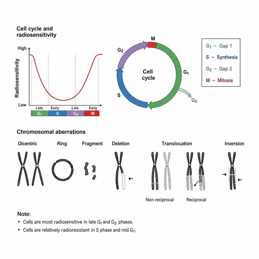
Which of these cells is most radio-resistant?
Which of the following is an anti-apoptotic gene?
Which of the following is most susceptible to ionizing radiation?
The following pattern of aberration occurs, when irradiation is done at which of the following phase of cell cycle?
Type of necrosis seen in case of burn:
Crumpled paper appearance of cells is a feature of-
Stain used in electron microscopy
Endotoxin shock is initiated by:
Practice by Chapter
Cell Injury and Cell Death
Practice Questions
Adaptations of Cellular Growth
Practice Questions
Accumulations and Deposits
Practice Questions
Acute and Chronic Inflammation
Practice Questions
Tissue Repair and Wound Healing
Practice Questions
Hemodynamic Disorders
Practice Questions
Genetic Disorders
Practice Questions
Environmental Pathology
Practice Questions
Nutritional Diseases
Practice Questions
Molecular Basis of Disease
Practice Questions
Want unlimited practice?
Get full access to all questions, explanations, and performance tracking.
Scan to download app