
General Pathology — MCQs

On this page
Identify the intracellular hyaline body.
Identify the structure marked in the image of cardiac myocyte. (AIIMS Nov 2018)
Ischemia-Reperfusion syndrome is characterized by:
Autosomal recessive disorders include all except :
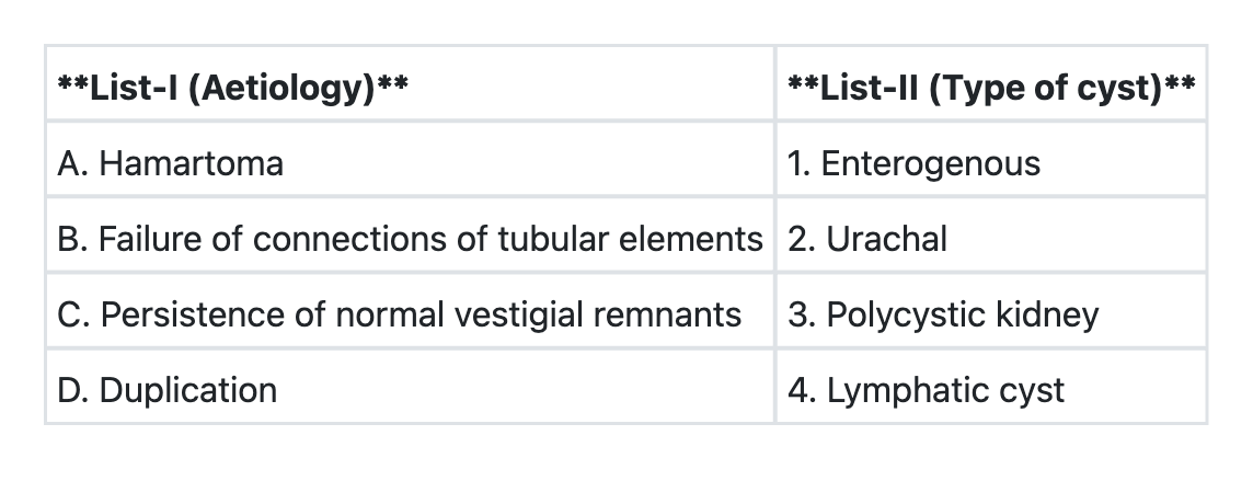
Match List-I with List-II and select the correct answer using the code given below the Lists:
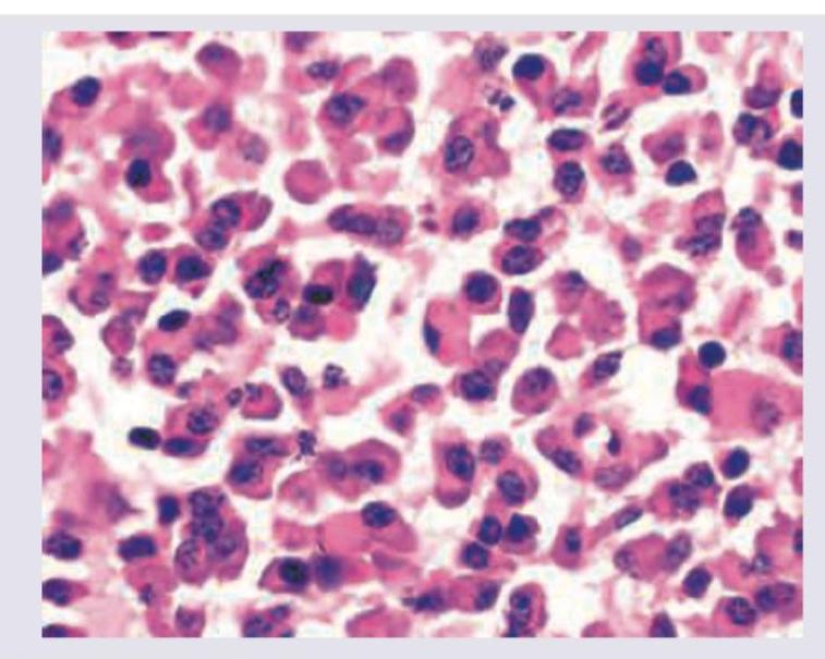
A patient presents with tingling sensation in both legs, polyuria, and weight loss. The bone marrow aspirate findings are shown in the image. What is the most likely diagnosis?
A child undergoes prophylactic irradiation as preparation for bone marrow transplantation (BMT) for treatment of acute lymphoblastic leukemia (ALL). Which of the following cell types will be least affected by the radiation?
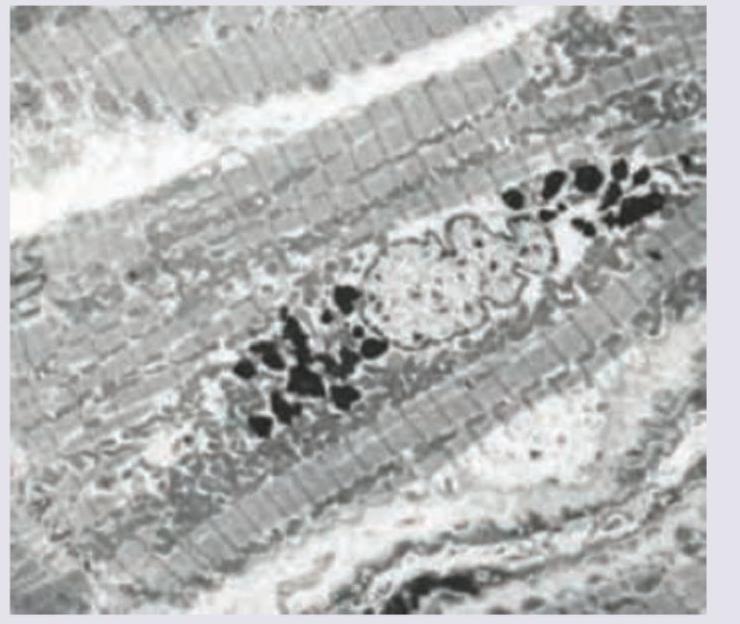
Iron in tissues is stained by:
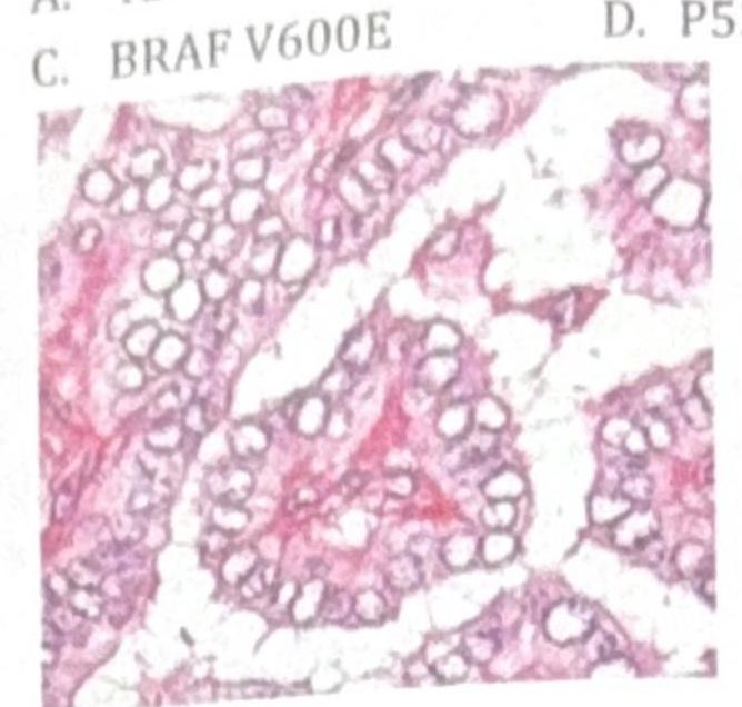
A baby is being evaluated for delayed developmental milestones. On examination, the child has hepatosplenomegaly. A microscopic image of the bone marrow evaluation is shown below. What is the most appropriate treatment?

Order of drawing blood in vacutainers should be in the following sequence to prevent contamination?
Practice by Chapter
Cell Injury and Cell Death
Practice Questions
Adaptations of Cellular Growth
Practice Questions
Accumulations and Deposits
Practice Questions
Acute and Chronic Inflammation
Practice Questions
Tissue Repair and Wound Healing
Practice Questions
Hemodynamic Disorders
Practice Questions
Genetic Disorders
Practice Questions
Environmental Pathology
Practice Questions
Nutritional Diseases
Practice Questions
Molecular Basis of Disease
Practice Questions
Want unlimited practice?
Get full access to all questions, explanations, and performance tracking.
Scan to download app