Autosomal recessive disorders include all except :
Which one of the following is due to the monosomy of X-chromosome?
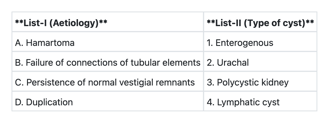
Match List-I with List-II and select the correct answer using the code given below the Lists:

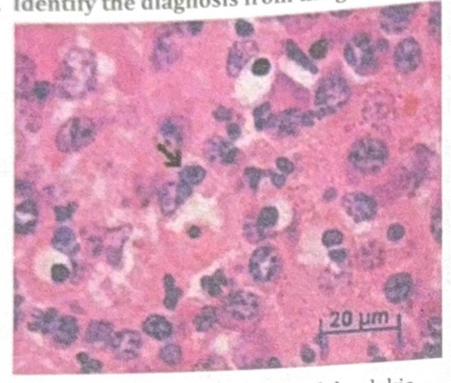
Identify the diagnosis from the given image

TTF-1 (Thyroid Transcription Factor-1) immunohistochemical marker is most commonly seen in which of the following?
A child undergoes prophylactic irradiation as preparation for bone marrow transplantation (BMT) for treatment of acute lymphoblastic leukemia (ALL). Which of the following cell types will be least affected by the radiation?
Iron in tissues is stained by:
A baby is being evaluated for delayed developmental milestones. On examination, the child has hepatosplenomegaly. A microscopic image of the bone marrow evaluation is shown below. What is the most appropriate treatment?

Abnormal accumulation of misfolded protein is seen in?
Order of drawing blood in vacutainers should be in the following sequence to prevent contamination?
Explanation: ***Retinoblastoma*** - Retinoblastoma is the correct answer because it is **NOT an autosomal recessive disorder**. - It follows **autosomal dominant** inheritance in hereditary cases (~40%), where a germline mutation in the RB1 gene is inherited. - Sporadic cases (~60%) result from somatic mutations in both RB1 alleles. - The **two-hit hypothesis** explains tumor development: both copies of the tumor suppressor RB1 gene must be inactivated. *Hirschsprung disease* - Hirschsprung disease has **complex multifactorial inheritance**, not autosomal recessive. - It involves multiple genes (RET, EDNRB, EDN3) with **incomplete penetrance and variable expressivity**. - However, it is not classified as a classic autosomal recessive disorder, making it technically debatable. - Characterized by **absence of ganglion cells** in the distal colon causing functional obstruction. *Albinism* - Oculocutaneous albinism (OCA types 1-4) is a classic **autosomal recessive** disorder [2]. - Results from mutations in genes involved in **melanin biosynthesis** (TYR, OCA2, TYRP1, SLC45A2) [2]. - Leads to reduced or absent pigmentation in skin, hair, and eyes [2]. *Sickle cell anaemia* - Sickle cell anemia is a well-established **autosomal recessive** hemoglobinopathy [1], [3]. - Caused by a point mutation (Glu6Val) in the **HBB gene** encoding beta-globin [3]. - Results in production of hemoglobin S (HbS), causing RBC sickling under hypoxic conditions [3]. **References:** [1] Kumar V, Abbas AK, et al.. Robbins and Cotran Pathologic Basis of Disease. 9th ed. Genetic Disorders, pp. 150-151. [2] Cross SS. Underwood's Pathology: A Clinical Approach. 6th ed. (Basic Pathology) introduces the student to key general principles of pathology, both as a medical science and as a clinical activity with a vital role in patient care. Part 2 (Disease Mechanisms) provides fundamental knowledge about the cellular and molecular processes involved in diseases, providing the rationale for their treatment. Part 3 (Systematic Pathology) deals in detail with specific diseases, with emphasis on the clinically important aspects., pp. 119-120. [3] Cross SS. Underwood's Pathology: A Clinical Approach. 6th ed. (Basic Pathology) introduces the student to key general principles of pathology, both as a medical science and as a clinical activity with a vital role in patient care. Part 2 (Disease Mechanisms) provides fundamental knowledge about the cellular and molecular processes involved in diseases, providing the rationale for their treatment. Part 3 (Systematic Pathology) deals in detail with specific diseases, with emphasis on the clinically important aspects., pp. 93-94.
Explanation: ***Turner's syndrome*** - Turner's syndrome is characterized by the **monosomy of the X chromosome** (genotype 45, XO), meaning an individual has only one X chromosome instead of the usual two [4]. - This chromosomal abnormality leads to a range of developmental issues, primarily affecting females, including **short stature**, **ovarian dysgenesis**, and a **webbed neck** [2]. *Klinefelter's syndrome* - This syndrome is due to the presence of an **extra X chromosome in males** (genotype 47, XXY), not a monosomy [1]. - It typically results in **testicular atrophy**, **gynecomastia**, and **tall stature** [1]. *Testicular feminization* - Also known as **Androgen Insensitivity Syndrome (AIS)**, this is a **genetic disorder** where individuals with XY chromosomes are unable to respond to androgens [3]. - It results in the development of female external genitalia, but it is due to a **receptor defect**, not a chromosomal monosomy [3]. *Adrenogenital syndrome* - This is primarily caused by **congenital adrenal hyperplasia (CAH)**, a group of genetic disorders affecting the adrenal glands' hormone production. - It is an **autosomal recessive disorder** involving enzyme deficiencies in cortisol synthesis, not a chromosomal monosomy. **References:** [1] Kumar V, Abbas AK, et al.. Robbins and Cotran Pathologic Basis of Disease. 9th ed. Genetic Disorders, pp. 174-175. [2] Kumar V, Abbas AK, et al.. Robbins and Cotran Pathologic Basis of Disease. 9th ed. Genetic Disorders, pp. 175-177. [3] Cross SS. Underwood's Pathology: A Clinical Approach. 6th ed. (Basic Pathology) introduces the student to key general principles of pathology, both as a medical science and as a clinical activity with a vital role in patient care. Part 2 (Disease Mechanisms) provides fundamental knowledge about the cellular and molecular processes involved in diseases, providing the rationale for their treatment. Part 3 (Systematic Pathology) deals in detail with specific diseases, with emphasis on the clinically important aspects., pp. 93-94. [4] Kumar V, Abbas AK, et al.. Robbins and Cotran Pathologic Basis of Disease. 9th ed. Genetic Disorders, pp. 173-174.
Explanation: ***A→4 B→3 C→2 D→1*** - A **hamartoma** is a benign, focal malformation resembling a neoplasm, composed of mature cells and tissues normally found in the organ from which it arises, but growing in a disorganized mass. A **lymphatic cyst** (or lymphangioma/cystic hygroma), though often confused with a true neoplasm, is a congenital malformation of the lymphatic system, a type of hamartoma [1]. - **Polycystic kidney disease** is characterized by the formation of numerous cysts in the kidneys. One of the theories for its pathogenesis involves the failure of connections between collecting tubules and nephrons during renal development, leading to isolated segments that dilate to form cysts [2], [3]. - The **urachus** is an embryonic remnant of the allantois, connecting the fetal bladder to the umbilicus. If the urachus fails to involute completely after birth, it can persist as a patent or partially patent structure, leading to various **urachal anomalies**, including urachal cysts. - **Duplication** can lead to the formation of an **enterogenous cyst**, which is a congenital cyst lined by typical gastrointestinal mucosa. These cysts arise from developmental errors during embryogenesis where portions of the primitive gut tube become duplicated or sequestered. *A→3 B→4 C→1 D→2* - This option incorrectly associates hamartoma with polycystic kidney and duplication with urachal cysts. - **Polycystic kidney** is primarily due to defects in tubular connections, not hamartomas, and **urachal cysts** are remnants of vestigial structures, not duplications. *A→3 B→1 C→4 D→2* - This option incorrectly associates hamartoma with polycystic kidney and persistence of normal vestigial remnants with lymphatic cyst. - **Polycystic kidney** is not a hamartoma, and **lymphatic cysts** are not typical vestigial remnants but rather developmental malformations of the lymphatic system [1]. *A→2 B→4 C→1 D→3* - This option incorrectly associates hamartoma with urachal cysts and persistence of normal vestigial remnants with enterogenous cysts. - **Urachal cysts** are vestigial remnants, not hamartomas. **Enterogenous cysts** are a result of duplication, not persistence of normal vestigial remnants. **References:** [1] Kumar V, Abbas AK, et al.. Robbins and Cotran Pathologic Basis of Disease. 9th ed. Diseases of Infancy and Childhood, pp. 481-482. [2] Cross SS. Underwood's Pathology: A Clinical Approach. 6th ed. Common Clinical Problems From Diseases Of The Urinary And Male Genital Tracts, pp. 544-545. [3] Kumar V, Abbas AK, et al.. Robbins and Cotran Pathologic Basis of Disease. 9th ed. The Kidney, pp. 951-952.
Explanation: ***Malakoplakia*** - The image displays characteristic **Michaelis-Gutmann bodies**, which are concentrically laminated calcified inclusions found within macrophages (**Von Hansemann cells**). These are pathognomic for malakoplakia. - **Von Hansemann cells** are large, foamy macrophages with abundant cytoplasm, also visible in the image, mixed with lymphocytes and plasma cells. *TB* - Tuberculosis (TB) typically presents with **granulomatous inflammation** characterized by caseating necrosis, epithelioid macrophages, Langhans giant cells, and lymphocytes [2]. These features are not apparent in the image. - While TB can involve macrophages, the distinct Michaelis-Gutmann bodies seen here are not a feature of tuberculous granulomas [1]. *BCC* - Basal cell carcinoma (BCC) is a malignant epithelial tumor characterized by nests of basaloid cells with peripheral palisading, clear clefts, and often stromal retraction. This biopsy shows inflammatory cells and calcified inclusions, not epithelial malignancy. - BCC would show atypical epithelial cells and features of a neoplastic process, which are distinctly different from the inflammatory infiltrate and inclusion bodies in the image. *Drug induced lesion* - Drug-induced lesions are highly variable and context-dependent, but they do not typically present with the specific histopathological features of Michaelis-Gutmann bodies within macrophages. - The image depicts a specific and recognizable inflammatory pattern with unique inclusions, which points to a distinct disease entity rather than a non-specific drug reaction. **References:** [1] Cross SS. Underwood's Pathology: A Clinical Approach. 6th ed. (Basic Pathology) introduces the student to key general principles of pathology, both as a medical science and as a clinical activity with a vital role in patient care. Part 2 (Disease Mechanisms) provides fundamental knowledge about the cellular and molecular processes involved in diseases, providing the rationale for their treatment. Part 3 (Systematic Pathology) deals in detail with specific diseases, with emphasis on the clinically important aspects., pp. 198-200. [2] Kumar V, Abbas AK, et al.. Robbins and Cotran Pathologic Basis of Disease. 9th ed. Infectious Diseases, p. 360.
Explanation: ***Lung adenocarcinoma*** - **TTF-1 (Thyroid Transcription Factor-1)** is a nuclear transcription factor that is highly expressed in adenocarcinomas of the lung. Positivity for TTF-1 is a key marker used in the diagnosis of primary lung adenocarcinoma, distinguishing it from other lung cancers and metastatic tumors. - While TTF-1 can also be positive in thyroid follicular and papillary carcinomas, its strong association with **lung adenocarcinoma** makes it a crucial diagnostic marker in this context, especially when differentiating between primary lung tumors and metastases or other lung cancer types. *Squamous Cell Carcinoma (SCC)* - **Squamous cell carcinoma of the lung** is generally **negative for TTF-1**. It typically expresses markers like p40 and CK5/6. - TTF-1 has very low sensitivity and specificity for squamous cell carcinoma, making it a poor choice for identifying this type of lung cancer. *Large cell lung cancer* - **Large cell lung carcinoma** is a diagnosis of exclusion and is typically **negative for TTF-1**, as well as other specific markers for adenocarcinoma or squamous cell carcinoma. - This type of cancer is characterized by large, anaplastic cells that lack features of other specific lung cancer types when viewed under a microscope. *Papillary thyroid carcinoma* - While **papillary thyroid carcinoma** is also **TTF-1 positive**, the question asks for the most common context in which TTF-1 is seen, and TTF-1 is a highly valuable marker for confirming a lung primary in the setting of lung masses. - TTF-1's utility in lung cancer diagnostics is particularly significant for differentiating primary lung adenocarcinomas from metastatic tumors and other lung cancer subtypes.
Explanation: ***Neurons*** - **Neurons** are highly differentiated cells with very low rates of cell division in adults. As radiation primarily targets rapidly dividing cells [4], **neurons are least susceptible** to radiation damage. - While high doses of radiation can eventually damage neurons, their **radioresistance** is significantly higher compared to rapidly proliferating tissues. *Spermatogonia* - **Spermatogonia** are germ cells that undergo continuous and rapid division to produce sperm, making them **highly sensitive to radiation** [2]. - Radiation exposure can lead to **sterility** due to the destruction of these rapidly dividing cells [2]. *Bone marrow* - The **bone marrow** contains hematopoietic stem cells that are responsible for the continuous production of blood cells, involving **rapid cell division** [3]. - It is one of the most **radiosensitive tissues** [1], and radiation exposure can lead to **myelosuppression** and pancytopenia. *Intestinal epithelial cells* - **Intestinal epithelial cells** have a high turnover rate due to their constant shedding and replacement [5], making them **very sensitive to radiation** [1]. - Radiation damage to these cells can cause **mucositis, nausea, vomiting, and diarrhea**. **References:** [1] Cross SS. Underwood's Pathology: A Clinical Approach. 6th ed. (Basic Pathology) introduces the student to key general principles of pathology, both as a medical science and as a clinical activity with a vital role in patient care. Part 2 (Disease Mechanisms) provides fundamental knowledge about the cellular and molecular processes involved in diseases, providing the rationale for their treatment. Part 3 (Systematic Pathology) deals in detail with specific diseases, with emphasis on the clinically important aspects., pp. 112-113. [2] Cross SS. Underwood's Pathology: A Clinical Approach. 6th ed. (Basic Pathology) introduces the student to key general principles of pathology, both as a medical science and as a clinical activity with a vital role in patient care. Part 2 (Disease Mechanisms) provides fundamental knowledge about the cellular and molecular processes involved in diseases, providing the rationale for their treatment. Part 3 (Systematic Pathology) deals in detail with specific diseases, with emphasis on the clinically important aspects., pp. 113-114. [3] Kumar V, Abbas AK, et al.. Robbins and Cotran Pathologic Basis of Disease. 9th ed. Inflammation and Repair, pp. 112-113. [4] Kumar V, Abbas AK, et al.. Robbins and Cotran Pathologic Basis of Disease. 9th ed. Central Nervous System Synapse, pp. 436-437. [5] Cross SS. Underwood's Pathology: A Clinical Approach. 6th ed. (Basic Pathology) introduces the student to key general principles of pathology, both as a medical science and as a clinical activity with a vital role in patient care. Part 2 (Disease Mechanisms) provides fundamental knowledge about the cellular and molecular processes involved in diseases, providing the rationale for their treatment. Part 3 (Systematic Pathology) deals in detail with specific diseases, with emphasis on the clinically important aspects., pp. 79-80.
Explanation: ***Prussian blue*** - The **Prussian blue reaction**, also known as **Perls' stain**, specifically detects **ferric iron (Fe³⁺)** in tissues [3]. - It forms a **bright blue precipitate** (ferric ferrocyanide) when ferric iron reacts with potassium ferrocyanide in an acidic solution. *Masson's trichrome* - This stain is primarily used to differentiate **collagen fibers** from muscle fibers, which appear blue/green and red, respectively [1]. - It does not specifically stain or identify iron deposits in tissues. *PAS* - The **Periodic acid-Schiff (PAS) stain** is used to identify **carbohydrates** such as glycogen, mucosubstances, and fungal cell walls, which appear magenta. - It is not used for the detection of iron. *Congo red* - **Congo red** is a specialized stain used for identifying **amyloid deposits**, which appear pink-red and show apple-green birefringence under polarized light [2]. - It is not used for staining iron in tissues. **References:** [1] Cross SS. Underwood's Pathology: A Clinical Approach. 6th ed. (Basic Pathology) introduces the student to key general principles of pathology, both as a medical science and as a clinical activity with a vital role in patient care. Part 2 (Disease Mechanisms) provides fundamental knowledge about the cellular and molecular processes involved in diseases, providing the rationale for their treatment. Part 3 (Systematic Pathology) deals in detail with specific diseases, with emphasis on the clinically important aspects., pp. 25-26. [2] Kumar V, Abbas AK, et al.. Robbins and Cotran Pathologic Basis of Disease. 9th ed. Diseases of the Immune System, pp. 268-269. [3] Cross SS. Underwood's Pathology: A Clinical Approach. 6th ed. Common Clinical Problems From Liver And Biliary System Disease, pp. 394-395.
Explanation: ***Biological therapy*** - The image likely shows a **Gaucher cell** (macrophage engorged with glucocerebroside, characterized by a "crinkled paper" or "crumpled tissue paper" cytoplasm) [1]. This is pathognomonic for **Gaucher disease**, which presents with **hepatosplenomegaly** and **developmental delay** in infants. - The standard treatment for Gaucher disease is **enzyme replacement therapy (ERT)**, which is a form of biological therapy replacing the deficient glucocerebrosidase enzyme [2]. *Macrolides for 4 months* - Macrolides are a class of **antibiotics** used to treat bacterial infections. - They are not indicated for lysosomal storage disorders like Gaucher disease, which is a **genetic metabolic disorder** [2]. *Steroid irrigation and antihistamines* - This treatment regimen is typically associated with allergic conditions or localized inflammatory responses, such as **allergic rhinitis** or **sinusitis**. - It bears no relevance to the systemic accumulation of lipids seen in Gaucher disease. *Repeat surgery* - While splenectomy was historically performed for massive splenomegaly in Gaucher disease, it does not address the underlying metabolic defect or systemic manifestations. - Furthermore, this case does not suggest a previous surgical intervention that requires repeating, and surgery is not the primary treatment for the metabolic disorder itself. **References:** [1] Kumar V, Abbas AK, et al.. Robbins and Cotran Pathologic Basis of Disease. 9th ed. Genetic Disorders, pp. 162-163. [2] Kumar V, Abbas AK, et al.. Robbins and Cotran Pathologic Basis of Disease. 9th ed. Genetic Disorders, p. 159.
Explanation: ***Creutzfeldt-Jakob disease*** - This is a neurodegenerative disease characterized by the accumulation of **abnormally folded prion proteins (PrPSc)** in the brain, leading to spongiform encephalopathy [1]. - The misfolding of normal cellular prion protein (PrPC) into its infectious and pathogenic form is central to the disease's pathology [2]. *Nephritic syndrome* - This syndrome is characterized by inflammation of the **glomeruli** in the kidneys, leading to hematuria, proteinuria, and hypertension. - It involves immune complex deposition and inflammation, not primarily the accumulation of misfolded proteins. *Sickle cell anemia* - This is a **genetic blood disorder** caused by a mutation in the beta-globin gene, leading to abnormal **hemoglobin S**. - While hemoglobin S can polymerize and deform red blood cells, it is not considered a disease of generalized misfolded protein accumulation in the same sense as prion diseases. *Megaloblastic anemia* - This condition is caused by impaired **DNA synthesis**, often due to **vitamin B12 or folate deficiency**, leading to large, immature red blood cells. - The pathology involves defective cell division and maturation, not the accumulation of misfolded proteins. **References:** [1] Kumar V, Abbas AK, et al.. Robbins and Cotran Pathologic Basis of Disease. 9th ed. The Central Nervous System, pp. 1284-1286. [2] Cross SS. Underwood's Pathology: A Clinical Approach. 6th ed. Common Clinical Manifestations Of Central And Peripheral Nervous System Disease, pp. 712-713.
Explanation: ***Blue, Red, Violet, Grey*** - This sequence follows the **CLSI (Clinical and Laboratory Standards Institute) order of draw** guidelines, which are critical for preventing **cross-contamination** between additives of different vacutainers. - The order starts with tubes for **coagulation studies** (blue top with sodium citrate), followed by serum tubes (red top), EDTA tubes (violet top for hematology), and finally glycolytic inhibitor tubes (grey top for glucose). - This prevents tissue thromboplastin contamination and anticoagulant carryover that could affect laboratory test results. *Grey, Blue, Red, Violet* - This order is incorrect as it places the **grey top tube** (containing fluoride/oxalate) first, which could contaminate subsequent tubes with its additives and affect tests. - The **blue top tube** for coagulation studies should come early in the sequence to minimize tissue thromboplastin contamination. *Blue, Violet, Red, Grey* - This sequence is incorrect because the **violet top tube** (EDTA) is placed before the **red top tube** (serum). - **EDTA contamination** can chelate calcium and other cations, interfering with chemistry tests performed on serum in the red top tube. *Red, Blue, Violet, Grey* - This order is incorrect as the **red top tube** is placed first, followed by the **blue top tube** (citrate). - The blue top tube should precede tubes with clot activators to **prevent tissue thromboplastin** from contaminating coagulation samples, which would lead to falsely shortened clotting times.
Cell Injury and Cell Death
Practice Questions
Adaptations of Cellular Growth
Practice Questions
Accumulations and Deposits
Practice Questions
Acute and Chronic Inflammation
Practice Questions
Tissue Repair and Wound Healing
Practice Questions
Hemodynamic Disorders
Practice Questions
Genetic Disorders
Practice Questions
Environmental Pathology
Practice Questions
Nutritional Diseases
Practice Questions
Molecular Basis of Disease
Practice Questions
Get full access to all questions, explanations, and performance tracking.
Start For Free