Which of the following is a change seen in irreversible cell injury?
Which of the following types of cell death involves activation of caspase enzymes?
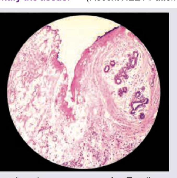
Identify the tissue shown in the histopathological image:

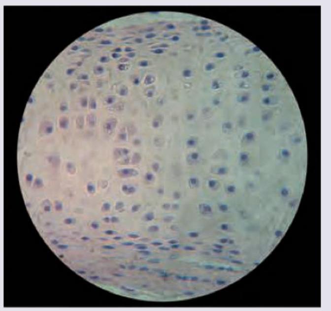
All are true about the cartilage shown in the figure except: (Recent NEET Pattern 2016-17)

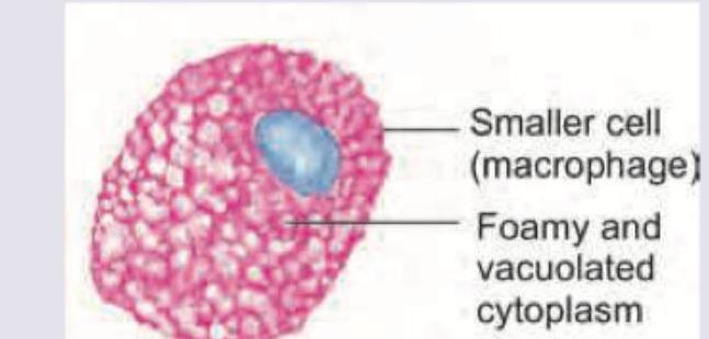
A 6-month-old child presents with loss of vision and regression of milestones. On examination hepatosplenomegaly and lymphadenopathy is seen. The bone marrow aspiration shows abnormal macrophage shown below. It stains positive for fat and negative for iron. Identify the cell.
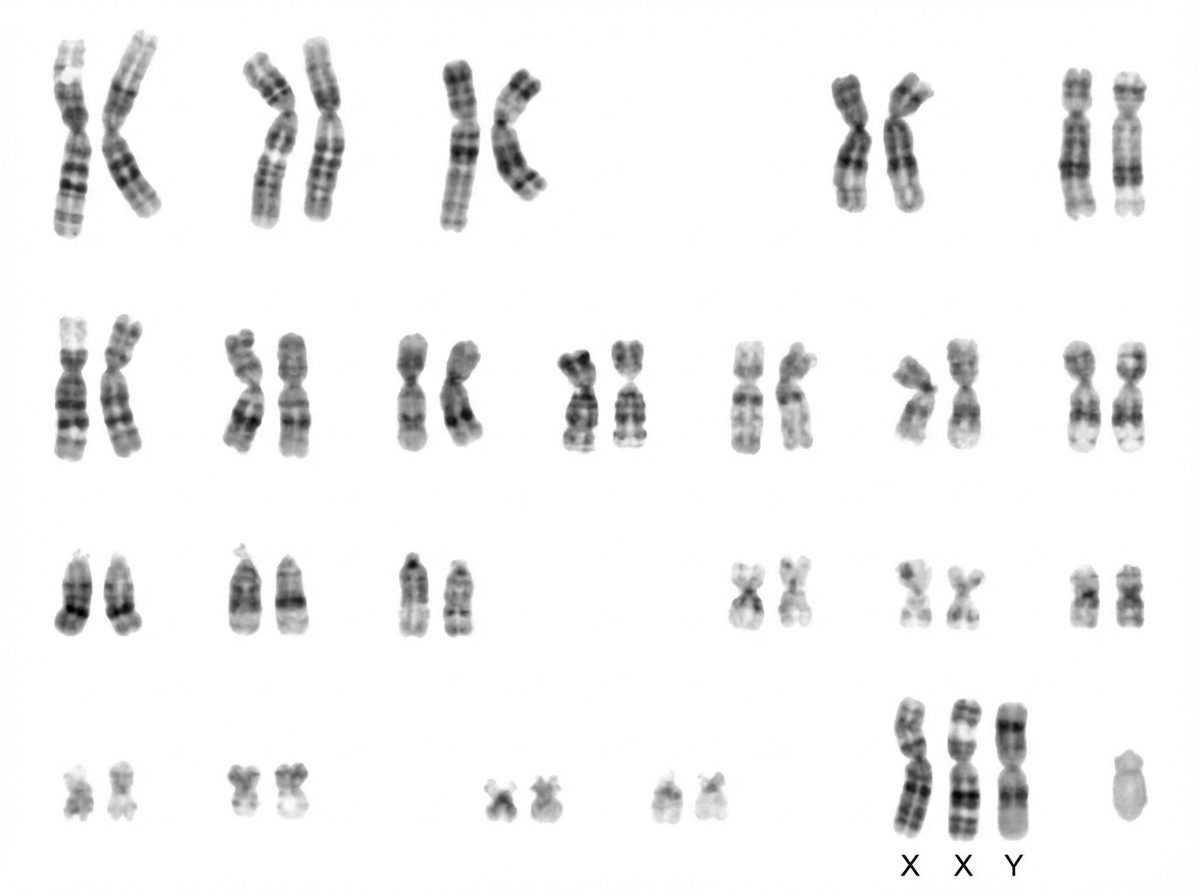
Comment on the diagnosis.

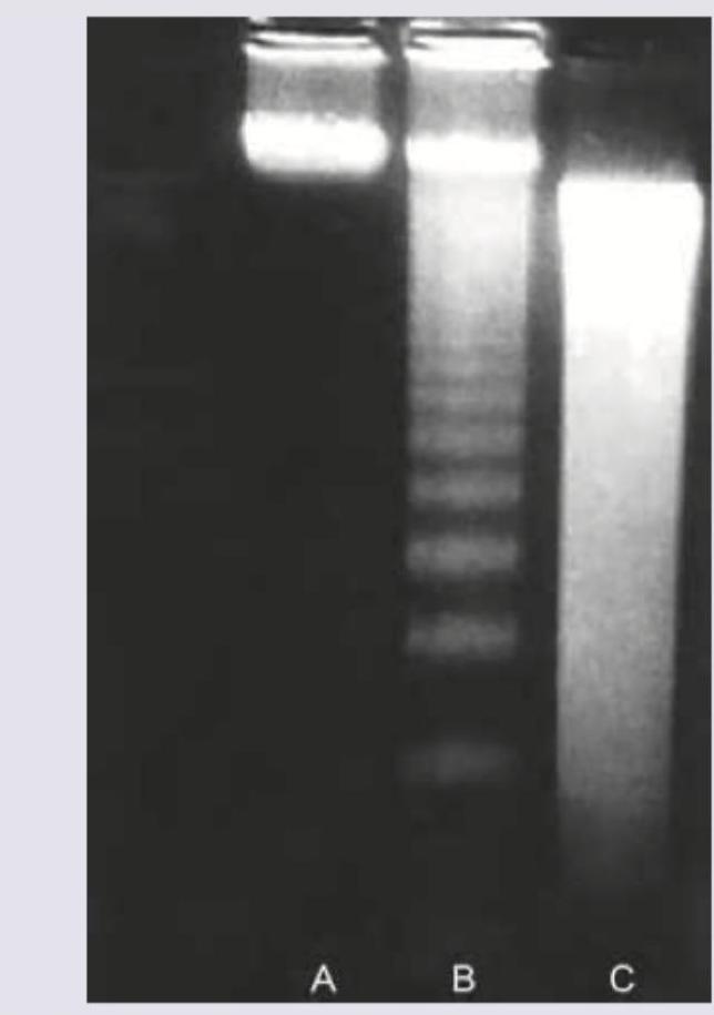
Agarose gel electrophoresis from DNA of a population of cells as seen under ultraviolet light is shown below. What is the correct explanation for the finding seen in the band labeled as "C"?

Identify the intracellular hyaline body.

Ischemia-Reperfusion syndrome is characterized by:
Which of the following are metabolic causes of splenic enlargement?
Explanation: ***Amorphous density of mitochondria***- This refers to the formation of large, irregular, dense calcium deposits within the mitochondrial matrix, which is a classic morphological feature indicating irreparable damage to the organelle [1].- The presence of these **amorphous densities** signifies severe mitochondrial dysfunction, including the complete loss of inner membrane potential and irreversible failure of **oxidative phosphorylation**, committing the cell to necrosis [2].*Bleb formation*- Small, transient surface blebs are common manifestations of **reversible** cell injury resulting from alterations in the cytoskeleton due to ATP depletion [1].- While uncontrolled blebbing is a feature preceding plasma membrane rupture in necrosis, its simple occurrence is not exclusively tied to irreversible injury, unlike profound mitochondrial changes [4].*Clumping of chromatin*- This initial condensation of nuclear material is characteristic of **reversible** cell injury, often due to decreased ATP causing a decrease in nuclear components and an acidic intracellular environment.- This reversible change must progress to irreversible nuclear hallmarks like **pyknosis** (irreversible condensation) or karyorrhexis/karyolysis to confirm cell death [4].*Cellular Swelling*- **Cellular swelling** (or hydropic change) is the most common manifestation of **reversible** cell injury [4], resulting from the failure of the ATP-dependent **Na+/K+ pump** [3].- This failure leads to the influx and accumulation of intracellular sodium and water, a condition that can be reversed if oxygenation and ATP synthesis are restored [3]. **References:** [1] Kumar V, Abbas AK, et al.. Robbins and Cotran Pathologic Basis of Disease. 9th ed. Cellular Responses to Stress and Toxic Insults: Adaptation, Injury, and Death, pp. 53-55. [2] Cross SS. Underwood's Pathology: A Clinical Approach. 6th ed. (Basic Pathology) introduces the student to key general principles of pathology, both as a medical science and as a clinical activity with a vital role in patient care. Part 2 (Disease Mechanisms) provides fundamental knowledge about the cellular and molecular processes involved in diseases, providing the rationale for their treatment. Part 3 (Systematic Pathology) deals in detail with specific diseases, with emphasis on the clinically important aspects., pp. 102-103. [3] Kumar V, Abbas AK, et al.. Robbins and Cotran Pathologic Basis of Disease. 9th ed. Cellular Responses to Stress and Toxic Insults: Adaptation, Injury, and Death, pp. 56-57. [4] Kumar V, Abbas AK, et al.. Robbins and Cotran Pathologic Basis of Disease. 9th ed. Cellular Responses to Stress and Toxic Insults: Adaptation, Injury, and Death, p. 53.
Explanation: ***Apoptosis and Pyroptosis*** - **Apoptosis** is the classic caspase-dependent programmed cell death pathway [1] - Involves **initiator caspases** (Caspase-8, Caspase-9) and **executioner caspases** (Caspase-3, -6, -7) - Characterized by cell shrinkage, chromatin condensation, and apoptotic body formation - **Pyroptosis** is an inflammatory form of programmed cell death that is strictly caspase-dependent [2] - Mediated by **inflammatory caspases** (Caspase-1, -4, -5, -11) [2] - Involves cleavage of Gasdermin-D leading to membrane pore formation - Triggered by inflammasome activation (particularly NLRP3) *Necrosis and Apoptosis* - **Necrosis** is caspase-independent and represents unregulated cell death from acute injury - Characterized by cell swelling, membrane rupture, and inflammatory response - Does not involve programmed activation of caspase enzymes *Apoptosis and necroptosis* - **Necroptosis** is a regulated form of necrotic cell death that occurs in a **caspase-independent** manner [2] - Executed through RIPK1-RIPK3-MLKL kinase pathway [2] - Typically occurs when caspase activity is inhibited or blocked *Apoptosis only* - Incomplete answer as **pyroptosis** is also inherently caspase-dependent - Pyroptosis uses inflammatory caspases (Caspase-1/4/5) distinct from apoptotic caspases - Both pathways represent distinct forms of caspase-mediated programmed cell death **References:** [1] Kumar V, Abbas AK, et al.. Robbins and Cotran Pathologic Basis of Disease. 9th ed. Cellular Responses to Stress and Toxic Insults: Adaptation, Injury, and Death, pp. 64-67. [2] Kumar V, Abbas AK, et al.. Robbins and Cotran Pathologic Basis of Disease. 9th ed. Cellular Responses to Stress and Toxic Insults: Adaptation, Injury, and Death, p. 71.
Explanation: ***Thymus*** - The image shows a **thymic cortical area** characterized by a high density of lymphocytes (thymocytes) and Hassall's corpuscles [2]. - **Hassall's corpuscles**, visible as pink-staining, whorled epithelial structures are pathognomonic for the thymus [1]. *Lymph node* - Lymph nodes contain distinct cortical and medullary regions with **lymphoid follicles**, germinal centers, and sinuses filled with lymphocytes and macrophages. - They lack the characteristic **Hassall's corpuscles** seen in the image. *Tonsil* - Tonsils are characterized by crypts and germinal centers within lymphoid follicles, often covered by **stratified squamous epithelium**. - While they are lymphoid organs, they do not contain **Hassall's corpuscles**. *Spleen* - The spleen has a distinct **red pulp** (involved in blood filtration) and **white pulp** (lymphoid tissue around arterioles). - It does not contain Hassall's corpuscles and its architecture is markedly different from the image provided. **References:** [1] Kumar V, Abbas AK, et al.. Robbins and Cotran Pathologic Basis of Disease. 9th ed. Diseases of White Blood Cells, Lymph Nodes, Spleen, and Thymus, p. 634. [2] Cross SS. Underwood's Pathology: A Clinical Approach. 6th ed. Common Clinical Problems From Diseases Of The Urinary And Male Genital Tracts, pp. 572-574.
Explanation: ***Present in intervertebral disks*** ✓ Correct Answer (FALSE Statement) - The image shows **hyaline cartilage**, characterized by isolated chondrocytes (or in isogenous groups) within lacunae and a homogenous, glassy matrix. - **Intervertebral discs** are primarily composed of **fibrocartilage** (annulus fibrosus) with a gelatinous nucleus pulposus, NOT hyaline cartilage. - This is the **FALSE statement**, making it the correct answer to this "EXCEPT" question. *Hyaline cartilage with chondrocytes in lacunae* (TRUE Statement) - This statement is **true** about the cartilage shown. - The image clearly depicts **chondrocytes** residing within small spaces called **lacunae** embedded in an extracellular matrix [1]. - The matrix around the chondrocytes appears relatively uniform, which is characteristic of **hyaline cartilage**. *Ossifies with ageing* (TRUE Statement) - This is **true** for hyaline cartilage, especially articular cartilage and the cartilage of the nasal septum, larynx, and trachea. - Over time, in a process known as **endochondral ossification**, hyaline cartilage can be replaced by bone, leading to a decrease in flexibility [1]. *Most abundant cartilage in body* (TRUE Statement) - This statement is **true**. **Hyaline cartilage** is the most prevalent type of cartilage in the human body. - It is found in various locations such as the **articular surfaces of joints**, the **trachea**, bronchi, larynx, costal cartilages, and the embryonic skeleton. **References:** [1] Kumar V, Abbas AK, et al.. Robbins and Cotran Pathologic Basis of Disease. 9th ed. Bones, Joints, and Soft Tissue Tumors, pp. 1195-1204.
Explanation: ***Niemann-Pick cell*** - The image shows a macrophage with a **foamy, vacuolated cytoplasm**, which is characteristic of a Niemann-Pick cell, resulting from the accumulation of **sphingomyelin**. [1] - The clinical presentation of a 6-month-old with **loss of vision, regression of milestones, hepatosplenomegaly, and lymphadenopathy** is highly consistent with Niemann-Pick disease. [1] *Gaucher cell* - A Gaucher cell typically has a **fibrillar or "crinkled paper" cytoplasm** appearance due to the accumulation of glucocerebroside, which is different from the foamy appearance seen here. - While Gaucher disease presents with hepatosplenomegaly, **neurological regression and vision loss in infancy** are more characteristic of Niemann-Pick disease type A in this age group. *Hemosiderosis* - Hemosiderosis involves the accumulation of **iron (hemosiderin)** within macrophages, which would stain positive for iron. The question states the cells stained **negative for iron**. - Clinically, hemosiderosis is primarily characterized by organ damage due to iron overload, not neurological regression or vision loss in infancy as described. *Hallervorden Spatz disease* - Hallervorden Spatz disease (now known as pantothenate kinase-associated neurodegeneration) is a neurological disorder characterized by **iron accumulation in the basal ganglia**. - It does not primarily present with generalized macrophage abnormalities like those seen here, nor with prominent hepatosplenomegaly or lymphadenopathy and the affected cells would contain iron, not fat. **References:** [1] Kumar V, Abbas AK, et al.. Robbins and Cotran Pathologic Basis of Disease. 9th ed. Genetic Disorders, pp. 161-162.
Explanation: ***Klinefelter syndrome*** - The **karyotype** shows an extra X chromosome (47, XXY), which is **diagnostic** of **Klinefelter syndrome** [1]. - Klinefelter syndrome is the most common sex chromosome disorder in males, occurring in approximately 1 in 500-1,000 live male births [1]. - Clinical features typically manifest at **puberty** and include **testicular atrophy**, **gynecomastia**, **tall stature with long limbs**, **decreased facial and body hair**, and **infertility** due to azoospermia [2]. - The karyotype is definitive for diagnosis. *Turner syndrome* - Turner syndrome is characterized by **monosomy X (45,XO)**, meaning the absence of one X chromosome, which is not what the karyotype shows [2]. - Clinical features include **short stature**, **webbed neck**, **lymphedema of hands and feet**, **shield chest**, and **ovarian dysgenesis** [2]. - The karyotype clearly shows XXY, not XO. *Down syndrome* - Down syndrome is caused by **trisomy 21**, an extra copy of chromosome 21 (47,XX+21 or 47,XY+21) [1]. - The karyotype shows an extra sex chromosome (X), not an extra chromosome 21. - Common features include **flat facial profile**, **upslanting palpebral fissures**, **single palmar crease**, **intellectual disability**, and **congenital heart defects** [1]. *Cri-du-chat syndrome* - Cri-du-chat syndrome results from a **deletion on the short arm of chromosome 5** (5p deletion). - The karyotype shows 47,XXY without any visible deletions on chromosome 5. - Characteristic features include a **high-pitched cry** resembling a cat's meow, **microcephaly**, **intellectual disability**, and **distinctive facial features**. **References:** [1] Cross SS. Underwood's Pathology: A Clinical Approach. 6th ed. (Basic Pathology) introduces the student to key general principles of pathology, both as a medical science and as a clinical activity with a vital role in patient care. Part 2 (Disease Mechanisms) provides fundamental knowledge about the cellular and molecular processes involved in diseases, providing the rationale for their treatment. Part 3 (Systematic Pathology) deals in detail with specific diseases, with emphasis on the clinically important aspects., pp. 92-93. [2] Kumar V, Abbas AK, et al.. Robbins and Cotran Pathologic Basis of Disease. 9th ed. Genetic Disorders, pp. 191-192.
Explanation: ***Apoptotic cells*** - Band C shows a characteristic **DNA ladder pattern** with discrete bands representing fragmentation into nucleosome-sized units (multiples of ~180-200 base pairs), which is pathognomonic of **apoptosis**. [1] - During apoptosis, **endonucleases** cleave DNA at internucleosomal linker regions, creating uniform fragments that migrate as distinct bands on agarose gel electrophoresis. *Predominantly necrotic cells* - **Necrotic cell DNA** undergoes random, non-specific degradation by cellular enzymes, resulting in a continuous **smear pattern** rather than discrete bands. - The **smear appearance** reflects DNA fragments of varying sizes distributed throughout the gel, unlike the organized pattern seen in band C. *Mixed population of normal and apoptotic cells* - A mixed population would show both **intact high molecular weight DNA** (remaining near the gel origin) and the **apoptotic ladder pattern** superimposed. - Band C displays a pure ladder pattern without evidence of intact DNA, indicating a homogeneous apoptotic population. *A population of viable cells* - **Viable cells** maintain intact genomic DNA that remains as a single **high molecular weight band** near the top of the gel. - The **fragmented ladder pattern** in band C is incompatible with viable cell DNA, which should show minimal degradation. **References:** [1] Kumar V, Abbas AK, et al.. Robbins and Cotran Pathologic Basis of Disease. 9th ed. Cellular Responses to Stress and Toxic Insults: Adaptation, Injury, and Death, pp. 63-64.
Explanation: ***Russell bodies*** - The image depicts **plasma cells** containing large, eosinophilic, and homogenous cytoplasmic inclusions, which are characteristic features of **Russell bodies**. - These bodies represent an accumulation of **excess immunoglobulins** within the dilated cisternae of the endoplasmic reticulum in plasma cells [1]. *Corpora amylacea* - These are small, concentrically layered, usually **basophilic bodies** found extracellularly in the prostate, brain, and lung. - They are composed of glycoproteins and often increase with age, not directly seen here as intracellular hyaline bodies in plasma cells. *Hyaline droplets* - These are typically **intracellular protein reabsorption droplets** found in epithelial cells, particularly renal tubular cells, when there is excessive proteinuria [1]. - They are generally smaller and more uniformly distributed compared to Russell bodies and are not pathognomonic for plasma cells. *Mallory Hyaline bodies* - Also known as Mallory bodies, these are **eosinophilic, rope-like cytoplasmic inclusions** found in the hepatocytes of patients with alcoholic liver disease or certain other liver conditions [1]. - They are composed of intermediate filaments and are distinct from the immunoglobulin inclusions seen in plasma cells. **References:** [1] Kumar V, Abbas AK, et al.. Robbins and Cotran Pathologic Basis of Disease. 9th ed. Cellular Responses to Stress and Toxic Insults: Adaptation, Injury, and Death, pp. 73-74.
Explanation: **Hypoxia and activation of inflammation** - **Ischemia-reperfusion injury** occurs when blood flow is restored to tissues previously deprived of oxygen, leading to further damage due to the rapid influx of oxygen and inflammatory mediators. - The initial **hypoxia** during ischemia triggers cellular changes, and subsequent reperfusion activates a robust **inflammatory response**, including the recruitment of neutrophils and the release of reactive oxygen species [1]. *Thrombo embolic angiopathy* - While thrombus formation can be a cause of ischemia, **thromboembolic angiopathy** itself is not the primary characteristic of the ischemia-reperfusion syndrome. - The syndrome's defining feature is the injury that occurs *after* the initial ischemic event, upon restoration of blood flow. *Build up of bicarbonate and Na+ ions* - Ischemia typically leads to a buildup of **lactic acid** and a decrease in pH (acidosis), not bicarbonate. - While electrolyte imbalances can occur, a specific buildup of bicarbonate and Na+ ions is not a hallmark characteristic of ischemia-reperfusion injury. *Acute mesenteric thrombosis* - **Acute mesenteric thrombosis** is a *cause* of mesenteric ischemia, not a general characteristic of the ischemia-reperfusion syndrome itself. - The syndrome describes the cellular and tissue injury that results from the initial ischemia and subsequent reperfusion, regardless of the underlying cause of ischemia. **References:** [1] Kumar V, Abbas AK, et al.. Robbins and Cotran Pathologic Basis of Disease. 9th ed. The Gastrointestinal Tract, pp. 786-787.
Explanation: ***Amyloid and Gaucher's disease*** - **Amyloidosis** is characterized by the extracellular deposition of insoluble abnormal **fibrillar proteins (amyloid)** in various organs, including the spleen, leading to its enlargement [2]. - **Gaucher's disease** is a **lysosomal storage disorder** where macrophages accumulate **glucocerebroside**, particularly in the spleen, liver, and bone marrow, causing significant **splenomegaly** [1]. *Porphyria and Felty's syndrome* - **Porphyria** is a group of metabolic disorders primarily affecting **heme synthesis**, leading to diverse symptoms, but **splenomegaly** is not a primary or prominent feature. - **Felty's syndrome** is a severe manifestation of **rheumatoid arthritis** combined with **splenomegaly** and **neutrogenia**, and while it causes splenomegaly, it's an **immune-mediated** condition rather than a primary metabolic storage disease. *Myelofibrosis and Weil's disease* - **Myelofibrosis** is a **myeloproliferative neoplasm** characterized by bone marrow fibrosis, extramedullary hematopoiesis (often in the spleen), and **splenomegaly**, but it is not a metabolic disorder [3]. - **Weil's disease** is a severe form of **leptospirosis**, an **infectious disease** that can cause hepatosplenomegaly, but it is not a metabolic cause. *Rickets and Still's disease* - **Rickets** is a bone disease caused by a **vitamin D deficiency** leading to impaired bone mineralization, and it does not typically cause **splenomegaly**. - **Still's disease** (Systemic Juvenile Idiopathic Arthritis in children, or Adult-Onset Still's Disease) is an **inflammatory disorder** characterized by fever, rash, joint pain, and can cause **splenomegaly** as part of a systemic inflammatory response, not due to a metabolic storage issue. **References:** [1] Kumar V, Abbas AK, et al.. Robbins and Cotran Pathologic Basis of Disease. 9th ed. Genetic Disorders, pp. 162-163. [2] Cross SS. Underwood's Pathology: A Clinical Approach. 6th ed. (Basic Pathology) introduces the student to key general principles of pathology, both as a medical science and as a clinical activity with a vital role in patient care. Part 2 (Disease Mechanisms) provides fundamental knowledge about the cellular and molecular processes involved in diseases, providing the rationale for their treatment. Part 3 (Systematic Pathology) deals in detail with specific diseases, with emphasis on the clinically important aspects., pp. 135-136. [3] Kumar V, Abbas AK, et al.. Robbins and Cotran Pathologic Basis of Disease. 9th ed. Diseases of White Blood Cells, Lymph Nodes, Spleen, and Thymus, pp. 631-632.
Cell Injury and Cell Death
Practice Questions
Adaptations of Cellular Growth
Practice Questions
Accumulations and Deposits
Practice Questions
Acute and Chronic Inflammation
Practice Questions
Tissue Repair and Wound Healing
Practice Questions
Hemodynamic Disorders
Practice Questions
Genetic Disorders
Practice Questions
Environmental Pathology
Practice Questions
Nutritional Diseases
Practice Questions
Molecular Basis of Disease
Practice Questions
Get full access to all questions, explanations, and performance tracking.
Start For Free