Which of the following statements regarding red infarcts is incorrect?
What is the molecular change typically seen in lysosomal storage disorders?
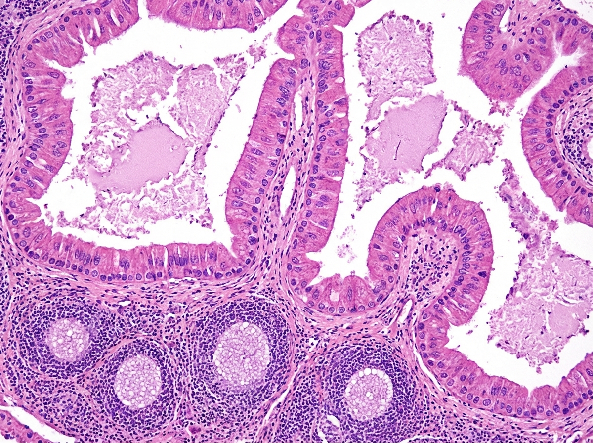
A histopathological image of a cystic lesion is shown. What is the most likely diagnosis?

What characterizes a granuloma?
A 24-year-old man is found to have an increased total body iron concentration. Biopsy of his liver shows large amounts of granular golden-brown pigment which stains blue with Prussian blue stain. The presence of which one of the following diseases best explains these findings?
Which of the following is true about stem cells?
Accumulation of which of the following substances causes fatty liver?
What is Turner hypoplasia?
Gene for retinoblastoma is located on which chromosome?
Which of the following is an example of metastatic calcification?
Explanation: **Explanation** In pathology, infarcts are classified based on their color into **Red (Hemorrhagic)** and **White (Anemic)**. **Why Option A is the correct (incorrect statement):** Red infarcts are typically associated with **venous occlusions** (e.g., testicular torsion) rather than arterial ones [1]. When an artery is occluded in a solid organ, it usually results in a white infarct [1]. While red infarcts can occur following an arterial occlusion, it only happens under specific conditions (like reperfusion or dual blood supply). Therefore, stating that red infarcts simply "occur in arterial occlusions" as a general rule is incorrect; arterial occlusion is the hallmark of **White Infarcts** [1]. **Analysis of other options:** * **Option B (Loose tissues):** Correct statement. In loose tissues (e.g., lungs), RBCs can easily seep into the necrotic area from adjacent intact vessels, making the infarct red [1]. * **Option C (Dual circulation):** Correct statement. Organs with dual blood supply (e.g., Lungs via pulmonary/bronchial arteries; Liver via hepatic artery/portal vein) allow blood from the unobstructed vessel to flow into the necrotic zone [1]. * **Option D (Venous congestion):** Correct statement. If a tissue is already congested due to sluggish venous outflow, the area is already packed with blood, leading to a hemorrhagic appearance upon infarction [1]. **NEET-PG High-Yield Pearls:** * **White Infarcts:** Occur in **solid organs** with **end-arterial circulation** (Heart, Spleen, Kidney) [1]. * **Red Infarcts:** Occur in **Lungs, Small Intestine, Brain** (due to collateral/dual supply) and **Testis/Ovary** (due to venous torsion) [1]. * **Reperfusion Injury:** Restoring blood flow to a previously white infarct (e.g., after angioplasty) can convert it into a red infarct [1]. * **Morphology:** All infarcts (except Brain) undergo **Coagulative Necrosis**. The brain undergoes **Liquefactive Necrosis** [2]. **References:** [1] Kumar V, Abbas AK, et al.. Robbins and Cotran Pathologic Basis of Disease. 9th ed. Hemodynamic Disorders, Thromboembolic Disease, and Shock, pp. 140-142. [2] Cross SS. Underwood's Pathology: A Clinical Approach. 6th ed. (Basic Pathology) introduces the student to key general principles of pathology, both as a medical science and as a clinical activity with a vital role in patient care. Part 2 (Disease Mechanisms) provides fundamental knowledge about the cellular and molecular processes involved in diseases, providing the rationale for their treatment. Part 3 (Systematic Pathology) deals in detail with specific diseases, with emphasis on the clinically important aspects., pp. 148-149.
Explanation: **Explanation:** **1. Why Option C is Correct:** Lysosomal Storage Disorders (LSDs) are a group of approximately 50 genetic diseases typically inherited in an **autosomal recessive** manner [1]. The fundamental molecular defect is a **mutation in genes encoding lysosomal acid hydrolases** [1]. These enzymes are responsible for the degradation of complex macromolecules (sphingolipids, mucopolysaccharides, etc.). When an enzyme is deficient or non-functional, the insoluble metabolite accumulates within the lysosomes, leading to cellular dysfunction and characteristic "storage" cells (e.g., Gaucher cells) [1]. **2. Why Other Options are Incorrect:** * **Option A:** Defective fusion of lysosomes and phagosomes is the hallmark of **Chédiak-Higashi syndrome**, not classic LSDs. * **Option B:** In LSDs, the substrate synthesis is normal; the pathology arises from a **failure of degradation**, leading to secondary accumulation [1]. * **Option C (Alternative mechanism):** While most LSDs involve hydrolase mutations, some involve defects in post-translational processing (e.g., **I-cell disease**, where there is a failure to create the Mannose-6-Phosphate tag) [1]. However, gene mutation of hydrolases remains the most common "typical" change. * **Option D:** Primary membrane defects are rare; the membrane usually becomes secondary damaged due to the accumulation of toxic metabolites. **High-Yield Clinical Pearls for NEET-PG:** * **Most Common LSD:** Gaucher Disease (Glucocerebrosidase deficiency) [1]. * **Only X-linked LSDs:** Fabry Disease and Hunter Syndrome (Mnemonic: *The **Hunter** is **Fab**ulous and aims for the **X***). * **Cherry Red Spot on Macula:** Seen in Tay-Sachs, Niemann-Pick, and Sandhoff disease. * **I-Cell Disease:** Defect in **N-acetylglucosaminyl-1-phosphotransferase**, leading to empty lysosomes and high levels of enzymes in the plasma. **References:** [1] Kumar V, Abbas AK, et al.. Robbins and Cotran Pathologic Basis of Disease. 9th ed. Genetic Disorders, pp. 159-161.
Explanation: ***Papillary cystadenoma lymphomatosum*** - Features characteristic **bilayered oncocytic epithelium** lining papillary projections within cystic spaces, creating a distinctive architectural pattern. - Contains **lymphoid stroma with germinal centers**, which is pathognomonic for Warthin's tumor and distinguishes it from other cystic lesions. *Dentigerous cyst* - Shows simple **squamous epithelial lining** derived from dental follicle, lacking the complex papillary architecture. - Associated with **unerupted teeth** and contains dental follicular tissue, not lymphoid stroma. *Odontogenic keratocyst* - Characterized by **parakeratinized stratified squamous epithelium** with a distinctive palisaded basal layer. - Lacks the **papillary projections** and **lymphoid component** seen in papillary cystadenoma lymphomatosum. *Unicystic ameloblastoma* - Features **ameloblastic epithelium** with characteristic **stellate reticulum** and reverse nuclear polarity. - Shows **odontogenic differentiation** patterns, not the oncocytic epithelium and lymphoid stroma of Warthin's tumor.
Explanation: **Explanation:** A **granuloma** is a hallmark of chronic granulomatous inflammation. It is defined as a microscopic focal collection of **activated macrophages**, which often transform into **epithelioid cells** (cells with abundant pink cytoplasm and indistinct borders resembling epithelial cells) [1], [2]. This process is primarily a Type IV hypersensitivity reaction driven by T-lymphocytes (CD4+ Th1 cells) secreting cytokines like IFN-γ to activate macrophages [3]. * **Why Option A is correct:** The core component of any granuloma is the activated macrophage. These may fuse to form **multinucleated giant cells** (e.g., Langhans giant cells) and are typically surrounded by a collar of lymphocytes and occasionally a rim of fibroblasts/connective tissue [1]. * **Why Option B is incorrect:** A collection of neutrophils characterizes **acute inflammation** or abscess formation, not granulomatous inflammation. * **Why Option C is incorrect:** Newly formed vessels (angiogenesis) along with fibroblasts and inflammatory cells define **granulation tissue**, which is a feature of the healing/repair process, not a granuloma. * **Why Option D is incorrect:** Eosinophilic infiltrates are typically seen in parasitic infections or allergic reactions (Type I hypersensitivity). **High-Yield Clinical Pearls for NEET-PG:** 1. **Caseating Granuloma:** Characterized by central "cheesy" necrosis; pathognomonic for **Tuberculosis**. 2. **Non-caseating Granuloma:** Seen in **Sarcoidosis**, Crohn’s disease, and Berylliosis [2]. 3. **Asteroid bodies and Schaumann bodies:** Cytoplasmic inclusions often found in the giant cells of Sarcoidosis. 4. **Stains:** Always use **Ziehl-Neelsen (ZN) stain** to rule out Mycobacterium tuberculosis in granulomatous lesions. **References:** [1] Kumar V, Abbas AK, et al.. Robbins and Cotran Pathologic Basis of Disease. 9th ed. Inflammation and Repair, p. 109. [2] Cross SS. Underwood's Pathology: A Clinical Approach. 6th ed. (Basic Pathology) introduces the student to key general principles of pathology, both as a medical science and as a clinical activity with a vital role in patient care. Part 2 (Disease Mechanisms) provides fundamental knowledge about the cellular and molecular processes involved in diseases, providing the rationale for their treatment. Part 3 (Systematic Pathology) deals in detail with specific diseases, with emphasis on the clinically important aspects., pp. 198-200. [3] Cross SS. Underwood's Pathology: A Clinical Approach. 6th ed. (Basic Pathology) introduces the student to key general principles of pathology, both as a medical science and as a clinical activity with a vital role in patient care. Part 2 (Disease Mechanisms) provides fundamental knowledge about the cellular and molecular processes involved in diseases, providing the rationale for their treatment. Part 3 (Systematic Pathology) deals in detail with specific diseases, with emphasis on the clinically important aspects., pp. 173-174.
Explanation: The clinical presentation and histological findings point towards **Hemochromatosis**, a disorder of iron overload. [1] **1. Why the Correct Answer is Right:** The "granular golden-brown pigment" described is **hemosiderin**, which represents intracellular aggregates of ferritin [3]. In cases of systemic iron overload (Hemochromatosis), iron accumulates in parenchymal cells of organs like the liver, pancreas, and heart. The **Prussian blue stain** (Perls’ reaction) is the gold standard for identifying iron; it reacts with ferric iron to produce a bright blue color [1], distinguishing it from other pigments like lipofuscin or bilirubin. **2. Analysis of Incorrect Options:** * **Systemic Amyloidosis (A):** Characterized by the extracellular deposition of misfolded proteins. On H&E stain, it appears as an amorphous eosinophilic material, but it stains **Congo Red positive** with apple-green birefringence under polarized light, not Prussian blue. * **Chronic Congestive Heart Failure (B):** While this can lead to "heart failure cells" (hemosiderin-laden macrophages) in the lungs due to local hemorrhage, it does not typically cause a massive increase in *total body iron concentration* or diffuse hepatic golden-brown pigmentation unless secondary to multiple transfusions. * **Advanced Atrophy (C):** Often associated with the accumulation of **Lipofuscin** (the "wear-and-tear" pigment). Lipofuscin is also golden-brown but is **Prussian blue negative** and represents lipid peroxidation. **Clinical Pearls for NEET-PG:** * **Classic Triad of Hemochromatosis:** Cirrhosis, Diabetes mellitus ("Bronze diabetes"), and Skin hyperpigmentation. * **Hereditary Hemochromatosis:** Most commonly due to a mutation in the **HFE gene** (C282Y) [2]. * **Staining Distinction:** * Iron = Prussian Blue (+) [1] * Lipofuscin = Prussian Blue (-) / PAS (+) * Bilirubin = Fouchet Stain (+) / Prussian Blue (-) **References:** [1] Kumar V, Abbas AK, et al.. Robbins and Cotran Pathologic Basis of Disease. 9th ed. Liver and Gallbladder, pp. 854-855. [2] Kumar V, Abbas AK, et al.. Robbins and Cotran Pathologic Basis of Disease. 9th ed. Liver and Gallbladder, p. 858. [3] Kumar V, Abbas AK, et al.. Robbins and Cotran Pathologic Basis of Disease. 9th ed. Cellular Responses to Stress and Toxic Insults: Adaptation, Injury, and Death, pp. 75-76.
Explanation: **Explanation:** **1. Why Option A is Correct:** Stem cells are characterized by their ability for self-renewal and potency to differentiate into various cell lineages [1]. During embryonic development, the **yolk sac** is the primary site of hematopoiesis (the "mesoblastic phase"). It contains **Hematopoietic Stem Cells (HSCs)** that originate from the extraembryonic mesoderm. These cells later migrate to the liver, spleen, and eventually the bone marrow [2]. Therefore, the yolk sac is a critical early reservoir for primitive stem cells. **2. Why Other Options are Incorrect:** * **Option B (Used in gene therapy):** While stem cells are a *vehicle* or target for gene therapy (e.g., modifying HSCs in SCID or Thalassemia), they are not the therapy itself. Gene therapy involves the delivery of nucleic acids into a patient's cells as a drug. This option is a clinical application, but it does not define a fundamental biological property of stem cells as accurately as their anatomical origin. * **Option C (Terminally differentiated cells):** This is the opposite of a stem cell. Terminally differentiated cells (like neurons or cardiac myocytes) have reached his or her final functional form and have lost the ability to divide or differentiate into other cell types [1]. Stem cells are **undifferentiated**. **3. NEET-PG High-Yield Pearls:** * **Potency Hierarchy:** Totipotent (Zygote) > Pluripotent (Embryonic Stem Cells - can form all three germ layers) > Multipotent (Adult stem cells like HSCs) > Unipotent [3]. * **Stem Cell Markers:** **CD34+** is the classic marker for Hematopoietic Stem Cells. * **The "Niche":** Stem cells reside in a specialized microenvironment called a "niche" which regulates their proliferation and differentiation via Wnt signaling [2]. * **Induced Pluripotent Stem Cells (iPSCs):** Somatic cells "reprogrammed" to a pluripotent state using transcription factors (Oct3/4, Sox2, Klf4, c-Myc). **References:** [1] Kumar V, Abbas AK, et al.. Robbins and Cotran Pathologic Basis of Disease. 9th ed. With Illustrations By, pp. 38-39. [2] Cross SS. Underwood's Pathology: A Clinical Approach. 6th ed. (Basic Pathology) introduces the student to key general principles of pathology, both as a medical science and as a clinical activity with a vital role in patient care. Part 2 (Disease Mechanisms) provides fundamental knowledge about the cellular and molecular processes involved in diseases, providing the rationale for their treatment. Part 3 (Systematic Pathology) deals in detail with specific diseases, with emphasis on the clinically important aspects., pp. 104-105. [3] Cross SS. Underwood's Pathology: A Clinical Approach. 6th ed. (Basic Pathology) introduces the student to key general principles of pathology, both as a medical science and as a clinical activity with a vital role in patient care. Part 2 (Disease Mechanisms) provides fundamental knowledge about the cellular and molecular processes involved in diseases, providing the rationale for their treatment. Part 3 (Systematic Pathology) deals in detail with specific diseases, with emphasis on the clinically important aspects., pp. 84-85.
Explanation: **Explanation:** **Fatty change (Steatosis)** refers to the abnormal accumulation of neutral fats within parenchymal cells [1]. It is most commonly seen in the liver because it is the central organ for lipid metabolism [2]. **Why Triglycerides are the Correct Answer:** The primary substance that accumulates in fatty liver is **Triglycerides** [1]. Under normal conditions, hepatocytes process free fatty acids (FFAs) into triglycerides, which are then complexed with apoproteins to form Very Low-Density Lipoproteins (VLDL) for secretion into the blood. Steatosis occurs when there is an imbalance in this pathway—either due to increased delivery of FFAs (as in obesity or diabetes), increased synthesis of fatty acids (alcohol consumption), or decreased synthesis/secretion of apoproteins (protein-energy malnutrition) [2]. This leads to the coalescence of triglycerides into large, clear cytoplasmic vacuoles [2]. **Analysis of Incorrect Options:** * **A & C (Cholesterol and Cholesterol Esters):** While these are lipids, their accumulation is associated with different pathologies such as **Atherosclerosis** (in smooth muscle cells/macrophages), **Xanthomas**, and **Cholesterolosis** (strawberry gallbladder), rather than classic fatty liver. * **D (Phospholipids):** These are structural components of cell membranes. Their accumulation is typically seen in lysosomal storage diseases (e.g., Niemann-Pick disease) where they form "myelin figures," but they do not characterize steatosis. **High-Yield Clinical Pearls for NEET-PG:** * **Most common cause:** In developed nations, the most common causes are Alcohol abuse and Non-Alcoholic Fatty Liver Disease (NAFLD) associated with Diabetes/Obesity [2]. * **Stains:** Since routine processing (alcohol/xylene) dissolves fat, triglycerides are best demonstrated on **frozen sections** using **Sudan IV** or **Oil Red O** (stains orange-red). * **Gross Appearance:** The liver becomes enlarged, yellow, greasy, and soft (Nutmeg liver appearance is seen in chronic passive congestion, not simple steatosis) [2]. **References:** [1] Kumar V, Abbas AK, et al.. Robbins and Cotran Pathologic Basis of Disease. 9th ed. Cellular Responses to Stress and Toxic Insults: Adaptation, Injury, and Death, p. 73. [2] Kumar V, Abbas AK, et al.. Robbins and Cotran Pathologic Basis of Disease. 9th ed. Liver and Gallbladder, pp. 848-854.
Explanation: **Turner Hypoplasia** (also known as Turner Tooth) is a specific type of enamel hypoplasia resulting from localized environmental factors affecting a developing tooth bud. ### **Explanation of the Correct Answer** **Option A** is correct because Turner hypoplasia occurs when a **local infection** (periapical periodontitis) or **trauma** to a deciduous (primary) tooth causes damage to the underlying permanent tooth germ. The inflammatory exudate or physical displacement of the primary tooth disturbs the ameloblasts (enamel-forming cells) of the permanent successor, leading to defects ranging from white/brown discoloration to severe pitting and irregularity of the enamel. It most commonly affects permanent maxillary incisors (due to trauma) and permanent premolars (due to infection of primary molars). ### **Analysis of Incorrect Options** * **Option B:** Systemic infections (like measles or chickenpox) typically cause **generalized** enamel hypoplasia, affecting all teeth developing at that specific chronological time, rather than a single localized tooth. * **Option C:** Congenital syphilis leads to specific dental anomalies known as **Hutchinson’s incisors** (notched, screwdriver-shaped) and **Mulberry molars** (globular occlusal surfaces), which are distinct from Turner hypoplasia. * **Option D:** Fluoride ingestion causes **Dental Fluorosis**, characterized by symmetric, bilateral mottling of enamel due to chronic ingestion of fluoride (>1 ppm) during tooth development [1]. ### **NEET-PG High-Yield Pearls** * **Most common site:** Permanent Premolars (due to the proximity of primary molar roots to the premolar bud). * **Clinical appearance:** Ranges from a simple yellowish-brown spot to "corrugated" enamel. * **Key Distinction:** Turner hypoplasia is **localized (focal)**, whereas systemic causes (syphilis, fluoride, vitamin D deficiency) result in **generalized** defects. **References:** [1] Kumar V, Abbas AK, et al.. Robbins and Cotran Pathologic Basis of Disease. 9th ed. The Lung, pp. 734-735.
Explanation: **Explanation:** The **RB1 gene**, the first tumor suppressor gene ever identified, is located on **Chromosome 13 (specifically at locus 13q14)** [1], [2]. This gene encodes the pRB protein, which plays a critical role in the cell cycle by inhibiting the transition from the G1 to the S phase. According to **Knudson’s "Two-Hit" Hypothesis**, both alleles of the RB1 gene must be inactivated for retinoblastoma to develop [1]. In familial cases, the first "hit" is inherited (germline), while in sporadic cases, both "hits" occur somatically [1], [2]. **Analysis of Incorrect Options:** * **Chromosome 1:** Associated with genes like *NRAS* and certain mutations in medullary thyroid carcinoma, but not the RB gene. * **Chromosome 10:** Home to the **PTEN** tumor suppressor gene (mutated in Cowden syndrome and glioblastomas) and the **RET** proto-oncogene (associated with MEN 2A/2B). * **Chromosome 5:** Contains the **APC gene** (5q21), which is mutated in Familial Adenomatous Polyposis (FAP). **High-Yield Clinical Pearls for NEET-PG:** * **Mechanism:** Hypophosphorylated (active) pRB binds to **E2F transcription factors**, preventing cell cycle progression. Hyperphosphorylation (inactivation) by Cyclin D-CDK4/6 complexes releases E2F, allowing the cell to enter the S phase. * **Associated Tumors:** Patients with germline RB1 mutations have a significantly increased risk of developing **Osteosarcoma** later in life. * **Histology:** Look for **Flexner-Wintersteiner rosettes** (specific for retinoblastoma). **References:** [1] Kumar V, Abbas AK, et al.. Robbins and Cotran Pathologic Basis of Disease. 9th ed. Neoplasia, p. 300. [2] Cross SS. Underwood's Pathology: A Clinical Approach. 6th ed. (Basic Pathology) introduces the student to key general principles of pathology, both as a medical science and as a clinical activity with a vital role in patient care. Part 2 (Disease Mechanisms) provides fundamental knowledge about the cellular and molecular processes involved in diseases, providing the rationale for their treatment. Part 3 (Systematic Pathology) deals in detail with specific diseases, with emphasis on the clinically important aspects., pp. 227-228.
Explanation: **Explanation:** Calcification is broadly classified into two types: **Dystrophic** and **Metastatic**. The fundamental difference lies in serum calcium levels and the state of the tissue involved. **Why Option C is Correct:** **Milk-alkali syndrome** is a classic example of **metastatic calcification**. Metastatic calcification occurs in **normal tissues** due to **hypercalcemia** (elevated serum calcium levels) [1]. Milk-alkali syndrome results from excessive ingestion of calcium and absorbable antacids, leading to hypercalcemia, metabolic alkalosis, and renal failure. Other causes include hyperparathyroidism, vitamin D toxicity, and bone resorption from malignancies [1]. **Why the Other Options are Incorrect:** * **A. Psammoma bodies:** These are laminated, concentric calcifications seen in papillary thyroid carcinoma, meningioma, and serous cystadenocarcinoma of the ovary [1]. They represent **dystrophic calcification** occurring in areas of cell death. * **B. Rheumatic heart disease:** Calcification of damaged heart valves in chronic rheumatic heart disease is a form of **dystrophic calcification**, as it occurs in injured/necrotic tissue despite normal serum calcium levels. * **D. Monckeberg’s medial calcific sclerosis:** This involves calcification of the tunica media of medium-sized muscular arteries. It is a form of **dystrophic calcification** associated with aging and does not narrow the vessel lumen. **NEET-PG High-Yield Pearls:** * **Dystrophic Calcification:** Normal serum calcium; occurs in necrotic/dead tissue (e.g., Atherosclerosis, Caseous necrosis in TB). * **Metastatic Calcification:** High serum calcium; occurs in normal tissue, especially those with an internal alkaline environment (Stomach, Kidneys, Lungs, Systemic arteries, and Pulmonary veins) [1]. * **Mnemonic for Metastatic sites:** "**K**ids **L**ove **S**weet **S**tuff" (**K**idney, **L**ungs, **S**tomach, **S**ystemic arteries). **References:** [1] Cross SS. Underwood's Pathology: A Clinical Approach. 6th ed. (Basic Pathology) introduces the student to key general principles of pathology, both as a medical science and as a clinical activity with a vital role in patient care. Part 2 (Disease Mechanisms) provides fundamental knowledge about the cellular and molecular processes involved in diseases, providing the rationale for their treatment. Part 3 (Systematic Pathology) deals in detail with specific diseases, with emphasis on the clinically important aspects., pp. 127-128, 134-135.
Cell Injury and Cell Death
Practice Questions
Adaptations of Cellular Growth
Practice Questions
Accumulations and Deposits
Practice Questions
Acute and Chronic Inflammation
Practice Questions
Tissue Repair and Wound Healing
Practice Questions
Hemodynamic Disorders
Practice Questions
Genetic Disorders
Practice Questions
Environmental Pathology
Practice Questions
Nutritional Diseases
Practice Questions
Molecular Basis of Disease
Practice Questions
Get full access to all questions, explanations, and performance tracking.
Start For Free