All of the following statements are false, except?
What is the most common type of tumor seen in Barrett's esophagus?
What is the single most important prognostic indicator of colorectal carcinoma?
Ulceration of Peyer's patches occurs in which infection?
Barrett's ulcer is due to:
Which of the following is a true statement about Barrett's Esophagus?
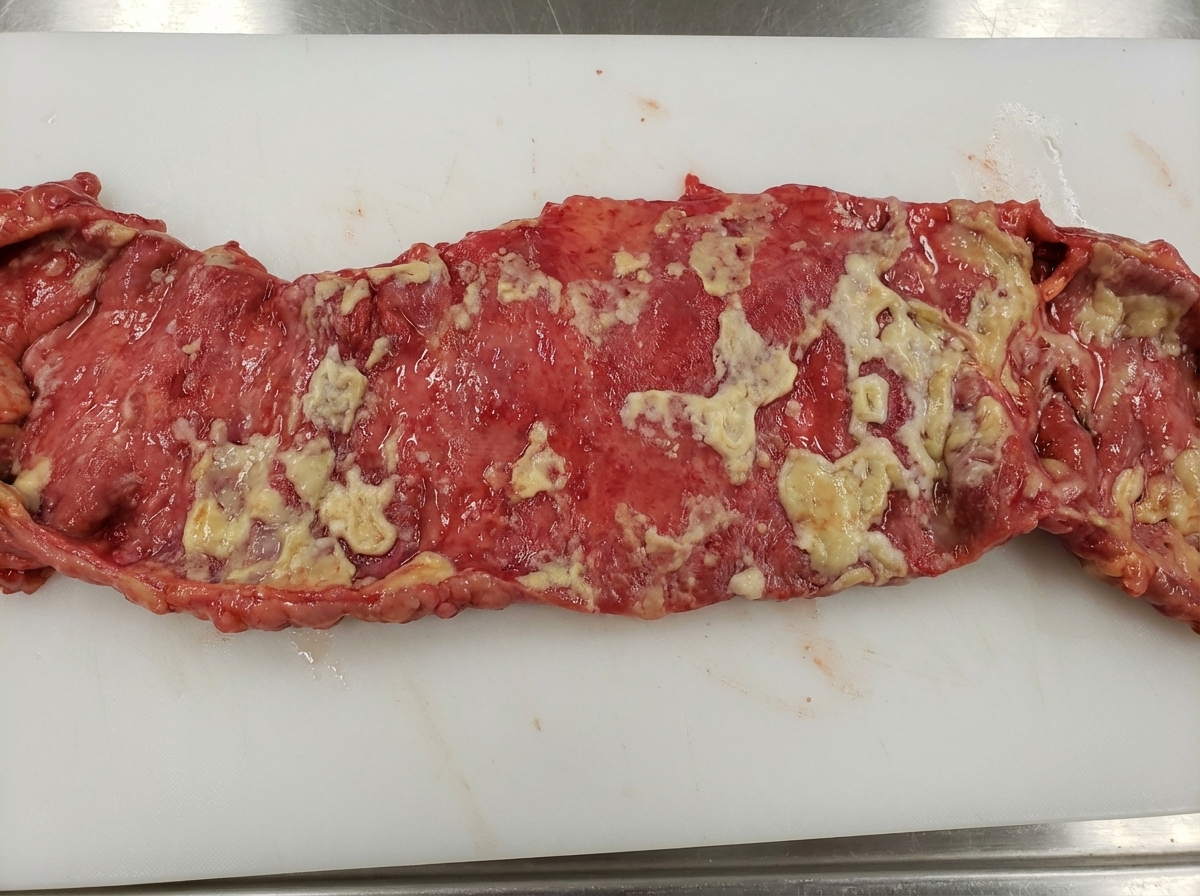
Linitis plastica is described as:
Alpha-1 antitrypsin in stool is indicative of which condition?
Which of the following statements about Barrett's esophagus are true?
Stress ulcers are caused by all of the following except?
Explanation: **Explanation:** **Correct Answer: C. Ulcerative colitis involves mucosa and submucosa** **Why it is correct:** Ulcerative colitis (UC) is a chronic inflammatory bowel disease characterized by **superficial inflammation**. Unlike Crohn’s disease, which is transmural, UC is typically limited to the **mucosa and the superficial submucosa** [1]. Histologically, it presents with crypt abscesses [1], crypt distortion, and inflammatory pseudopolyps, but the deeper muscularis propria is usually spared except in severe cases like toxic megacolon [2]. **Analysis of Incorrect Options:** * **A. Collagenous colitis is seen in children:** This is false. Collagenous colitis (a subtype of Microscopic Colitis) typically affects **middle-aged and elderly women**. It presents with chronic watery diarrhea and is characterized by a thick subepithelial collagen band (>10 µm). * **B. Ulcerative colitis is an acute granulomatous disease:** This is false. UC is a **non-granulomatous** disease [2]. The presence of non-caseating granulomas is a hallmark feature of **Crohn’s disease**, not UC [3]. * **D. Necrotizing enterocolitis (NEC) is an acute granulomatous disease:** This is false. NEC is an **ischemic/inflammatory** necrosis of the bowel seen primarily in premature infants. It is characterized by transmural necrosis and **pneumatosis intestinalis** (gas in the bowel wall), not granulomas. **High-Yield NEET-PG Pearls:** * **UC Distribution:** Always involves the rectum and spreads proximally in a **continuous** fashion (no skip lesions) [2]. * **Lead Pipe Appearance:** Loss of haustrations on barium enema due to chronic inflammation. * **Malignancy Risk:** UC carries a higher risk of adenocarcinoma compared to Crohn’s, especially with long-standing pancolitis. * **Extraintestinal Manifestation:** Primary Sclerosing Cholangitis (PSC) is strongly associated with UC (p-ANCA positive). **References:** [1] Kumar V, Abbas AK, et al.. Robbins and Cotran Pathologic Basis of Disease. 9th ed. The Gastrointestinal Tract, pp. 807-809. [2] Cross SS. Underwood's Pathology: A Clinical Approach. 6th ed. Common Clinical Problems From Alimentary System Disease, pp. 367-368. [3] Cross SS. Underwood's Pathology: A Clinical Approach. 6th ed. Common Clinical Problems From Alimentary System Disease, pp. 366-367.
Explanation: **Explanation:** **Barrett’s Esophagus (BE)** is a condition characterized by **intestinal metaplasia**, where the normal stratified squamous epithelium of the lower esophagus is replaced by simple columnar epithelium with goblet cells [3]. This change occurs as a protective response to chronic gastroesophageal reflux disease (GERD). **Why Adenocarcinoma is correct:** Adenocarcinoma arises from glandular tissue. Since Barrett’s esophagus involves the transformation of the esophageal lining into a glandular (intestinal) type, it serves as the primary precursor lesion for **Esophageal Adenocarcinoma** [1][3]. Chronic inflammation leads to a progression from metaplasia to low-grade dysplasia, high-grade dysplasia, and finally, invasive adenocarcinoma. **Why other options are incorrect:** * **Squamous cell carcinoma (SCC):** While SCC is the most common esophageal cancer worldwide, it typically arises from the native squamous epithelium and is associated with smoking and alcohol, not Barrett’s esophagus [1][2]. * **Melanoma:** Primary esophageal melanoma is extremely rare and arises from melanocytes, not metaplastic glandular tissue. * **Basal cell carcinoma:** This is a skin cancer and does not occur as a primary tumor of the esophageal mucosa. **High-Yield Clinical Pearls for NEET-PG:** * **Definition:** Metaplasia must show **Goblet cells** on histology to be diagnosed as Barrett's in many classifications [3]. * **Location:** Adenocarcinoma typically occurs in the **distal third** of the esophagus, whereas SCC is more common in the **middle third** [1][2]. * **Endoscopy:** Barrett’s appears as "salmon-pink" velvety tongues extending upward from the GE junction. * **Risk Factor:** The risk of developing adenocarcinoma in patients with BE is approximately 30 to 40 times higher than the general population [3]. **References:** [1] Kumar V, Abbas AK, et al.. Robbins and Cotran Pathologic Basis of Disease. 9th ed. The Gastrointestinal Tract, pp. 765-766. [2] Kumar V, Abbas AK, et al.. Robbins and Cotran Pathologic Basis of Disease. 9th ed. The Gastrointestinal Tract, pp. 766-767. [3] Cross SS. Underwood's Pathology: A Clinical Approach. 6th ed. Common Clinical Problems From Alimentary System Disease, pp. 348-349.
Explanation: ### Explanation The prognosis of colorectal carcinoma (CRC) is primarily determined by the **extent of the tumor at the time of diagnosis**, which is formalised through **TNM Staging** [1], [2]. **1. Why "Extent of Tumor" is correct:** In oncology, "extent" refers to the anatomical spread of the cancer. For CRC, this includes the depth of local invasion into the bowel wall (T stage), the presence of regional lymph node metastasis (N stage), and distant metastasis (M stage) [2]. Staging is the most reliable predictor of survival outcomes [1]. For instance, a tumor confined to the mucosa (Stage 0) has a 5-year survival rate of >90%, whereas Stage IV (distant metastasis) drops significantly. **2. Why other options are incorrect:** * **CEA (Carcinoembryonic Antigen) titres:** While CEA is a useful tumor marker, it is **not used for diagnosis or primary prognosis**. Its chief clinical utility is in **monitoring for recurrence** after surgical resection and assessing response to therapy. * **Degree of atypia (Grading):** Histological grading (well, moderately, or poorly differentiated) provides some prognostic information, but it is subjective and far less predictive of survival than the anatomical stage. * **Size of tumor:** Unlike some other cancers (like breast or renal cell carcinoma), the physical size of a colorectal tumor does not correlate well with its metastatic potential or prognosis. A large exophytic mass may be less dangerous than a small, deeply infiltrating ulcerative lesion. **Clinical Pearls for NEET-PG:** * **Most common site:** Sigmoid colon (overall), though the incidence of right-sided (proximal) colon cancer is increasing. * **Most common site of metastasis:** Liver (via portal circulation). * **Dukes’ Classification:** An older staging system for CRC, now largely replaced by the TNM system [2]. * **Molecular Pathways:** Remember the **Chromosomal Instability Pathway** (APC gene - most common) and the **Microsatellite Instability Pathway** (DNA mismatch repair genes). **References:** [1] Cross SS. Underwood's Pathology: A Clinical Approach. 6th ed. Common Clinical Problems From Alimentary System Disease, pp. 374-375. [2] Cross SS. Underwood's Pathology: A Clinical Approach. 6th ed. (Basic Pathology) introduces the student to key general principles of pathology, both as a medical science and as a clinical activity with a vital role in patient care. Part 2 (Disease Mechanisms) provides fundamental knowledge about the cellular and molecular processes involved in diseases, providing the rationale for their treatment. Part 3 (Systematic Pathology) deals in detail with specific diseases, with emphasis on the clinically important aspects., pp. 236-237.
Explanation: **Explanation:** The correct answer is **Salmonella infection (Typhoid fever)**. **Salmonella typhi** and **paratyphi** have a specific tropism for lymphoid tissue. After ingestion, the bacteria invade the intestinal mucosa and proliferate within the **Peyer’s patches** (lymphoid follicles) of the terminal ileum [1]. This leads to marked hyperplasia, followed by necrosis and sloughing of the overlying mucosa, resulting in characteristic **longitudinal ulcers** that follow the long axis of the bowel. This is a high-yield distinction from other types of intestinal ulceration. **Why other options are incorrect:** * **Amoebiasis (Entamoeba histolytica):** Typically causes **"flask-shaped" ulcers** with narrow necks and broad bases. These ulcers are usually found in the colon (cecum and rectosigmoid) rather than Peyer's patches. * **Crohn’s disease:** Characterized by **transmural inflammation**, "aphthous" ulcers that progress to **"cobblestone" appearance**, and non-caseating granulomas. It is not specifically localized to lymphoid follicles. * **Clostridium difficile:** Causes **Pseudomembranous colitis**. It is characterized by "volcano-like" eruptions of purulent exudate (fibrin, mucus, and neutrophils) forming a yellow-green membrane over the colonic mucosa, rather than discrete ulceration of Peyer's patches. **NEET-PG High-Yield Pearls:** * **Typhoid Ulcers:** Longitudinal, located in the ileum; risk of perforation is highest in the 3rd week [1]. * **Tubercular Ulcers:** Transverse (circumferential) due to spread via lymphatics. * **Widal Test:** Measures antibodies against O and H antigens; usually becomes positive in the 2nd week of Typhoid fever. * **Microscopy:** Look for **"Typhoid cells"** (erythrophagocytic macrophages) in the liver, spleen, and bone marrow [1]. **References:** [1] Cross SS. Underwood's Pathology: A Clinical Approach. 6th ed. Common Clinical Problems From Alimentary System Disease, pp. 362-363.
Explanation: **Explanation:** **Barrett’s esophagus** is a complication of chronic **Gastroesophageal Reflux Disease (GERD)**. The fundamental mechanism is **Reflux Esophagitis**, where the persistent irritation of the esophageal lining by gastric acid and bile leads to intestinal metaplasia [1]. In this process, the normal stratified squamous epithelium is replaced by simple columnar epithelium with goblet cells to better withstand the acidic environment [1]. A **Barrett’s ulcer** is a specific type of deep peptic ulcer that develops within this metaplastic columnar segment due to the continued corrosive action of the refluxed gastric contents. **Analysis of Options:** * **Reflux Esophagitis (Correct):** This is the primary driver [2]. The chronic inflammation (esophagitis) caused by acid reflux triggers the metaplastic change and subsequent ulceration. * **Ischemia (Incorrect):** While ischemia causes ulcers in the stomach (e.g., Stress ulcers or Curling’s ulcers in burn patients), it is not the etiology of Barrett’s ulcer. * **Ectopic Gastric Mucosa (Incorrect):** This refers to a "Inlet Patch," which is a congenital anomaly usually found in the upper third of the esophagus. Barrett’s is an *acquired* metaplastic process in the lower third. * **Bile Reflux (Incorrect):** While bile can contribute to the severity of mucosal damage in GERD, "Reflux Esophagitis" is the broader, more definitive clinical diagnosis encompassing the pathophysiology of Barrett's. **High-Yield Pearls for NEET-PG:** * **Definition:** Metaplasia of Squamous to Columnar epithelium (specifically requiring **Goblet cells** for diagnosis in many classifications) [1]. * **Location:** Always occurs in the **distal esophagus**. * **Pre-malignant Potential:** Barrett’s esophagus is the single most important risk factor for **Esophageal Adenocarcinoma** (increasing risk by 30-40 times) [1]. * **Endoscopy:** Appears as "salmon-pink" velvety tongues of mucosa extending upwards from the gastroesophageal junction. **References:** [1] Cross SS. Underwood's Pathology: A Clinical Approach. 6th ed. Common Clinical Problems From Alimentary System Disease, pp. 348-349. [2] Kumar V, Abbas AK, et al.. Robbins and Cotran Pathologic Basis of Disease. 9th ed. The Gastrointestinal Tract, pp. 761-762.
Explanation: **Explanation:** **Barrett’s Esophagus (BE)** is a classic example of **adaptive metaplasia** occurring in the distal esophagus. 1. **Why Option D is Correct:** The primary driver of BE is **Chronic Gastroesophageal Reflux Disease (GERD)**. Persistent exposure to gastric acid and bile salts causes chronic mucosal injury. In response, the normal stratified squamous epithelium undergoes metaplasia, transforming into **columnar epithelium with goblet cells** (intestinal metaplasia), which is more resistant to acidic environments [1]. 2. **Why Other Options are Incorrect:** * **Option A:** BE is highly **premalignant**. It follows a predictable progression: Metaplasia → Low-grade dysplasia → High-grade dysplasia → Adenocarcinoma [2]. * **Option B:** BE is a precursor to **Adenocarcinoma**, not squamous cell carcinoma (SCC) [1]. SCC is typically associated with smoking and alcohol, whereas BE-related adenocarcinoma is associated with obesity and reflux [3]. * **Option C:** While difficult to manage, BE is **potentially reversible** with aggressive medical or surgical intervention (e.g., Proton Pump Inhibitors or fundoplication) and can be treated via endoscopic ablation. **NEET-PG High-Yield Pearls:** * **Definition:** Intestinal metaplasia (presence of **Goblet cells**) is the histological hallmark and a prerequisite for diagnosis [1]. * **Endoscopy:** Appears as "salmon-pink," velvety tongues of mucosa extending upward from the gastroesophageal junction. * **Risk Factor:** The single most important risk factor for **Esophageal Adenocarcinoma** [1]. * **Surveillance:** Patients require periodic endoscopic biopsy to monitor for dysplasia [2]. **References:** [1] Cross SS. Underwood's Pathology: A Clinical Approach. 6th ed. Common Clinical Problems From Alimentary System Disease, pp. 348-349. [2] Kumar V, Abbas AK, et al.. Robbins and Cotran Pathologic Basis of Disease. 9th ed. The Gastrointestinal Tract, pp. 764-765. [3] Kumar V, Abbas AK, et al.. Robbins and Cotran Pathologic Basis of Disease. 9th ed. The Gastrointestinal Tract, pp. 766-767.
Explanation: **Explanation:** **Linitis plastica** (also known as "leather bottle stomach") is a classic morphological presentation of **Diffuse-type Gastric Adenocarcinoma** (Lauren classification) [1]. **Why Option C is correct:** In this condition, the tumor cells (often **signet ring cells**) infiltrate the stomach wall extensively without forming a discrete mass [1]. This diffuse infiltration triggers a massive **desmoplastic reaction** (fibrosis), leading to marked thickening and rigidity of the entire gastric wall. Grossly, the stomach loses its distensibility and resembles a rigid leather pouch, hence the name [1]. **Why other options are incorrect:** * **Option A:** Lichen planus is a chronic inflammatory mucocutaneous condition; it has no pathological relationship with linitis plastica. * **Option B:** Linitis plastica is a highly aggressive **malignancy** with a poor prognosis, not a benign lesion. * **Option D:** While "itis" usually denotes inflammation, linitis plastica is a neoplastic process. Although the stomach wall is thickened, it is due to cancer and fibrosis, not acute inflammatory cell infiltration. **High-Yield Facts for NEET-PG:** * **Microscopy:** Characterized by **Signet Ring Cells** (nucleus pushed to the periphery by a large mucin vacuole) [1]. * **Genetics:** Often associated with mutations in the **CDH1 gene**, which encodes the cell adhesion protein **E-cadherin** [1]. * **Spread:** Frequently involves the entire stomach and can metastasize to the ovaries (**Krukenberg tumor**). * **Radiology:** Barium meal shows a rigid, narrow, "flask-shaped" stomach with absent mucosal folds. **References:** [1] Kumar V, Abbas AK, et al.. Robbins and Cotran Pathologic Basis of Disease. 9th ed. The Gastrointestinal Tract, pp. 779-780.
Explanation: **Explanation:** **Alpha-1 antitrypsin (A1AT)** is a plasma protein synthesized by the liver with a molecular weight similar to albumin. Unlike other plasma proteins, A1AT is highly resistant to degradation by digestive enzymes (proteolysis) within the intestinal lumen. 1. **Why Option A is correct:** In **Protein-losing enteropathy (PLE)**, there is an excessive loss of serum proteins into the gastrointestinal tract due to mucosal injury or lymphatic obstruction. Because A1AT is not digested or reabsorbed, its presence in the stool serves as a reliable endogenous marker for intestinal protein loss. Measuring the **fecal clearance of A1AT** is considered the gold standard for diagnosing PLE. 2. **Why other options are incorrect:** * **Chronic & Acute Pancreatitis (B & C):** These conditions involve exocrine insufficiency or inflammation. While fecal elastase or chymotrypsin levels are used to assess pancreatic function, A1AT is not a marker for pancreatic pathology. * **Whipple Disease (D):** While Whipple disease *can* cause PLE as a secondary complication (due to lymphatic obstruction), A1AT in the stool specifically indicates the **mechanism** (protein loss) rather than the specific **etiology** (Tropheryma whipplei infection) [1]. **High-Yield Clinical Pearls for NEET-PG:** * **Endogenous Marker:** A1AT is the preferred marker for PLE because it is neither secreted nor absorbed in the gut and remains stable at low pH. * **Limitation:** Fecal A1AT clearance cannot be used if the patient has gastric acid hypersecretion (e.g., Zollinger-Ellison syndrome), as A1AT can be degraded at a pH below 3.0. * **Differential Diagnosis for PLE:** Menetrier’s disease, Celiac disease, Intestinal lymphangiectasia, and Crohn’s disease. **References:** [1] Kumar V, Abbas AK, et al.. Robbins and Cotran Pathologic Basis of Disease. 9th ed. The Gastrointestinal Tract, pp. 798-799.
Explanation: **Explanation:** Barrett’s Esophagus (BE) is a complication of chronic Gastroesophageal Reflux Disease (GERD) [1]. It is defined by **intestinal metaplasia**, where the normal stratified squamous epithelium of the esophagus is replaced by non-ciliated columnar epithelium with **Goblet cells** [1]. * **Why Option B is correct:** 1. **Metaplasia:** This is the hallmark of BE (Squamous to Columnar) [1]. 2. **Leads to Adenocarcinoma:** BE is a well-established pre-malignant condition [1]. Patients have a 30- to 100-fold increased risk of developing esophageal adenocarcinoma [3]. 3. **Long Esophageal Segment:** While "Short-segment BE" exists (<3cm), classic Barrett's typically involves a significant portion of the distal esophagus (Long-segment BE is ≥3cm), visible endoscopically as "salmon-pink" tongues of mucosa. 4. **Peptic Ulcer & Paraesophageal Hernia:** While a **Sliding Hiatal Hernia** is strongly associated with GERD and BE, a *Paraesophageal* hernia is not a characteristic feature. Similarly, while "Barrett’s ulcers" can occur, "Peptic ulcer" typically refers to gastric or duodenal pathology. * **Why other options are wrong:** Options A, C, and D are incorrect because they either falsely exclude the risk of adenocarcinoma or the involvement of long segments, or they incorrectly include paraesophageal hernia as a defining feature. **High-Yield Clinical Pearls for NEET-PG:** * **Diagnosis:** Requires both endoscopic evidence (Z-line displacement) and histological confirmation of **Goblet cells** [1], [2]. * **Most common site:** Distal 1/3rd of the esophagus [3]. * **Screening:** Periodic endoscopy with biopsies (Seattle Protocol) is required to monitor for dysplasia [1], [2]. * **Molecular Marker:** Overexpression of **p53** and **p16** is often seen in the progression to malignancy. **References:** [1] Cross SS. Underwood's Pathology: A Clinical Approach. 6th ed. Common Clinical Problems From Alimentary System Disease, pp. 348-349. [2] Kumar V, Abbas AK, et al.. Robbins and Cotran Pathologic Basis of Disease. 9th ed. The Gastrointestinal Tract, pp. 764-765. [3] Kumar V, Abbas AK, et al.. Robbins and Cotran Pathologic Basis of Disease. 9th ed. The Gastrointestinal Tract, pp. 765-766.
Explanation: **Explanation:** Stress ulcers are acute gastric mucosal lesions that develop in patients under severe physiological stress [1]. The correct answer is **Penicillin therapy**, as it is an antibiotic and does not have a known pathophysiological mechanism for inducing gastric ulceration. **Why the other options are causes of Stress Ulcers:** * **Burns (Option A):** Severe burns lead to **Curling’s ulcers**. These occur due to reduced plasma volume and subsequent mucosal ischemia, which compromises the protective gastric barrier [1]. * **Cortisol therapy (Option B):** Exogenous corticosteroids (and high endogenous cortisol) increase gastric acid secretion and decrease the synthesis of protective prostaglandins and mucus, predisposing the mucosa to injury. * **Pulmonary insufficiency (Option C):** Severe respiratory distress or mechanical ventilation causes systemic hypoxia. This leads to splanchnic vasoconstriction and mucosal ischemia, a primary driver of stress-related mucosal disease (SRMD) [1]. **High-Yield Clinical Pearls for NEET-PG:** 1. **Curling’s Ulcer:** Associated with severe **burns** (Think: *Curling* iron causes burns). Usually found in the duodenum. 2. **Cushing’s Ulcer:** Associated with **CNS injury** or increased intracranial pressure. These are caused by vagal overstimulation leading to hypersecretion of gastric acid [1]. (Think: *Cushing* the brain). 3. **Location:** Unlike chronic peptic ulcers, stress ulcers are typically **multiple**, small, and located primarily in the **stomach** (fundus and body). 4. **Pathogenesis:** The common denominator is usually **mucosal ischemia** (except in Cushing’s, where it is hyperacidity) [1]. **References:** [1] Cross SS. Underwood's Pathology: A Clinical Approach. 6th ed. Common Clinical Problems From Alimentary System Disease, pp. 353-354.
Oral Cavity and Esophageal Pathology
Practice Questions
Gastritis and Peptic Ulcer Disease
Practice Questions
Inflammatory Bowel Disease
Practice Questions
Malabsorption Syndromes
Practice Questions
Vascular Disorders of Intestine
Practice Questions
Diverticular Disease
Practice Questions
Intestinal Obstruction
Practice Questions
Gastrointestinal Infections
Practice Questions
Polyps and Neoplasms
Practice Questions
Appendiceal Pathology
Practice Questions
Get full access to all questions, explanations, and performance tracking.
Start For Free