A small intestinal biopsy is specific for which of the following conditions?
Early gastric carcinoma is indicated by what finding?
The mother of a 4-year-old child notes blood when laundering his underwear. Physical examination reveals a rectal mass. On proctoscopy, there is a smooth-surfaced, pedunculated, 1.5-cm polyp. It is excised and microscopically shows cystically dilated crypts filled with mucin and inflammatory debris, but no dysplasia. What is the most likely diagnosis?
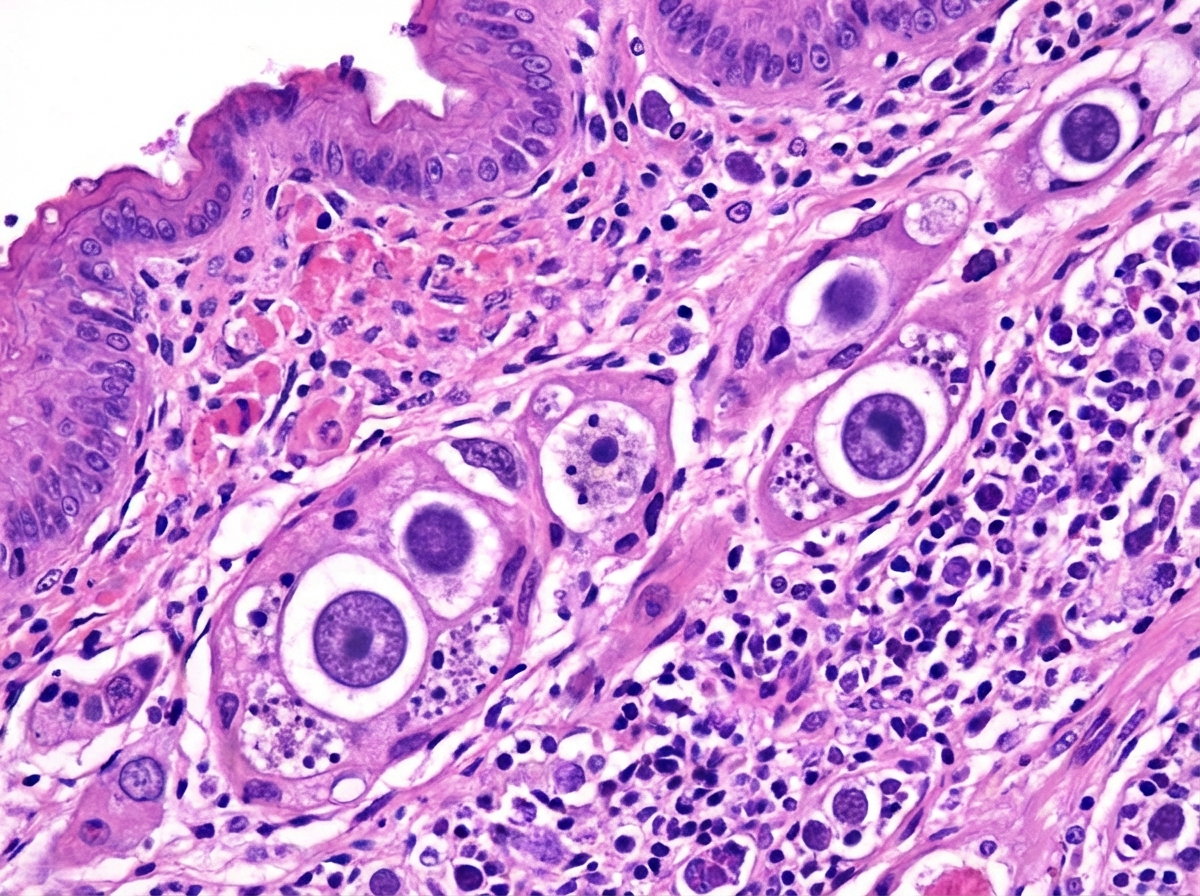
A 40-year-old immunocompromised patient develops multiple ulcers in the esophagus leading to painful swallowing. The biopsy from the esophagus is shown below. What is the diagnosis?

Which of the following statements about ulcerative colitis (UC) and colon carcinoma is FALSE?
Which one of the following conditions commonly predisposes to colonic carcinoma?
Jejunal biopsy is diagnostic in which of the following conditions?
A 70-year-old man presents with fatigue, weight loss, abdominal pain, and blood in the stools. A complete blood count reveals anemia with hemoglobin of 10 g/dL. A colonoscopy and subsequent colonic biopsy reveal adenocarcinoma. Which of the following is the most likely predisposing lesion or disorder that led to this malignancy?
The APC gene is involved in which of the following conditions?
Collar Button Ulcer is a feature of which condition?
Explanation: In gastrointestinal pathology, the specificity of a biopsy refers to whether the histological findings are pathognomonic (diagnostic on their own) or merely suggestive of a disease. **Why Whipple’s Disease is the Correct Answer:** Whipple’s disease is caused by the bacterium *Tropheryma whipplei*. A small intestinal biopsy is considered **specific** because it reveals a unique, diagnostic feature: the presence of **PAS-positive, diastase-resistant macrophages** in the lamina propria containing rod-shaped bacilli [1]. These findings, especially when confirmed by electron microscopy or PCR, are definitive for the diagnosis [1]. **Why Other Options are Incorrect:** * **Celiac Sprue:** While biopsy is essential, the findings (villous atrophy, crypt hyperplasia, and increased intraepithelial lymphocytes) are **nonspecific** [2, 4]. Similar patterns can be seen in tropical sprue, cow’s milk allergy, or viral gastroenteritis. Diagnosis requires correlation with serology (anti-ttG) and clinical response to a gluten-free diet [3]. * **Tropical Sprue:** The histological changes are virtually identical to Celiac disease but often involve the entire small intestine [5]. It is a diagnosis of exclusion based on travel history and response to antibiotics/folate. * **All of the above:** Incorrect because only Whipple’s disease provides a pathognomonic histological marker. **High-Yield Clinical Pearls for NEET-PG:** * **Whipple’s Disease Mnemonic:** Remember the **"4 Ms"**: **M**alabsorption, **M**esenteric lymphadenopathy, **M**igratory polyarthritis, and **M**acrophages (PAS+). * **Celiac Disease:** Most specific serological marker is **Anti-Endomysial Antibody (EMA)**; most sensitive is **Anti-tissue Transglutaminase (tTG)** [3]. * **Biopsy Site:** In Celiac disease, biopsies must be taken from the **second part of the duodenum** or beyond, as the bulb may show non-specific changes [2]. **References:** [1] Kumar V, Abbas AK, et al.. Robbins and Cotran Pathologic Basis of Disease. 9th ed. The Gastrointestinal Tract, pp. 798-799. [2] Cross SS. Underwood's Pathology: A Clinical Approach. 6th ed. Common Clinical Problems From Alimentary System Disease, pp. 361-362. [3] Cross SS. Underwood's Pathology: A Clinical Approach. 6th ed. Common Clinical Problems From Alimentary System Disease, pp. 360-361. [4] Kumar V, Abbas AK, et al.. Robbins and Cotran Pathologic Basis of Disease. 9th ed. The Gastrointestinal Tract, pp. 789-790. [5] Kumar V, Abbas AK, et al.. Robbins and Cotran Pathologic Basis of Disease. 9th ed. The Gastrointestinal Tract, pp. 790-791.
Explanation: **Explanation:** The definition of **Early Gastric Carcinoma (EGC)** is strictly anatomical and is based on the depth of invasion, regardless of the presence or absence of regional lymph node metastasis [1]. 1. **Why Option A is Correct:** By definition, EGC is a carcinoma limited to the **mucosa and submucosa** [1]. This classification is crucial because these lesions carry a significantly better prognosis (5-year survival rate >90%) compared to advanced gastric cancer [2]. Even if perigastric lymph nodes are involved (which occurs in about 10-15% of cases), it is still classified as "early" as long as the muscularis propria is not breached [1]. 2. **Why Other Options are Incorrect:** * **Option B:** Once a lesion involves the **muscularis propria** (muscular wall) or deeper layers, it is classified as **Advanced Gastric Carcinoma** [1]. * **Option C:** A "cauliflower" or fungating growth is a macroscopic description typically associated with the **Exophytic/Polypoid** type of advanced gastric cancer [3]. * **Option D:** **Linitis Plastica** (leather bottle stomach) refers to a diffuse, thickening of the stomach wall caused by an infiltrating **Signet Ring Cell Carcinoma** (Lauren’s Diffuse type) [3]. This represents an advanced stage with a very poor prognosis. **High-Yield NEET-PG Pearls:** * **Most common site:** The lesser curvature of the antrum/pylorus [3]. * **Classification:** EGC is often classified using the **Japanese Endoscopic Classification** (Type I: Protruded, Type II: Superficial, Type III: Excavated). * **Prognosis:** The most important prognostic factor in gastric cancer is the **depth of invasion** and the status of **lymph node metastasis** [1]. * **Screening:** Countries like Japan have high survival rates due to mass endoscopic screening detecting EGC early. **References:** [1] Cross SS. Underwood's Pathology: A Clinical Approach. 6th ed. Common Clinical Problems From Alimentary System Disease, pp. 355-356. [2] Cross SS. Underwood's Pathology: A Clinical Approach. 6th ed. Common Clinical Problems From Alimentary System Disease, pp. 349-350. [3] Kumar V, Abbas AK, et al.. Robbins and Cotran Pathologic Basis of Disease. 9th ed. The Gastrointestinal Tract, p. 779.
Explanation: ### Explanation **1. Why Juvenile Polyp is Correct:** The clinical presentation and histopathology are classic for a **Juvenile Polyp** (also known as a Retention Polyp) [1]. These are the most common type of gastrointestinal polyps in children (typically <5 years old) [1]. * **Clinical Presentation:** They usually present as a solitary, smooth-surfaced, pedunculated mass in the rectum, often causing painless rectal bleeding [2] or "prolapse" during defecation. * **Histopathology:** The hallmark is **cystically dilated glands (crypts)** filled with mucin, surrounded by an expanded, inflamed lamina propria [2]. Crucially, these are **hamartomatous** lesions, meaning they lack dysplasia and have no malignant potential when solitary. **2. Why Incorrect Options are Wrong:** * **A. Familial Adenomatous Polyposis (FAP):** Characterized by hundreds to thousands of **adenomatous** polyps, typically developing before age 30 [3]. Histologically, these show epithelial dysplasia (crowded, dark, cigar-shaped nuclei) [4], which is absent in this case. * **B. Gardner Syndrome:** A variant of FAP. While it involves colonic polyps, it also presents with extra-intestinal manifestations like osteomas, epidermal cysts, and desmoid tumors [1]. * **C. Lynch Syndrome (HNPCC):** This is caused by germline mutations in DNA mismatch repair genes [3]. It typically presents in young adults (not 4-year-olds) with right-sided colon cancer, not pedunculated hamartomatous polyps [3]. **3. High-Yield Clinical Pearls for NEET-PG:** * **Solitary vs. Syndromic:** A solitary juvenile polyp carries no cancer risk. However, **Juvenile Polyposis Syndrome** (multiple polyps, SMAD4 or BMPR1A mutations) carries a significant risk of gastric and colonic adenocarcinoma [1]. * **Microscopic "Buzzword":** "Swiss-cheese appearance" due to the numerous dilated cystic spaces [2]. * **Most common site:** Rectum (often self-amputate due to torsion of the stalk). * **Differential:** Peutz-Jeghers Syndrome (also hamartomatous but shows a "Christmas tree" branching pattern of smooth muscle) [1]. **References:** [1] Kumar V, Abbas AK, et al.. Robbins and Cotran Pathologic Basis of Disease. 9th ed. The Gastrointestinal Tract, p. 813. [2] Kumar V, Abbas AK, et al.. Robbins and Cotran Pathologic Basis of Disease. 9th ed. The Gastrointestinal Tract, pp. 813-814. [3] Kumar V, Abbas AK, et al.. Robbins and Cotran Pathologic Basis of Disease. 9th ed. The Gastrointestinal Tract, pp. 821-822. [4] Cross SS. Underwood's Pathology: A Clinical Approach. 6th ed. Common Clinical Problems From Alimentary System Disease, pp. 371-372.
Explanation: ***CMV*** - **CMV esophagitis** shows characteristic **owl-eye intranuclear inclusions** in endothelial and stromal cells on histopathology, which is pathognomonic for CMV infection. - Common in **immunocompromised patients** (HIV, transplant recipients) presenting with **odynophagia** and multiple deep esophageal ulcers. *Candida* - Histopathology shows **pseudohyphae** and **budding yeast forms** invading the superficial squamous epithelium, not intranuclear inclusions. - Typically presents with **white plaques** on endoscopy rather than deep ulcerative lesions described in this case. *Eosinophilic esophagitis* - Characterized by **eosinophilic infiltration** (>15 eosinophils per high-power field) in the epithelium with **microabscesses**. - Usually occurs in **atopic individuals** with food allergies, not typically in immunocompromised patients with infectious etiology. *Herpes* - Shows **multinucleated giant cells** with **Cowdry A intranuclear inclusions** specifically in the **squamous epithelium**, not in stromal cells. - **HSV esophagitis** typically causes **vesicular lesions** that progress to shallow ulcers, different from the deep ulcerative pattern of CMV.
Explanation: **Explanation:** The risk of **Colorectal Carcinoma (CRC)** is a significant long-term complication of Ulcerative Colitis (UC). Understanding the nuances of this risk is crucial for NEET-PG. [1] **Why Option B is the Correct (False) Statement:** The cumulative risk of developing adenocarcinoma in UC patients is significantly higher than 5% over time. It is estimated at approximately **2% after 10 years, 8% after 20 years, and up to 18% after 30 years** of disease. Therefore, stating the overall risk is "less than 5%" is clinically inaccurate. **Analysis of Other Options:** * **Option A:** The risk of malignancy is directly proportional to the **duration** and **extent** of the disease (pancolitis carries the highest risk). [1] * **Option C:** In the general population, colon cancer is more common; however, in patients with UC, there is a higher predilection for **rectal involvement** and synchronous (multiple) tumors compared to sporadic cases. [2] * **Option D:** The "Dysplasia-Carcinoma Sequence" in UC is the rationale for surveillance. High-grade (severe) dysplasia is a strong indicator of concurrent or imminent invasive cancer, making **total proctocolectomy** the standard surgical indication. **High-Yield Clinical Pearls for NEET-PG:** * **Surveillance:** Screening colonoscopies should begin **8–10 years** after the onset of symptoms. [1] * **Backwash Ileitis:** Patients with pancolitis and backwash ileitis have a higher risk of developing CRC. * **Molecular Pathogenesis:** Unlike sporadic CRC (APC mutation first), UC-associated CRC often shows **p53 mutations early** and APC mutations late. * **Protective Factor:** Regular use of **5-ASA (Mesalamine)** and folic acid may reduce the risk of progression to dysplasia. **References:** [1] Kumar V, Abbas AK, et al.. Robbins and Cotran Pathologic Basis of Disease. 9th ed. The Gastrointestinal Tract, pp. 807-809. [2] Cross SS. Underwood's Pathology: A Clinical Approach. 6th ed. Common Clinical Problems From Alimentary System Disease, pp. 373-374.
Explanation: **Explanation:** The risk of developing **Colorectal Carcinoma (CRC)** is significantly elevated in patients with **Ulcerative Colitis (UC)** [1]. The underlying mechanism is chronic mucosal inflammation, which leads to a "dysplasia-carcinoma sequence." Unlike sporadic CRC (which follows the APC-adenoma-carcinoma pathway), UC-associated CRC often arises from flat, non-adenomatous dysplastic lesions and is frequently multifocal [1]. **Why Ulcerative Colitis is the correct answer:** The risk of malignancy in UC depends on: 1. **Duration of disease:** Risk increases significantly after 8–10 years of involvement. 2. **Extent of disease:** Pancolitis carries a much higher risk than left-sided colitis [1]. 3. **Severity of inflammation:** Frequent flares and histological inflammation increase the risk. **Analysis of Incorrect Options:** * **Crohn’s Disease:** While Crohn’s also increases the risk of CRC, the risk is statistically lower than in UC (partly because Crohn's often involves the small bowel or is segmental). UC remains the "classic" pre-malignant inflammatory condition for the colon in exam contexts. * **Diverticular Disease:** This is a structural abnormality (herniation of mucosa through the muscularis) caused by high intraluminal pressure. It is associated with inflammation (diverticulitis) but is **not** a pre-malignant condition. * **Ischaemic Colitis:** This results from decreased blood flow (usually at watershed areas like the splenic flexure). It causes acute or chronic injury but does not predispose to neoplastic transformation. **High-Yield NEET-PG Pearls:** * **Surveillance:** For UC patients, colonoscopic surveillance is recommended starting 8 years after diagnosis. * **Molecular Pathogenesis:** In UC-associated CRC, **p53 mutations** occur early, while **APC mutations** occur late (the opposite of sporadic CRC). * **Protective Factor:** Regular use of 5-ASA (Mesalamine) and surveillance reduces the risk of progression to carcinoma. **References:** [1] Kumar V, Abbas AK, et al.. Robbins and Cotran Pathologic Basis of Disease. 9th ed. The Gastrointestinal Tract, pp. 807-809.
Explanation: **Explanation:** **Whipple’s Disease (Correct Answer):** Jejunal biopsy is considered diagnostic for Whipple’s disease because it reveals pathognomonic histological features [1]. The hallmark is the presence of **PAS-positive, diastase-resistant macrophages** in the lamina propria [1]. these macrophages contain the causative organism, *Tropheryma whipplei*. Electron microscopy (showing rod-shaped bacilli) can confirm the diagnosis, but the characteristic infiltration on a mucosal biopsy is the gold standard for identification. **Analysis of Incorrect Options:** * **Giardiasis:** While a biopsy may show trophozoites attached to the epithelium, it is **not** the diagnostic method of choice. Diagnosis is typically made via stool microscopy (cysts/trophozoites) or the more sensitive stool antigen test (ELISA). * **Tropical Sprue:** Biopsy shows non-specific changes such as villous atrophy and crypt hyperplasia. These features overlap significantly with Celiac disease; therefore, biopsy is suggestive but not "diagnostic" on its own without clinical and geographical correlation. * **Radiation Enteritis:** Diagnosis is primarily clinical, based on a history of radiation therapy. Biopsy shows non-specific acute inflammation or chronic fibrosis and vascular changes (endarteritis obliterans), which are not unique enough to be definitively diagnostic. **NEET-PG High-Yield Pearls:** * **Whipple’s Disease Triad:** Malabsorption, migratory polyarthritis, and abdominal pain [1]. * **Rule of "M" for Whipple's:** **M**acrophages, **M**alabsorption, **M**ens (more common in males), and **M**iddle-aged. * **PAS Stain:** Stains the glycoprotein cell wall of the actinomycete *T. whipplei*. * **Differential for PAS+ Macrophages in Gut:** Must exclude *Mycobacterium avium-intracellulare* (MAI) in HIV patients using an Acid-Fast (AFB) stain; Whipple’s is AFB negative [1]. **References:** [1] Kumar V, Abbas AK, et al.. Robbins and Cotran Pathologic Basis of Disease. 9th ed. The Gastrointestinal Tract, pp. 798-799.
Explanation: **Explanation:** The clinical presentation of weight loss, anemia, and occult blood in an elderly patient is highly suggestive of **Colorectal Carcinoma (CRC)**. In the general population, the vast majority of sporadic CRCs arise via the **Adenoma-Carcinoma Sequence**, where a benign epithelial polyp transforms into a malignancy over several years [1], [3]. **Why Tubular Adenoma is correct:** Tubular adenomas are the most common type of neoplastic polyps [1]. While most individual tubular adenomas do not become malignant, they are so prevalent in the aging population that they represent the most frequent precursor lesion for sporadic colorectal adenocarcinoma [1]. The risk of progression increases with size (>2 cm), high-grade dysplasia, and villous architecture [2]. **Why other options are incorrect:** * **Familial Adenomatous Polyposis (FAP):** While FAP carries a 100% risk of CRC, it typically presents in much younger patients (20s–30s) with thousands of polyps [3]. It is a rare genetic syndrome, not the "most likely" cause in a 70-year-old. * **Hyperplastic Polyp:** These are generally considered non-neoplastic, small, and located in the rectosigmoid. They do not have significant malignant potential [3]. * **Hereditary Nonpolyposis Colorectal Cancer (HNPCC/Lynch Syndrome):** This involves the DNA mismatch repair (MMR) pathway. While it is the most common *inherited* CRC syndrome, it still accounts for only 3-5% of all CRCs, making sporadic adenomas a more statistically likely precursor [3]. **High-Yield Pearls for NEET-PG:** * **Molecular Pathways:** Sporadic CRC usually follows the **APC/Wnt pathway** (Chromosomal Instability), while HNPCC follows the **Microsatellite Instability (MSI) pathway** [3]. * **Morphology:** "Villous" adenomas have a higher malignant potential than "Tubular" adenomas ("Villous is Villainous") [2]. * **Right vs. Left:** Right-sided (proximal) cancers often present with iron deficiency anemia (occult bleeding), while left-sided (distal) cancers present with "pencil-thin" stools or obstruction. **References:** [1] Cross SS. Underwood's Pathology: A Clinical Approach. 6th ed. Common Clinical Problems From Alimentary System Disease, pp. 371-373. [2] Kumar V, Abbas AK, et al.. Robbins and Cotran Pathologic Basis of Disease. 9th ed. The Gastrointestinal Tract, pp. 815-817. [3] Kumar V, Abbas AK, et al.. Robbins and Cotran Pathologic Basis of Disease. 9th ed. The Gastrointestinal Tract, pp. 821-822.
Explanation: **Explanation:** The **APC (Adenomatous Polyposis Coli)** gene is a critical tumor suppressor gene located on chromosome **5q21**. It plays a central role in the **Wnt signaling pathway** [1]. Under normal conditions, the APC protein facilitates the degradation of **β-catenin** [2]. When the APC gene is mutated or lost, β-catenin accumulates and translocates to the nucleus, where it activates the transcription of genes (like *MYC* and *Cyclin D1*) that promote cellular proliferation [2]. 1. **Why A is Correct:** The "Adenoma-Carcinoma Sequence" is the classic pathway for the development of most sporadic colorectal cancers. A mutation in the APC gene is considered the **"first hit"** or the gatekeeping event in this sequence. Furthermore, germline mutations in APC lead to **Familial Adenomatous Polyposis (FAP)**, characterized by thousands of colonic polyps and a 100% risk of colorectal carcinoma if left untreated [1]. 2. **Why Incorrect Options are Wrong:** * **Gastric Carcinoma:** Primarily associated with *CDH1* mutations (Diffuse type) or *TP53* and *HER2/neu* amplification (Intestinal type). * **Gastric Lymphoma:** Most commonly MALToma, which is strongly associated with *H. pylori* infection and chromosomal translocations like t(11;18). * **Esophageal Adenocarcinoma:** Typically arises from Barrett’s esophagus due to chronic GERD; key molecular changes involve *TP53* and *p16/INK4a* inactivation. **High-Yield Clinical Pearls for NEET-PG:** * **APC Gene Location:** Chromosome 5q21 (Mnemonic: **5** letters in "**APPLE**" for APC). * **Gardner Syndrome:** FAP + Osteomas (mandible) + Soft tissue tumors (Desmoid tumors) + Dental abnormalities. * **Turcot Syndrome:** FAP + CNS tumors (Medulloblastoma). * **Vogelstein Model:** The sequence of mutations in CRC is **APC → KRAS → TP53** [2]. **References:** [1] Kumar V, Abbas AK, et al.. Robbins and Cotran Pathologic Basis of Disease. 9th ed. The Gastrointestinal Tract, pp. 821-822. [2] Kumar V, Abbas AK, et al.. Robbins and Cotran Pathologic Basis of Disease. 9th ed. Neoplasia, pp. 304-305.
Explanation: **Explanation:** **Ulcerative Colitis (UC)** is the correct answer. The "Collar Button Ulcer" is a classic radiological and pathological feature of UC. It occurs when the inflammatory process causes deep ulceration that penetrates through the mucosa and spreads laterally into the submucosa [1]. On a barium enema, the narrow neck (at the mucosa) and the wide base (in the submucosa) resemble a shirt stud or a collar button. **Analysis of Incorrect Options:** * **Crohn’s Disease:** Characterized by "Cobblestone appearance" due to linear, fissuring ulcers and "Creeping fat." It involves transmural inflammation, but the specific collar-button morphology is not a hallmark. * **Ischaemic Colitis:** Typically presents with "Thumbprinting" on imaging, which represents submucosal edema and hemorrhage, usually at splenic flexure (Griffith’s point). * **Sigmoid Volvulus:** A surgical emergency characterized by a "Coffee bean sign" or "Omega sign" on X-ray due to the twisting of the sigmoid colon. **NEET-PG High-Yield Pearls for Ulcerative Colitis:** 1. **Distribution:** Always involves the rectum (Rectal sparing is seen in Crohn's) and spreads proximally in a continuous fashion [1]. 2. **Microscopy:** Crypt abscesses (neutrophils in crypt lumens) and Crypt distortions. 3. **Lead Pipe Colon:** Loss of haustrations in chronic cases due to mucosal thickening and longitudinal muscle contraction. 4. **Pseudopolyps:** Formed by regenerating mucosal islands amidst areas of ulceration. 5. **Complications:** Toxic megacolon and a significantly higher risk of Adenocarcinoma compared to Crohn's. 6. **Serology:** p-ANCA positive (whereas Crohn's is often ASCA positive). **References:** [1] Cross SS. Underwood's Pathology: A Clinical Approach. 6th ed. Common Clinical Problems From Alimentary System Disease, pp. 367-368.
Oral Cavity and Esophageal Pathology
Practice Questions
Gastritis and Peptic Ulcer Disease
Practice Questions
Inflammatory Bowel Disease
Practice Questions
Malabsorption Syndromes
Practice Questions
Vascular Disorders of Intestine
Practice Questions
Diverticular Disease
Practice Questions
Intestinal Obstruction
Practice Questions
Gastrointestinal Infections
Practice Questions
Polyps and Neoplasms
Practice Questions
Appendiceal Pathology
Practice Questions
Get full access to all questions, explanations, and performance tracking.
Start For Free