
Gastrointestinal Pathology — MCQs

On this page
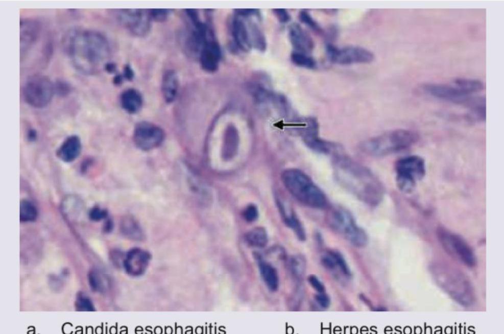
A 40-year-old immunocompromised patient presents with complaints of dysphagia. UGI endoscopy shows multiple ulcers in distal esophagus. Biopsy was performed and histopathology is shown below. Diagnosis is:
What is the most common type of tumour of Vermiform Appendix?
Melanosis coli, which occurs due to long term consumption of stimulant laxatives, presents as brown discolouration of colonic mucosa due to deposition of which one of the following pigments?
Which of the following statements are correct with regard to Familial Adenomatous Polyposis ? 1. It is associated with mutation of APC gene located on the long arm of chromosome 5. 2. It is inherited as an autosomal recessive condition. 3. It is associated with 100% lifetime risk for development of Colorectal carcinoma. 4. Congenital hypertrophy of retinal pigment epithelium is present in half of the cases of familial adenomatous polyposis. Select the correct answer using the code given below :
Which one of the following is correct regarding Gastrointestinal Stromal Tumour (GIST)?
The following disorders are predisposing conditions for carcinoma of the colon except
The following statements regarding small bowel tuberculosis are correct except
Which one of the following is not a premalignant condition for colon cancer?
Acinic cell carcinoma is found in
In Crohn’s disease all are true except:
Practice by Chapter
Oral Cavity and Esophageal Pathology
Practice Questions
Gastritis and Peptic Ulcer Disease
Practice Questions
Inflammatory Bowel Disease
Practice Questions
Malabsorption Syndromes
Practice Questions
Vascular Disorders of Intestine
Practice Questions
Diverticular Disease
Practice Questions
Intestinal Obstruction
Practice Questions
Gastrointestinal Infections
Practice Questions
Polyps and Neoplasms
Practice Questions
Appendiceal Pathology
Practice Questions
Want unlimited practice?
Get full access to all questions, explanations, and performance tracking.
Scan to download app