A 55-year-old man presents with recurrent epigastric pain. Upper GI endoscopy and gastric biopsy reveal a neoplastic, lymphocytic infiltrate invading glandular tissue. Giemsa staining is positive for Helicobacter pylori. Which of the following is the most likely diagnosis?
Which of the following is FALSE about hypertrophic gastropathy?
The greatest volume of gastric secretion occurs during which phase?
Peptic ulcer is caused by which of the following microorganisms?
What is the most common site for diverticula?
Which anomaly is seen in the following clinical condition?
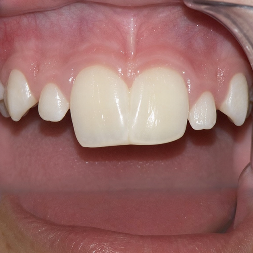
A child presents with generalized "freckling" affecting the buccal mucosa, lips, palms, soles, and sun-unexposed skin. Which of the following additional findings would most likely be present?
Leukoplakia appears white due to which of the following mechanisms?
Which of the following are true about celiac disease?
Linear enamel caries is also known as:
Explanation: **Explanation:** The clinical presentation and biopsy findings are classic for **Extranodal Marginal Zone B-cell Lymphoma of Mucosa-Associated Lymphoid Tissue (MALToma)**. 1. **Why it is correct:** MALToma is the most common primary gastric lymphoma [2]. It is strongly associated with chronic **Helicobacter pylori** infection, which triggers a persistent immune response leading to the formation of organized lymphoid tissue (MALT) in the gastric mucosa [2]. The hallmark histological feature is the presence of **lymphoepithelial lesions**, where neoplastic B-cells (marginal zone cells) infiltrate and destroy gastric glandular epithelium [2]. Giemsa staining is the gold standard for identifying *H. pylori* in tissue sections. 2. **Why other options are incorrect:** * **Burkitt Lymphoma:** Characterized by a "starry-sky" appearance and $t(8;14)$ translocation involving the *MYC* gene. It is highly aggressive and not typically associated with *H. pylori*. * **Follicular Lymphoma:** Defined by a nodular growth pattern and $t(14;18)$ translocation involving *BCL2*. It rarely presents as a primary gastric lesion. * **Mantle Cell Lymphoma:** Associated with $t(11;14)$ and Cyclin D1 overexpression. While it can involve the GI tract (Lymphomatous Polyposis), it does not form the specific lymphoepithelial lesions seen in MALToma [1]. **High-Yield Clinical Pearls for NEET-PG:** * **Pathogenesis:** Chronic inflammation $\rightarrow$ Polyclonal B-cell hyperplasia $\rightarrow$ Monoclonal B-cell proliferation (MALToma) [2]. * **Genetics:** Most common translocation is **$t(11;18)(q21;q21)$**, involving the *API2-MLT* fusion. This translocation predicts **resistance** to *H. pylori* eradication therapy. * **Treatment:** Early-stage MALToma often regresses completely with **triple therapy** (antibiotics + PPI) to eradicate *H. pylori*. * **Markers:** MALTomas are typically CD19+, CD20+, and CD5 negative. [2, 3] **References:** [1] Kumar V, Abbas AK, et al.. Robbins and Cotran Pathologic Basis of Disease. 9th ed. The Gastrointestinal Tract, pp. 780-781. [2] Cross SS. Underwood's Pathology: A Clinical Approach. 6th ed. Common Clinical Problems From Alimentary System Disease, pp. 356-357.
Explanation: **Explanation:** Hypertrophic gastropathy refers to a group of uncommon conditions characterized by giant enlargement of the gastric rugae due to epithelial hyperplasia. **Why Option D is the Correct Answer (The False Statement):** MALTomas (Mucosa-Associated Lymphoid Tissue lymphomas) are B-cell neoplasms strongly associated with chronic **_Helicobacter pylori_ infection**, which triggers chronic inflammation and lymphoid follicle formation [1]. Hypertrophic gastropathies, such as Menetrier’s disease, are characterized by **epithelial hyperplasia** (driven by growth factors like TGF-alpha), not lymphoid proliferation. Therefore, they do not typically produce MALTomas. **Analysis of Incorrect Options:** * **Option A:** Hypertrophic gastropathy is the umbrella term for **Menetrier’s disease** (foveolar hyperplasia) and **Zollinger-Ellison Syndrome** (parietal cell hyperplasia due to gastrinoma) [2]. * **Option B:** Menetrier’s disease is considered a **pre-malignant condition**; the risk of gastric adenocarcinoma is increased in these patients, necessitating close surveillance [2]. * **Option C:** On endoscopy, the massively enlarged gastric folds resemble the gyri of the brain, a classic finding described as **"cerebriform rugacity."** **High-Yield Clinical Pearls for NEET-PG:** * **Menetrier’s Disease:** Associated with over-expression of **TGF-alpha**. It presents with the triad of giant gastric folds, **hypochlorhydria** (due to loss of parietal cells), and **protein-losing enteropathy** (leading to edema/hypoalbuminemia) [2]. * **Zollinger-Ellison Syndrome:** Characterized by hypergastrinemia, leading to **hyperplasia of parietal cells** and doubling of oxyntic mucosal thickness, resulting in peptic ulcers. * **Key Distinction:** Menetrier’s affects the **body and fundus** (sparing the antrum), whereas ZES involves generalized hyperplasia due to the trophic effects of gastrin [2]. **References:** [1] Cross SS. Underwood's Pathology: A Clinical Approach. 6th ed. Common Clinical Problems From Alimentary System Disease, pp. 356-357. [2] Kumar V, Abbas AK, et al.. Robbins and Cotran Pathologic Basis of Disease. 9th ed. The Gastrointestinal Tract, pp. 775-776.
Explanation: The secretion of gastric acid occurs in three distinct phases, categorized by the location of the stimulus. **Correct Answer: B. Gastric phase** The **gastric phase** accounts for approximately **50-60%** of the total gastric acid secretion. It is triggered by the arrival of food in the stomach, which causes **distension** (activating vagovagal and local enteric reflexes) and the presence of **peptides/amino acids** (stimulating G-cells to release Gastrin) [1]. This phase provides the greatest volume of secretion because the physical presence of food provides a sustained stimulus for both hormonal and neural pathways. **Explanation of Incorrect Options:** * **A. Cephalic phase:** This phase accounts for about **30%** of secretion. It is triggered by the sight, smell, or thought of food via the Vagus nerve. While it prepares the stomach, it does not produce the bulk of the volume. * **C. Intestinal phase:** This phase accounts for only **5-10%** of secretion. It begins when chyme enters the duodenum. While initially stimulatory (via intestinal gastrin), it quickly becomes inhibitory (via secretin and CCK) to prevent over-acidification of the duodenum. * **D. Deglutition:** This refers to the act of swallowing. While it is part of the digestive process, it is a mechanical action and not a physiological "phase" of gastric acid secretion. **High-Yield Facts for NEET-PG:** * **Gastrin** is the most potent stimulator of the gastric phase [1]. * **Vagus nerve** is the primary mediator of the cephalic phase (blocked by vagotomy). * **Sham feeding** is an experimental method used to study the cephalic phase. * **pH < 1.5** in the antrum inhibits gastrin release (negative feedback) [1]. **References:** [1] Cross SS. Underwood's Pathology: A Clinical Approach. 6th ed. Common Clinical Problems From Alimentary System Disease, pp. 349-352.
Explanation: **Explanation:** **Helicobacter pylori (Option A)** is the correct answer. It is a gram-negative, spiral-shaped, urease-positive bacterium that colonizes the gastric mucosa [2]. It is the most common cause of peptic ulcer disease (PUD), accounting for approximately 70% of gastric ulcers and over 90% of duodenal ulcers [1]. *H. pylori* causes mucosal damage through the production of ammonia (via urease), inflammatory cytokines, and virulence factors like **CagA** (Cytotoxin-associated gene A) and **VacA** (Vacuolating cytotoxin). **Why other options are incorrect:** * **Campylobacter jejuni (Option B):** A common cause of bacterial gastroenteritis and bloody diarrhea. It is famously associated with the post-infectious complication **Guillain-Barré Syndrome**, but not peptic ulcers. * **Pneumocystis carinii (Option C):** Now reclassified as *Pneumocystis jirovecii*, this is a fungus that causes opportunistic pneumonia (PCP) in immunocompromised patients, particularly those with HIV/AIDS. * **Cryptosporidium (Option D):** An acid-fast protozoan parasite that causes self-limiting watery diarrhea in healthy individuals and chronic, severe diarrhea in AIDS patients. **High-Yield Clinical Pearls for NEET-PG:** * **Gold Standard Diagnosis:** Endoscopic biopsy followed by a **Rapid Urease Test (RUT)** or Histopathology (Warthin-Starry silver stain or Giemsa stain) [2]. * **Non-invasive Screening:** Urea Breath Test (using C13 or C14) is the investigation of choice for confirming eradication. * **Location:** *H. pylori* primarily colonizes the **Antrum** of the stomach [2]. * **Malignancy Risk:** Chronic *H. pylori* infection is a major risk factor for **Gastric Adenocarcinoma** and **MALToma** (Mucosa-Associated Lymphoid Tissue lymphoma). **References:** [1] Cross SS. Underwood's Pathology: A Clinical Approach. 6th ed. Common Clinical Problems From Alimentary System Disease, pp. 353-354. [2] Kumar V, Abbas AK, et al.. Robbins and Cotran Pathologic Basis of Disease. 9th ed. The Gastrointestinal Tract, pp. 770-771.
Explanation: **Explanation:** **Diverticulosis** refers to the herniation of mucosa and submucosa through the muscular layers of the colon (pseudodiverticula). **1. Why Sigmoid Colon is the Correct Answer:** The sigmoid colon is the most common site (involved in >90% of cases) due to **Laplace’s Law**. This law states that pressure is inversely proportional to the radius ($P = T/r$). Since the sigmoid has the smallest diameter of the colon, it generates the highest intraluminal pressures [1]. Additionally, the sigmoid acts as a "reservoir" where stool is most dehydrated and firm, requiring high-pressure contractions to move it forward [1]. These high pressures force the mucosa through weak points in the muscularis propria—specifically where the **vasa recta** (nutrient arteries) penetrate the muscle wall. **2. Analysis of Incorrect Options:** * **B. Ileum:** True diverticula (like Meckel’s) can occur here, but acquired pulsion diverticula are rare in the small intestine compared to the colon. * **C & D. Ascending and Transverse Colon:** While diverticula can occur here (more common in Asian populations), they are statistically less frequent than sigmoid involvement in global and Indian contexts. **High-Yield Clinical Pearls for NEET-PG:** * **Pathogenesis:** Associated with **low-fiber diets**, which decrease stool bulk and increase the pressure required for peristalsis [1]. * **Complications:** The most common cause of massive **painless lower GI bleeding** in the elderly [1]. If inflamed (**Diverticulitis**), it presents as "Left-sided appendicitis." * **Imaging:** Contrast CT is the gold standard for acute diverticulitis; Colonoscopy is contraindicated in the acute phase due to perforation risk. * **Associated Condition:** **Saint’s Triad** (Diverticulosis + Hiatus Hernia + Gallstones). **References:** [1] Cross SS. Underwood's Pathology: A Clinical Approach. 6th ed. Common Clinical Problems From Alimentary System Disease, pp. 370-371.
Explanation: **Explanation:** The clinical condition associated with this question is likely **Gardner Syndrome** (a variant of Familial Adenomatous Polyposis), which is a high-yield topic in GI Pathology. Gardner Syndrome is characterized by the triad of intestinal polyposis, soft tissue tumors (desmoids) [1], and skeletal/dental anomalies. **Why Dental Fusion is the Correct Answer:** Dental fusion occurs when two separate tooth buds join together during development, resulting in a single large or "double" tooth. In the context of syndromic GI pathology, dental fusion is a recognized dental anomaly. It is distinguished by a reduced number of teeth in the dental arch (unless fused with a supernumerary tooth) and the presence of two distinct pulp chambers on radiography. **Analysis of Incorrect Options:** * **B. Gemination:** This occurs when a single tooth bud attempts to divide into two. Unlike fusion, the total tooth count remains normal, and there is usually only one shared root canal. * **C. Concrescence:** This is a form of fusion where two fully formed teeth are joined only by **cementum**. It typically occurs due to trauma or crowding after root formation is complete. * **D. Supernumerary teeth:** While these (especially **Hyperdontia**) are the *most common* dental finding in Gardner Syndrome, the specific morphological anomaly described by "fusion" refers to the physical joining of units, which is a distinct developmental pathology. **NEET-PG Clinical Pearls:** * **Gardner Syndrome Triad:** FAP (100% risk of colon cancer) + Osteomas (mandible/skull) + Soft tissue tumors (Epidermoid cysts, Desmoid tumors) [1]. * **Dental Markers:** Impacted teeth, odontomas, and supernumerary teeth are often the *first* signs of Gardner syndrome, appearing before intestinal polyps. * **Radiographic Tip:** In fusion, the number of teeth is **decreased**; in gemination, the number of teeth is **normal**. **References:** [1] Cross SS. Underwood's Pathology: A Clinical Approach. 6th ed. Common Clinical Problems From Osteoarticular And Connective Tissue Disease, pp. 691-692.
Explanation: ### Explanation **Diagnosis: Peutz-Jeghers Syndrome (PJS)** The clinical presentation of generalized "freckling" (mucocutaneous hyperpigmentation) specifically involving the **buccal mucosa**, lips, and digits is the hallmark of **Peutz-Jeghers Syndrome** [1]. This is an autosomal dominant condition caused by a mutation in the **STK11 (LKB1)** gene on chromosome 19 [2]. **1. Why the Correct Answer is Right:** In PJS, the characteristic gastrointestinal finding is the presence of multiple **hamartomatous polyps**. While these polyps are most common in the small intestine, they frequently occur in the colon and stomach [1]. These polyps are histologically distinct, featuring a "Christmas tree" branching pattern of smooth muscle fibers. **2. Why the Incorrect Options are Wrong:** * **Options B, C, and D (Desmoid tumors, Epidermoid cysts, and Osteomas):** These are the classic extra-intestinal manifestations of **Gardner Syndrome** (a variant of Familial Adenomatous Polyposis/FAP). While Gardner Syndrome also presents with colonic polyps, it lacks the characteristic mucocutaneous melanocytic pigmentation seen in this child. **Clinical Pearls for NEET-PG:** * **Inheritance:** Autosomal Dominant; STK11 gene mutation [2]. * **Polyp Type:** Hamartomatous (not premalignant themselves, but associated with a high risk of visceral cancers) [1]. * **Cancer Risk:** Patients have a significantly increased risk of colorectal, pancreatic, breast, lung, and ovarian (Sertoli cell tumors) cancers [1]. * **Common Complication:** Intussusception (the polyps act as lead points). * **Distinction:** Unlike FAP, where polyps are adenomatous and number in the thousands, PJS polyps are hamartomatous and fewer in number. **References:** [1] Kumar V, Abbas AK, et al.. Robbins and Cotran Pathologic Basis of Disease. 9th ed. The Gastrointestinal Tract, p. 813. [2] Kumar V, Abbas AK, et al.. Robbins and Cotran Pathologic Basis of Disease. 9th ed. The Gastrointestinal Tract, pp. 813-814.
Explanation: **Explanation:** The term **Leukoplakia** is a clinical diagnosis defined by the WHO as "a white patch or plaque that cannot be characterized clinically or pathologically as any other disease" [1]. **Why Option A is correct:** The characteristic white appearance of leukoplakia is primarily due to **hyperkeratosis** (thickening of the stratum corneum). In the moist environment of the oral cavity, this thickened **cornified layer imbibes (absorbs) water**, causing it to swell and become opaque [2]. This hydrated keratin layer prevents the underlying vascularity of the connective tissue from showing through, resulting in a white clinical appearance rather than the normal pinkish-red hue of the oral mucosa. **Why other options are incorrect:** * **Option B:** While epithelial thickening (acanthosis) often co-exists with leukoplakia, thickness alone does not change the color to white unless the superficial keratin layer is altered [2]. * **Option C:** The underlying connective tissue is actually what gives the mucosa its normal pink color due to blood vessels. In leukoplakia, this tissue is masked by the superficial keratin, not the cause of the whiteness. **High-Yield NEET-PG Pearls:** * **Histopathology:** Shows hyperkeratosis, acanthosis, and varying degrees of epithelial dysplasia [2]. * **Risk Factors:** Tobacco (most common), alcohol, and HPV (strains 16 and 18) [1]. * **Malignant Transformation:** Approximately 3–25% of leukoplakias progress to Squamous Cell Carcinoma (SCC) [1]. * **Erythroplakia:** A red, velvety patch. It is much less common than leukoplakia but has a significantly **higher risk** of malignant transformation (>50%). **References:** [1] Cross SS. Underwood's Pathology: A Clinical Approach. 6th ed. Common Clinical Problems From Alimentary System Disease, pp. 344-345. [2] Kumar V, Abbas AK, et al.. Robbins and Cotran Pathologic Basis of Disease. 9th ed. The Female Genital Tract, pp. 1000-1002.
Explanation: **Explanation:** Celiac disease is an immune-mediated enteropathy triggered by the ingestion of gluten in genetically susceptible individuals (HLA-DQ2/DQ8) [1], [2]. The diagnosis is histopathologically characterized by the **Marsh Classification**. **Why Crypt Hyperplasia is the Correct Answer:** In Celiac disease, the chronic inflammatory response leads to increased enterocyte turnover. As the surface villi are destroyed (atrophy), the intestinal crypts undergo **compensatory hyperplasia** (elongation and increased mitotic activity) to replace the lost cells [1]. This is a hallmark feature used to grade the severity of the disease. **Analysis of Other Options:** * **Villous Atrophy (Option A):** While villous atrophy is a classic feature of Celiac disease, it is often "subtotal" or "total." In the context of many PG exams, if a question asks for a specific regenerative response to injury, **Crypt Hyperplasia** is the definitive proliferative change [1]. (Note: In many clinical scenarios, A, B, and C are all seen; however, if forced to choose the most specific regenerative marker, crypt hyperplasia is key). * **Infiltration of Lymphocytes (Option C):** Specifically, **Increased Intraepithelial Lymphocytes (IELs)**—usually >25 per 100 enterocytes—is the earliest histological change (Marsh Stage 1) [1]. "Infiltration of lymphocytes" is a broad term; IELs are the specific diagnostic requirement. * **Cryptitis (Option D):** This refers to neutrophils within the crypt epithelium and is a hallmark of **Inflammatory Bowel Disease (IBD)**, such as Ulcerative Colitis, rather than Celiac disease. **High-Yield Clinical Pearls for NEET-PG:** * **Gold Standard Diagnosis:** Duodenal biopsy (showing IELs, crypt hyperplasia, and villous atrophy). * **Serology:** Anti-tissue Transglutaminase (tTG) IgA is the screening drug of choice; Anti-Endomysial Antibody (EMA) is the most specific [2]. * **Site:** Most severe in the **distal duodenum and proximal jejunum** (highest concentration of dietary gluten) [2]. * **Associated Malignancy:** Enteropathy-associated T-cell lymphoma (EATL). **References:** [1] Kumar V, Abbas AK, et al.. Robbins and Cotran Pathologic Basis of Disease. 9th ed. The Gastrointestinal Tract, pp. 789-790. [2] Cross SS. Underwood's Pathology: A Clinical Approach. 6th ed. Common Clinical Problems From Alimentary System Disease, pp. 360-362.
Explanation: **Explanation:** **Linear Enamel Caries**, also known as **Odontoclasia** or Beltrami’s disease, is a specific type of dental caries characterized by a rapid, destructive process affecting the labial surfaces of the maxillary anterior teeth [1]. It typically presents as a horizontal line of decay across the cervical third of the tooth. It is most commonly seen in children (primary dentition) and is often associated with poor oral hygiene, malnutrition, or specific environmental factors [1]. **Analysis of Options:** * **A. Odontoclasia (Correct):** This is the synonymous term for linear enamel caries. The term "odontoclasia" refers to the progressive destruction of the tooth structure, specifically starting as a white or brown line of decalcification that eventually leads to the fracture of the crown. * **B. Occult Caries:** Also known as "hidden caries," these are lesions that are not clinically visible during a visual exam but are detected radiographically [2]. They occur beneath an apparently intact enamel surface. * **C. Fluoride Bomb:** This is a clinical phenomenon where the enamel surface remains hard and intact due to high fluoride intake, while a massive carious lesion (decay) hollows out the dentin underneath [2]. It is essentially a type of occult caries. **High-Yield Clinical Pearls for NEET-PG:** * **Nursing Bottle Caries:** Another form of rampant caries in children, but unlike linear caries, it is specifically linked to prolonged bottle feeding with sugary liquids. * **Radiation Caries:** A rapid form of decay following radiotherapy for head and neck cancers, primarily due to xerostomia (reduced salivary flow). * **Key Site:** Linear enamel caries most frequently involves the **maxillary incisors**, following the neonatal line. **References:** [1] Cross SS. Underwood's Pathology: A Clinical Approach. 6th ed. Common Clinical Problems From Alimentary System Disease, pp. 343-344. [2] Kumar V, Abbas AK, et al.. Robbins and Cotran Pathologic Basis of Disease. 9th ed. The Lung, pp. 734-735.
Oral Cavity and Esophageal Pathology
Practice Questions
Gastritis and Peptic Ulcer Disease
Practice Questions
Inflammatory Bowel Disease
Practice Questions
Malabsorption Syndromes
Practice Questions
Vascular Disorders of Intestine
Practice Questions
Diverticular Disease
Practice Questions
Intestinal Obstruction
Practice Questions
Gastrointestinal Infections
Practice Questions
Polyps and Neoplasms
Practice Questions
Appendiceal Pathology
Practice Questions
Get full access to all questions, explanations, and performance tracking.
Start For Free