Which of the following is NOT a predisposing cause for carcinoma of the stomach?
Which of the following is NOT true about solitary rectal ulcer syndrome?
A 63-year-old man undergoes a screening colonoscopy and is found to have a polyp in his sigmoid colon. Which type of polyp is most associated with malignancy?
Where does ulcerative colitis typically begin?
Zollinger-Ellison syndrome is not caused by tumors from which of the following organs?
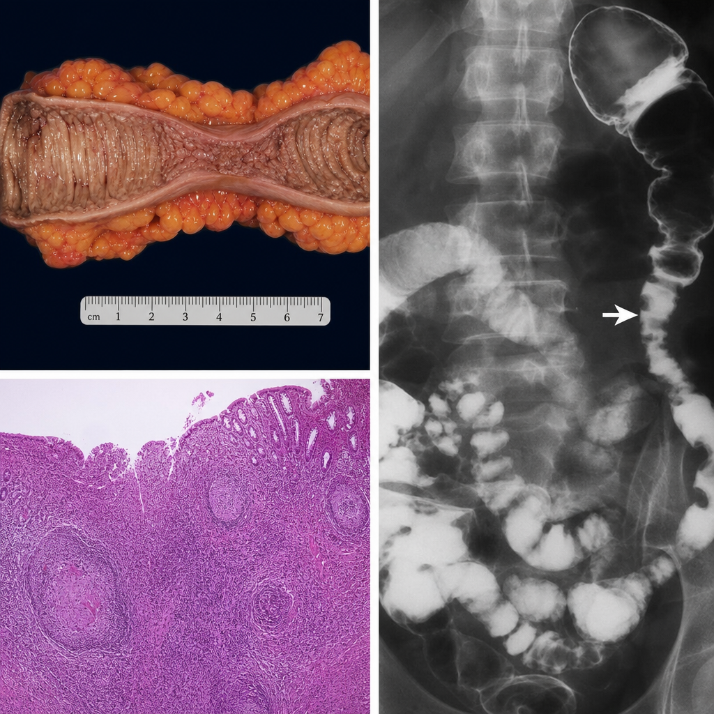
What is the most common site of MALToma?
What type of polyps are characteristic of Peutz-Jeghers syndrome?
What is the most common type of carcinoma of the right colon?
Which inflammatory bowel disease is characterized by transmural involvement and skip lesions?
Which of the following autosomal dominant disorders is characterized by the formation of multiple gastrointestinal polyps along with pigmented lesions around the oral cavity?
Explanation: **Explanation:** The development of gastric adenocarcinoma (specifically the intestinal type) follows a well-defined precancerous cascade known as **Correa’s Pathway**. This pathway involves a progression from chronic inflammation to atrophy, metaplasia, dysplasia, and finally, carcinoma [1]. **Why Hyperplastic Polyps are the correct answer:** Hyperplastic polyps are the most common type of gastric polyp (associated with chronic gastritis). They are considered **non-neoplastic** and have a negligible risk of malignant transformation (usually <1%). In contrast, **Adenomatous polyps** are true neoplastic precursors with a high risk of malignancy, with transformation reported in up to 75% of larger lesions [1]. **Analysis of other options:** * **Chronic Gastric Atrophy:** This leads to a loss of parietal cells and a decrease in acid secretion (hypochlorhydria), allowing for the colonization of nitrate-reducing bacteria [2]. These bacteria convert dietary nitrates into carcinogenic N-nitroso compounds. * **Intestinal Metaplasia (Grade III):** Grade III (or Type III/Incomplete) metaplasia is characterized by the presence of colonic-type epithelium (sulfomucins). It is a highly specific precursor lesion and carries the highest risk of progression to adenocarcinoma [1]. * **Pernicious Anemia:** This is an autoimmune condition causing destruction of parietal cells (Autoimmune Metaplastic Atrophic Gastritis). It leads to profound achlorhydria and hypergastrinemia, significantly increasing the risk of both gastric adenocarcinoma and carcinoid tumors [2]. **High-Yield Pearls for NEET-PG:** * **H. pylori:** The most common cause of chronic gastritis and the #1 risk factor for gastric cancer [2]. * **Menetrier Disease:** A hypertrophic gastropathy associated with an increased risk of adenocarcinoma. * **Blood Group A:** Epidemiologically associated with an increased risk of gastric cancer. * **Lauren Classification:** Divides gastric cancer into **Intestinal** (associated with environmental factors/precursors) and **Diffuse** (associated with CDH1 mutation/Signet ring cells). **References:** [1] Cross SS. Underwood's Pathology: A Clinical Approach. 6th ed. Common Clinical Problems From Alimentary System Disease, pp. 354-355. [2] Cross SS. Underwood's Pathology: A Clinical Approach. 6th ed. Common Clinical Problems From Alimentary System Disease, pp. 351-352.
Explanation: **Explanation:** Solitary Rectal Ulcer Syndrome (SRUS) is a chronic, non-neoplastic inflammatory condition caused by **impaired relaxation of the anorectal sphincter** and **pelvic floor dyssynergia** [1]. This leads to chronic mechanical trauma and mucosal ischemia during defecation [1]. **Why Option C is the correct answer:** The hallmark histological feature of SRUS is **fibromuscular obliteration** of the lamina propria. Instead of a dense inflammatory infiltrate (like lymphocytes or plasma cells seen in IBD), the lamina propria is replaced by **bundles of smooth muscle and collagen** (fibrosis) radiating between the crypts. Therefore, "lamina propria infiltration with lymphocytes" is incorrect and characteristic of other inflammatory conditions. **Analysis of Incorrect Options:** * **A. Increased muscle layer proliferation:** True. Chronic straining causes the muscularis mucosae to hypertrophy and send extensions upward into the lamina propria (fibromuscular hyperplasia). * **B. Crypt distortion:** True. The chronic mechanical injury and subsequent healing lead to architectural changes, including branching and distortion of the colonic crypts. * **D. Subepithelial fibrosis:** True. Ischemic injury and chronic trauma trigger fibroblast activity, leading to significant collagen deposition (fibrosis) beneath the epithelium. **NEET-PG High-Yield Pearls:** * **Clinical Triad:** Rectal bleeding, mucoid discharge, and chronic straining during defecation [1]. * **Misnomer:** Despite the name, the lesion is not always "solitary" and not always an "ulcer" (it can appear as a polypoid mass or erythematous mucosa). * **Key Histology:** "Fibromuscular obliteration" of the lamina propria is the pathognomonic buzzword. * **Differential Diagnosis:** Must be distinguished from inflammatory bowel disease (IBD) and rectal adenocarcinoma. **References:** [1] Kumar V, Abbas AK, et al.. Robbins and Cotran Pathologic Basis of Disease. 9th ed. The Gastrointestinal Tract, pp. 811-813.
Explanation: **Explanation:** The risk of malignancy in a colonic adenoma is determined by three main factors: **histological architecture, size, and the degree of dysplasia.** [1, 2] **1. Why Villous Adenoma is Correct:** Adenomas are classified based on their growth pattern. **Villous adenomas** (characterized by long, finger-like projections) carry the highest risk of harboring invasive carcinoma, estimated at **30–40%**. [1] This is significantly higher than other types because villous architecture is often associated with larger size and more severe epithelial dysplasia. [2] **2. Analysis of Incorrect Options:** * **Tubular Adenoma (Option A):** These are the most common type (approx. 90%) but have the lowest malignant potential (about 5%). [1] They are typically small and pedunculated. [1] * **Tubulovillous Adenoma (Option C):** These contain 25–75% villous architecture. [1] Their malignant risk is intermediate between tubular and villous adenomas. * **1 cm Polyp (Option D):** While size is a critical predictor (polyps >2 cm have a 40-50% risk of malignancy), a "1 cm polyp" is relatively small. [2] Histology (Villous) is a stronger independent predictor of malignancy than a 1 cm size threshold. **High-Yield Clinical Pearls for NEET-PG:** * **The "Rule of 3" for Malignancy Risk:** Risk increases if the polyp is **>2 cm**, has **Villous** histology, or shows **High-grade dysplasia**. [2] * **Villous Adenoma Presentation:** These may present with **secretory diarrhea** (hypokalemia and protein loss) due to the large surface area secreting mucus. * **Hyperplastic Polyps:** These are the most common non-neoplastic polyps and have virtually no malignant potential (usually found in the rectosigmoid). * **Molecular Pathway:** Most colorectal cancers arise via the **Adenoma-Carcinoma Sequence** involving mutations in *APC*, *KRAS*, and *p53*. **References:** [1] Cross SS. Underwood's Pathology: A Clinical Approach. 6th ed. Common Clinical Problems From Alimentary System Disease, pp. 371-372. [2] Kumar V, Abbas AK, et al.. Robbins and Cotran Pathologic Basis of Disease. 9th ed. The Gastrointestinal Tract, pp. 815-817.
Explanation: **Explanation:** Ulcerative Colitis (UC) is a chronic inflammatory bowel disease characterized by continuous mucosal inflammation. The hallmark of UC is its predictable anatomical progression: it **always involves the rectum** (proctitis) and extends proximally in a continuous, symmetrical fashion without "skip lesions" [1]. * **Why Rectum is Correct:** In virtually 100% of cases, the disease originates in the rectum [1]. From there, it may remain localized or spread proximally to involve the sigmoid, descending, or the entire colon (pancolitis). * **Why other options are incorrect:** * **Transverse and Descending Colon:** While these areas are frequently involved, they are affected as a result of proximal spread from the rectum, rather than being the primary site of origin. * **Caecum:** This is the most proximal part of the large intestine. While it is involved in pancolitis, it is not the starting point. (Note: A "caecal patch" can sometimes be seen in distal UC, but the rectum remains the primary site). **High-Yield NEET-PG Pearls:** 1. **Continuity:** Unlike Crohn’s disease (which is patchy/transmural), UC is **continuous** and limited to the **mucosa and submucosa** [1]. 2. **Backwash Ileitis:** In severe pancolitis, the terminal ileum may show superficial inflammation; this is the only time UC "crosses" the ileocecal valve [1]. 3. **Microscopy:** Look for **Crypt Abscesses** (neutrophils in crypt lumens) and crypt distortion. 4. **Clinical Sign:** Lead-pipe appearance on barium enema due to loss of haustrations. 5. **Risk:** UC carries a higher risk of **Toxic Megacolon** and **Adenocarcinoma** compared to Crohn’s. **References:** [1] Cross SS. Underwood's Pathology: A Clinical Approach. 6th ed. Common Clinical Problems From Alimentary System Disease, pp. 367-368.
Explanation: **Zollinger-Ellison Syndrome (ZES)** is characterized by the triad of gastric acid hypersecretion, severe peptic ulceration, and non-beta cell islet tumors (gastrinomas) [1]. The pathophysiology involves the ectopic secretion of **gastrin**, which stimulates parietal cells to produce excessive hydrochloric acid [3]. 1. **Why Colon is the correct answer:** Gastrinomas are neuroendocrine tumors (NETs). While they can occur in various locations, they are **not** associated with the colon. The colon does not typically harbor the neuroendocrine cells capable of transforming into gastrin-secreting tumors. 2. **Why other options are incorrect:** * **Pancreas (Option A):** Historically considered the most common site, pancreatic gastrinomas are often large and frequently malignant. * **Duodenum (Option D):** Currently recognized as the most common site for gastrinomas (up to 70% of cases) [1]. These are often small, multicentric, and slow-growing. * **Ovary (Option B):** Rare ectopic sites for gastrinomas include the ovary (specifically within cystic teratomas), liver, and stomach. **NEET-PG High-Yield Pearls:** * **The Gastrinoma Triangle (Passaro’s Triangle):** 90% of gastrinomas are found here. Boundaries: Junction of cystic and common bile duct, junction of 2nd and 3rd parts of the duodenum, and the neck/body of the pancreas [1]. * **Association:** Approximately 25% of ZES cases are associated with **Multiple Endocrine Neoplasia Type 1 (MEN1)** [2]. * **Diagnosis:** Best initial test is fasting serum gastrin levels (>1000 pg/mL is diagnostic). The most specific provocative test is the **Secretin Stimulation Test** (gastrin levels rise in ZES but fall in normal individuals). * **Clinical Feature:** Refractory peptic ulcers and chronic watery diarrhea (due to low pH inactivating pancreatic enzymes). **References:** [1] Kumar V, Abbas AK, et al.. Robbins and Cotran Pathologic Basis of Disease. 9th ed. The Endocrine System, pp. 1124-1125. [2] Kumar V, Abbas AK, et al.. Robbins and Cotran Pathologic Basis of Disease. 9th ed. The Gastrointestinal Tract, pp. 776-777. [3] Cross SS. Underwood's Pathology: A Clinical Approach. 6th ed. Common Clinical Problems From Alimentary System Disease, pp. 353-354.
Explanation: **Explanation:** **MALToma (Mucosa-Associated Lymphoid Tissue lymphoma)** is a type of extranodal marginal zone B-cell lymphoma. Under normal physiological conditions, the stomach lacks organized lymphoid tissue. However, chronic inflammation—most commonly due to **_Helicobacter pylori_ infection**—induces the formation of acquired MALT in the gastric mucosa [1]. Chronic antigenic stimulation leads to the proliferation of B-cells, eventually resulting in neoplastic transformation. * **Stomach (Correct):** It is the **most common site** for extranodal lymphomas, accounting for approximately 50% of all cases. Within the GI tract, the stomach is the site of origin for about 85% of MALTomas [1]. * **Neck & Mediastinum (Incorrect):** While nodal lymphomas (like Hodgkin or Diffuse Large B-cell Lymphoma) frequently involve cervical or mediastinal lymph nodes, MALToma is specifically an **extranodal** entity. * **Vertebra (Incorrect):** Bone involvement is characteristic of Multiple Myeloma or metastatic disease, not MALToma. **High-Yield Clinical Pearls for NEET-PG:** * **Association:** Strongly linked to **_H. pylori_** (80% of cases) [1]. Eradication of the bacteria with antibiotics can lead to complete regression of the tumor in early stages. * **Cytogenetics:** The most common translocation is **t(11;18)(q21;q21)**, involving the *API2-MLT* gene fusion. This translocation is typically associated with resistance to _H. pylori_ eradication therapy. * **Histology:** Characterized by **lymphoepithelial lesions** (invasion of gastric glands by neoplastic B-cells). * **Markers:** CD19+, CD20+, CD22+, and **CD5- / CD10-** (helps differentiate from Mantle cell and Follicular lymphoma). **References:** [1] Cross SS. Underwood's Pathology: A Clinical Approach. 6th ed. Common Clinical Problems From Alimentary System Disease, pp. 356-357.
Explanation: **Explanation:** **Peutz-Jeghers Syndrome (PJS)** is an autosomal dominant disorder characterized by the development of multiple **Hamartomatous polyps** throughout the gastrointestinal tract, most commonly in the small intestine [1]. These polyps are non-neoplastic malformations consisting of disorganized native tissue. Histologically, they show a characteristic "Christmas tree" branching pattern of smooth muscle (arising from the muscularis mucosae) covered by normal-appearing intestinal epithelium [1]. **Analysis of Options:** * **Option B (Correct):** Hamartomatous polyps are the hallmark of PJS [1]. They are caused by a germline mutation in the **STK11 (LKB1)** tumor suppressor gene on chromosome 19p13 [1]. * **Option A (Incorrect):** Villous adenomas are neoplastic epithelial polyps with a high risk of malignant transformation, typically associated with the sporadic adenoma-carcinoma sequence or Familial Adenomatous Polyposis (FAP). * **Option C (Incorrect):** Hyperplastic polyps are the most common non-neoplastic polyps in the colon, resulting from decreased epithelial cell turnover. They are not the defining feature of PJS. **High-Yield Clinical Pearls for NEET-PG:** * **Classic Triad:** Mucocutaneous hyperpigmentation (melanotic macules on lips, buccal mucosa, and digits) + Gastrointestinal hamartomatous polyps + Increased risk of visceral cancers [1]. * **Cancer Risk:** Patients have a significantly increased risk of colorectal, pancreatic, breast, lung, ovarian (Sertoli cell tumors), and uterine cancers [1]. * **Common Complication:** Intussusception is a frequent surgical emergency in these patients as the large polyps act as lead points. **References:** [1] Kumar V, Abbas AK, et al.. Robbins and Cotran Pathologic Basis of Disease. 9th ed. The Gastrointestinal Tract, pp. 813-814.
Explanation: **Explanation:** The morphological presentation of colorectal carcinoma varies significantly based on its anatomical location. **1. Why Ulcerative is correct:** While right-sided (proximal) colon cancers are classically described as **exophytic or fungating** masses in many textbooks, clinical and pathological data frequently categorize the most common macroscopic growth pattern as **ulcerative**. These lesions typically present as an irregular, deeply excavated ulcer with raised, everted edges. Because the right colon has a large caliber and the fecal matter is liquid, these tumors rarely cause obstruction early on; instead, they tend to bleed chronically, leading to iron-deficiency anemia. **2. Analysis of Incorrect Options:** * **Stenosing (Option A):** This is the hallmark of **left-sided (distal) colon cancer**. These lesions grow circumferentially, creating a "napkin-ring" or "apple-core" appearance, leading to early bowel obstruction. * **Tubular (Option C):** This refers to a microscopic growth pattern (histology) rather than a macroscopic/gross type of carcinoma [2]. * **Fungating (Option D):** While common in the right colon, fungating (polypoid/cauliflower-like) masses are often the precursor or a co-existing feature, but the **ulcerative** form is statistically documented as the most frequent presentation in surgical pathology. **Clinical Pearls for NEET-PG:** * **Right-sided tumors:** Present with **anemia**, occult blood, and weight loss [1]. They are often associated with **Microsatellite Instability (MSI)** and the BRAF mutation [1]. * **Left-sided tumors:** Present with **altered bowel habits** and obstruction. They are associated with the **CIN (Chromosomal Instability) pathway** (APC, KRAS, p53). * **Most common site:** The rectum and sigmoid colon remain the most frequent sites overall, though the incidence of right-sided (proximal) colon cancer is increasing. **References:** [1] Kumar V, Abbas AK, et al.. Robbins and Cotran Pathologic Basis of Disease. 9th ed. The Gastrointestinal Tract, pp. 819-821. [2] Cross SS. Underwood's Pathology: A Clinical Approach. 6th ed. Common Clinical Problems From Alimentary System Disease, pp. 371-372.
Explanation: ### Explanation **Crohn’s Disease (Option A)** is the correct answer because it is characterized by **transmural inflammation**, meaning the pathology involves all layers of the bowel wall (mucosa, submucosa, muscularis propria, and serosa) [1]. A hallmark endoscopic and gross finding is the presence of **skip lesions**, where areas of sharply demarcated disease are separated by segments of normal-appearing mucosa [1]. #### Analysis of Incorrect Options: * **Ulcerative Colitis (Option B):** Unlike Crohn’s, inflammation in UC is typically limited to the **mucosa and submucosa** [4]. It involves the rectum and extends proximally in a **continuous** fashion without skip lesions [4]. * **Shigella infection (Option C):** This causes acute infectious enterocolitis. While it causes significant mucosal damage and ulcers, it does not present with the chronic, transmural, or skip-pattern architecture seen in IBD. * **Clostridium infection (Option D):** *C. difficile* typically causes **Pseudomembranous colitis**, characterized by yellow-white plaques on the colonic mucosa. It is an acute toxin-mediated process, not a chronic transmural granulomatous disease. #### NEET-PG High-Yield Pearls: * **Microscopy:** Crohn’s disease features **non-caseating granulomas** (in 35% of cases) and lymphoid aggregates [3]. * **Gross Morphology:** Look for "cobblestone" appearance, "creeping fat" (mesenteric fat wrapping around the bowel), and "string sign of Kantor" on barium studies due to strictures [2]. * **Complications:** Because it is transmural, Crohn’s frequently leads to **fistulas, sinuses, and strictures**, whereas UC is more prone to **Toxic Megacolon** [1]. * **Site:** Crohn's can affect any part of the GIT (mouth to anus), but the **terminal ileum** is the most common site [1]. **References:** [1] Cross SS. Underwood's Pathology: A Clinical Approach. 6th ed. Common Clinical Problems From Alimentary System Disease, pp. 365-366. [2] Cross SS. Underwood's Pathology: A Clinical Approach. 6th ed. Common Clinical Problems From Alimentary System Disease, pp. 366-367. [3] Kumar V, Abbas AK, et al.. Robbins and Cotran Pathologic Basis of Disease. 9th ed. The Gastrointestinal Tract, pp. 806-807. [4] Cross SS. Underwood's Pathology: A Clinical Approach. 6th ed. Common Clinical Problems From Alimentary System Disease, pp. 367-368.
Explanation: **Explanation:** **Peutz-Jeghers Syndrome (PJS)** is the correct answer. It is an autosomal dominant condition caused by a germline mutation in the **STK11 (LKB1)** gene on chromosome 19 [1]. It is classically defined by the triad of: 1. **Hamartomatous polyps:** Multiple, characteristic "arborizing" polyps most commonly found in the small intestine [1]. 2. **Mucocutaneous hyperpigmentation:** Dark blue-to-brown macules (melanocytic spots) located around the mouth, lips, buccal mucosa, palms, and soles [1]. 3. **Increased Cancer Risk:** Significant risk for both GI (colorectal, pancreatic) and extra-GI malignancies (breast, ovary, testis) [1]. **Why the other options are incorrect:** * **Rotor Syndrome:** A benign autosomal recessive condition causing conjugated hyperbilirubinemia due to impaired hepatic storage of bilirubin. It does not involve polyps or pigmentation. * **Gardner Syndrome:** A variant of Familial Adenomatous Polyposis (FAP). While it features GI polyps (adenomatous, not hamartomatous), it is characterized by extra-intestinal manifestations like osteomas, epidermal cysts, and desmoid tumors, rather than perioral pigmentation. * **Cowden Disease:** Part of the PTEN hamartoma tumor syndrome [1]. It features GI hamartomas but is distinguished by macrocephaly, trichilemmomas (skin tumors), and a high risk of thyroid and endometrial cancer [1]. **High-Yield Clinical Pearls for NEET-PG:** * **Histology:** PJS polyps show a "Christmas tree" appearance due to branching frameworks of smooth muscle (arborization). * **Intussusception:** The most common surgical complication of PJS polyps in young patients. * **STK11:** Remember this gene; it is a tumor suppressor that regulates cell polarity and metabolism [1]. **References:** [1] Kumar V, Abbas AK, et al.. Robbins and Cotran Pathologic Basis of Disease. 9th ed. The Gastrointestinal Tract, pp. 813-814.
Oral Cavity and Esophageal Pathology
Practice Questions
Gastritis and Peptic Ulcer Disease
Practice Questions
Inflammatory Bowel Disease
Practice Questions
Malabsorption Syndromes
Practice Questions
Vascular Disorders of Intestine
Practice Questions
Diverticular Disease
Practice Questions
Intestinal Obstruction
Practice Questions
Gastrointestinal Infections
Practice Questions
Polyps and Neoplasms
Practice Questions
Appendiceal Pathology
Practice Questions
Get full access to all questions, explanations, and performance tracking.
Start For Free