Cobblestone appearance is seen in which of the following conditions?
E-cadherin mutation is seen in which type of gastric carcinoma classification?
All of the following are true about Barrett's esophagus, EXCEPT:
A 58-year-old woman undergoes routine colonoscopy. A 2-cm submucosal nodule is identified in the appendix. Biopsy of the nodule shows nests of cells with round, uniform nuclei. Electron microscopy reveals numerous neuroendocrine granules in the cytoplasm. This patient's neoplastic disease is associated with which of the following clinical features?
What is the immuno-marker for GIST?
A salivary gland tumor that histologically shows a double layer of epithelial cells based on a reactive lymphoid stroma is:
A 71-year-old male presents with dysphagia and is found to have a 5-cm mass that is located in the middle third of the esophagus and extends into adjacent lung tissue. A biopsy from this mass would most likely reveal?
The presence of anti-Saccharomyces cerevisiae antibody is a surrogate marker of which of the following conditions?
Which of the following is true about celiac disease?
Gardner's syndrome is a rare hereditary disorder involving the colon. It is characterized by
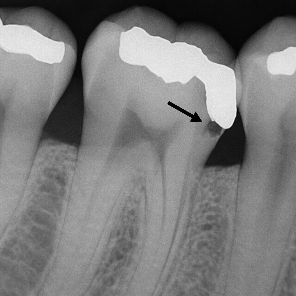
Explanation: **Explanation:** **Crohn's Disease (Correct Answer):** The "cobblestone appearance" is a classic macroscopic hallmark of Crohn’s disease [1]. It results from the presence of deep, linear, longitudinal, and transverse ulcers (fissures) that intersect, leaving islands of relatively normal, edematous, and inflamed intervening mucosa [1]. This creates an irregular, bumpy surface resembling a cobblestone street. Because Crohn's is a transmural inflammatory process, this pattern is often accompanied by "creeping fat" and bowel wall thickening [1]. **Incorrect Options:** * **Ulcerative Colitis:** Characterized by continuous, superficial mucosal inflammation starting from the rectum [3]. Instead of cobblestoning, it features **pseudopolyps** (islands of regenerating mucosa) and a "lead pipe" appearance on imaging due to loss of haustrations [1], [3]. * **Appendicitis:** Typically presents with neutrophilic infiltration of the muscularis propria. While it involves inflammation, it does not exhibit the specific patchy, fissured pattern required to form a cobblestone appearance. * **Carcinoma Rectum:** Usually presents as an exophytic mass, an annular "apple-core" lesion, or a malignant ulcer with everted edges, rather than a diffuse cobblestone pattern. **High-Yield Clinical Pearls for NEET-PG:** * **Skip Lesions:** Crohn’s is characterized by discontinuous involvement (skip lesions), whereas UC is continuous [1], [3]. * **Granulomas:** Non-caseating granulomas are seen in 40-60% of Crohn’s cases (pathognomonic in the GI tract) but are absent in UC [1], [2]. * **String Sign of Kantor:** Seen on barium swallow in Crohn's due to terminal ileal narrowing [1]. * **Transmural vs. Mucosal:** Crohn’s involves all layers (transmural), leading to fistulas and strictures; UC is limited to the mucosa and submucosa [1], [3]. **References:** [1] Cross SS. Underwood's Pathology: A Clinical Approach. 6th ed. Common Clinical Problems From Alimentary System Disease, pp. 365-367. [2] Kumar V, Abbas AK, et al.. Robbins and Cotran Pathologic Basis of Disease. 9th ed. The Gastrointestinal Tract, pp. 806-807. [3] Cross SS. Underwood's Pathology: A Clinical Approach. 6th ed. Common Clinical Problems From Alimentary System Disease, pp. 367-368.
Explanation: **Explanation:** The correct answer is **Lauren diffuse type**. Gastric adenocarcinoma is most commonly classified using the **Lauren Classification**, which divides tumors into Intestinal and Diffuse types based on their morphology and molecular pathogenesis [1]. **1. Why Lauren Diffuse Type is correct:** The hallmark of the diffuse type is a **loss of cell-cell adhesion** [1]. This is primarily due to a germline or somatic mutation in the **CDH1 gene**, which encodes **E-cadherin**, a surface glycoprotein responsible for intercellular adhesion [1]. The loss of E-cadherin leads to the characteristic "signet ring cell" morphology, where cells do not form glands but instead infiltrate the gastric wall individually or in small clusters, often resulting in *Linitis Plastica* (leather bottle stomach) [1]. **2. Why other options are incorrect:** * **Intestinal type (Option D):** This type is associated with environmental factors (H. pylori, smoking, diet) and follows the "Correa pathway" (gastritis → metaplasia → dysplasia). It is characterized by cohesive cells forming gland-like structures and is linked to mutations in **APC** or **β-catenin**, rather than E-cadherin [1]. * **Borrmann Classification (Options A & C):** This is a **macroscopic/gross** classification (Type I-IV) based on the physical appearance of the tumor (e.g., polypoid, ulcerative, infiltrative). It does not describe the molecular or genetic basis of the tumor. **Clinical Pearls for NEET-PG:** * **CDH1 Mutation:** Associated with **Hereditary Diffuse Gastric Cancer (HDGC)** syndrome and an increased risk of **Lobular Carcinoma of the breast**. * **Signet Ring Cells:** Nucleus is pushed to the periphery by a large mucin vacuole; cells stain positive with **PAS** or **Mucicarmine**. * **Virchow’s Node:** Left supraclavicular lymph node involvement. * **Sister Mary Joseph Nodule:** Periumbilical metastasis (common in intestinal type). * **Krukenberg Tumor:** Bilateral ovarian metastasis (common in diffuse type). **References:** [1] Kumar V, Abbas AK, et al.. Robbins and Cotran Pathologic Basis of Disease. 9th ed. The Gastrointestinal Tract, pp. 779-780.
Explanation: **Explanation:** **Barrett’s Esophagus (BE)** is a complication of chronic Gastroesophageal Reflux Disease (GERD) characterized by **metaplasia**. The normal stratified squamous epithelium of the lower esophagus is replaced by **non-ciliated columnar epithelium with goblet cells** (intestinal metaplasia) to better withstand acidic injury [1]. **Why Option D is the correct answer (The Exception):** Esophageal varices are dilated submucosal veins in the lower esophagus, typically caused by **portal hypertension** (secondary to liver cirrhosis) [3]. There is no direct pathophysiological link between Barrett’s esophagus (a mucosal metaplastic process) and esophageal varices (a vascular/hemodynamic process). **Analysis of other options:** * **Option A:** BE is a direct consequence of long-standing GERD, where chronic acid exposure triggers the adaptive change in the epithelium [1]. * **Option B:** It is a well-established **premalignant condition**, significantly increasing the risk of **Esophageal Adenocarcinoma** [1], [4]. * **Option C:** The hallmark of BE is the presence of **intestinal-type epithelium**, specifically identified by the presence of **Goblet cells** on histology [1]. **High-Yield Clinical Pearls for NEET-PG:** * **Gold Standard Diagnosis:** Endoscopy (showing "salmon-pink" velvety tongues of mucosa) + Biopsy (showing Goblet cells) [1], [2]. * **Metaplasia Type:** Squamous to Columnar metaplasia [1]. * **Risk Factor:** Most common in white males, smokers, and those with central obesity. * **Malignancy Risk:** BE leads to Adenocarcinoma; whereas smoking/alcohol (without BE) usually leads to Squamous Cell Carcinoma. * **Management:** Periodic endoscopic surveillance with biopsy is mandatory to check for dysplasia [2]. **References:** [1] Cross SS. Underwood's Pathology: A Clinical Approach. 6th ed. Common Clinical Problems From Alimentary System Disease, pp. 348-349. [2] Kumar V, Abbas AK, et al.. Robbins and Cotran Pathologic Basis of Disease. 9th ed. The Gastrointestinal Tract, pp. 764-765. [3] Kumar V, Abbas AK, et al.. Robbins and Cotran Pathologic Basis of Disease. 9th ed. The Gastrointestinal Tract, pp. 763-764. [4] Kumar V, Abbas AK, et al.. Robbins and Cotran Pathologic Basis of Disease. 9th ed. The Gastrointestinal Tract, pp. 765-766.
Explanation: ### Explanation **Diagnosis: Carcinoid Tumor of the Appendix** The clinical presentation describes a **Carcinoid tumor**, the most common neoplasm of the appendix [1]. The biopsy findings (nests of uniform cells with round nuclei) and electron microscopy (neuroendocrine granules) are classic hallmarks of neuroendocrine tumors (NETs) [1],[2]. **1. Why "Flushing and wheezing" is correct:** Carcinoid tumors secrete vasoactive substances, primarily **Serotonin (5-HT)**, as well as histamine and bradykinin. When these tumors metastasize to the liver (or bypass portal circulation), these substances enter the systemic circulation, leading to **Carcinoid Syndrome** [1]. The classic triad includes: * **Cutaneous flushing** (vasodilation) * **Wheezing** (bronchoconstriction) * **Diarrhea** (increased GI motility) **2. Why other options are incorrect:** * **A. Congestive heart failure:** While carcinoid syndrome can cause **Carcinoid Heart Disease** (typically right-sided endocardial fibrosis leading to tricuspid regurgitation or pulmonary stenosis), it does not typically present as generalized congestive heart failure unless valvular damage is severe and chronic. Flushing and wheezing are more immediate systemic markers. * **C. Muscular dystrophy:** This is a genetic primary muscle disorder with no association with neuroendocrine tumors. * **D. Progressive systemic sclerosis (Scleroderma):** While this involves fibrosis, it is an autoimmune connective tissue disease unrelated to serotonin-secreting tumors. **Clinical Pearls for NEET-PG:** * **Most common site for Carcinoid:** Appendix (overall), but the Small Intestine (Ileum) is the most common site to cause Carcinoid Syndrome [3]. * **Diagnosis:** Elevated **24-hour urinary 5-HIAA** (metabolite of serotonin). * **Histology:** "Salt and pepper" chromatin; Immunohistochemistry (IHC) markers: **Chromogranin A** and **Synaptophysin** [1]. * **Rule of 1/3s:** 1/3 are multiple, 1/3 are in the ileum, 1/3 are metastatic, and 1/3 have a second malignancy. **References:** [1] Kumar V, Abbas AK, et al.. Robbins and Cotran Pathologic Basis of Disease. 9th ed. The Gastrointestinal Tract, pp. 781-782. [2] Kumar V, Abbas AK, et al.. Robbins and Cotran Pathologic Basis of Disease. 9th ed. The Lung, p. 727. [3] Kumar V, Abbas AK, et al.. Robbins and Cotran Pathologic Basis of Disease. 9th ed. The Gastrointestinal Tract, pp. 780-781.
Explanation: **Explanation:** **Gastrointestinal Stromal Tumor (GIST)** is the most common mesenchymal tumor of the gastrointestinal tract. It originates from the **Interstitial Cells of Cajal (ICC)**, which serve as the electrical pacemakers of the gut. **1. Why CD 117 is correct:** Approximately 95% of GISTs express **CD 117**, which is the protein product of the **c-KIT proto-oncogene** [1]. This gene encodes a receptor tyrosine kinase. Mutations in c-KIT lead to constitutive activation of the kinase, driving uncontrolled cell proliferation [1]. Identifying CD 117 via immunohistochemistry (IHC) is the gold standard for diagnosis and is crucial because it predicts responsiveness to targeted therapy with **Imatinib** (a tyrosine kinase inhibitor) [1]. **2. Why the other options are incorrect:** * **CD 4:** A marker primarily found on T-helper cells, monocytes, and macrophages; it is used in the evaluation of lymphomas and HIV progression. * **CD 100 (Sema4D):** Involved in immune cell signaling and axonal guidance; it has no diagnostic relevance for GIST. * **CD 45:** Also known as **Leukocyte Common Antigen (LCA)**. It is a pan-leukocyte marker used to identify tumors of hematopoietic origin (lymphomas) and differentiate them from carcinomas or sarcomas. **High-Yield Facts for NEET-PG:** * **DOG1 (Discovered on GIST-1):** The most sensitive and specific marker for GIST, especially useful in CD 117-negative cases. * **Genetics:** Most cases involve **c-KIT** mutations; a subset involves **PDGFRA** mutations [1]. * **Morphology:** Most GISTs show a **spindle cell** pattern (70%), followed by epithelioid types. * **Location:** The **stomach** (60%) is the most common site, followed by the small intestine. **References:** [1] Kumar V, Abbas AK, et al.. Robbins and Cotran Pathologic Basis of Disease. 9th ed. The Gastrointestinal Tract, pp. 782-784.
Explanation: **Explanation:** The correct answer is **Warthin tumor** (also known as Papillary Cystadenoma Lymphomatosum). **1. Why Warthin Tumor is correct:** The histological hallmark of Warthin tumor is a unique **bi-layered (double layer) epithelium** arranged in papillary folds, projecting into cystic spaces [1]. * **Inner layer:** Tall, columnar, eosinophilic cells (oncocytes) with granular cytoplasm. * **Outer layer:** Cuboidal or flattened basal cells. * **Stroma:** This epithelium sits atop a dense **reactive lymphoid stroma**, often containing germinal centers [1]. This is a classic "high-yield" description for NEET-PG. **2. Why other options are incorrect:** * **Pleomorphic adenoma:** Characterized by a mixture of epithelial/myoepithelial cells and a **mesenchymal-like stroma** (myxoid, chondroid, or osteoid) [2]. It lacks the lymphoid component and the specific double-layered oncocytic pattern. * **Mucoepidermoid carcinoma:** Composed of a mixture of three cell types: squamous (epidermoid), mucus-secreting, and intermediate cells [3]. It does not feature a reactive lymphoid stroma. * **Acinic cell tumor:** Shows cells with granular basophilic cytoplasm resembling normal serous acinar cells, typically arranged in solid or microcystic patterns, without a prominent lymphoid background. **3. NEET-PG High-Yield Pearls for Warthin Tumor:** * **Location:** Almost exclusively occurs in the **Parotid gland** (often in the tail). * **Demographics:** Strong association with **smoking**; it is the most common salivary tumor to be **bilateral or multifocal** (10%). * **Imaging:** Shows high uptake on **Technetium-99m pertechnetate scan** (Hot tumor). * **Gender:** Historically more common in males, though the gap is narrowing due to smoking trends. **References:** [1] Kumar V, Abbas AK, et al.. Robbins and Cotran Pathologic Basis of Disease. 9th ed. Head and Neck, p. 753. [2] Kumar V, Abbas AK, et al.. Robbins and Cotran Pathologic Basis of Disease. 9th ed. Head and Neck, pp. 751-753. [3] Kumar V, Abbas AK, et al.. Robbins and Cotran Pathologic Basis of Disease. 9th ed. Head and Neck, pp. 753-755.
Explanation: ### Explanation **Correct Answer: D. Infiltrating sheets of cells forming keratin** The clinical presentation of a 71-year-old male with a large (5-cm) mass in the **middle third** of the esophagus that is locally invasive (extending into the lung) is highly characteristic of **Squamous Cell Carcinoma (SCC)** of the esophagus. 1. **Why it is correct:** Squamous cell carcinoma is the most common primary malignancy of the middle third of the esophagus [1]. Histologically, SCC is characterized by nests and sheets of malignant epithelial cells showing squamous differentiation, evidenced by **intercellular bridges** and **keratin pearls** (infiltrating sheets of cells forming keratin) [1]. 2. **Why the others are wrong:** * **Option A & B:** These describe benign tumors (Chondroma and Leiomyoma). While Leiomyoma is the most common benign esophageal tumor, a 5-cm invasive mass in an elderly patient with dysphagia is overwhelmingly likely to be malignant. * **Option C:** This describes **Adenocarcinoma**. While Adenocarcinoma is now more common in the West, it typically arises in the **distal third** of the esophagus, usually secondary to Barrett’s esophagus (metaplasia due to GERD) [1]. ### NEET-PG High-Yield Pearls * **Location Matters:** * **Middle Third:** Most common site for Squamous Cell Carcinoma [1]. * **Lower Third:** Most common site for Adenocarcinoma [1]. * **Risk Factors for SCC:** Smoking, alcohol, achalasia cardia, caustic injury, and Plummer-Vinson syndrome. * **Risk Factors for Adenocarcinoma:** Chronic GERD, Barrett’s esophagus (intestinal metaplasia), and obesity. * **Spread:** The esophagus lacks a serosa, facilitating early local extension into adjacent structures like the trachea (tracheoesophageal fistula), aorta, or lungs [1]. * **Lymphatic Drainage:** Middle third typically drains to the tracheobronchial and mediastinal nodes. **References:** [1] Kumar V, Abbas AK, et al.. Robbins and Cotran Pathologic Basis of Disease. 9th ed. The Gastrointestinal Tract, pp. 766-767.
Explanation: **Explanation:** The correct answer is **Crohn’s disease**. **Anti-Saccharomyces cerevisiae antibodies (ASCA)** are directed against mannan, a component of the cell wall of the yeast *Saccharomyces cerevisiae*. These antibodies are highly specific markers for Crohn’s disease (CD). In the context of Inflammatory Bowel Disease (IBD), ASCA is typically found in 60–70% of CD patients but is rarely present in Ulcerative Colitis (UC). **Analysis of Options:** * **Crohn’s disease (Correct):** ASCA is the characteristic serological marker. It is particularly associated with ileal involvement and fibrostenosing behavior. Crohn's disease is characterized by patchy segmental transmural chronic granulomatous inflammation [1]. * **Ulcerative colitis:** The surrogate marker for UC is **p-ANCA** (Perinuclear Anti-Neutrophil Cytoplasmic Antibody), found in 60–80% of cases. ASCA is usually negative in UC [1]. * **Celiac disease:** This is characterized by antibodies against gluten-related proteins. High-yield markers include **Anti-tissue Transglutaminase (tTG) IgA** (best screening test) and **Anti-Endomysial antibodies (EMA)** (most specific). * **Tropical sprue:** This is a malabsorption syndrome caused by chronic small bowel infection. Diagnosis is based on clinical history and biopsy showing villous atrophy; it does not have specific serological markers like ASCA. **High-Yield Clinical Pearls for NEET-PG:** * **ASCA (+) / p-ANCA (-):** Highly suggestive of Crohn’s Disease. * **ASCA (-) / p-ANCA (+):** Highly suggestive of Ulcerative Colitis. * **Transmural inflammation** and **Non-caseating granulomas** are the hallmark pathological features of Crohn’s disease [1]. * ASCA levels in Crohn’s disease often correlate with the severity of the disease and the likelihood of requiring surgery. **References:** [1] Cross SS. Underwood's Pathology: A Clinical Approach. 6th ed. Common Clinical Problems From Alimentary System Disease, pp. 365-367.
Explanation: **Explanation:** Celiac disease (Gluten-sensitive enteropathy) is an immune-mediated inflammatory disorder triggered by the ingestion of gluten in genetically predisposed individuals (HLA-DQ2/DQ8) [1, 2]. The pathogenesis involves a T-cell mediated response against gliadin, leading to characteristic histological changes in the small intestine, primarily the duodenum and proximal jejunum [1, 3]. The diagnosis relies on the **Marsh Classification**, which highlights the three features mentioned in the options: 1. **Villous Atrophy (Option A):** Chronic inflammation leads to the blunting and eventual flattening of the intestinal villi [1, 3]. This significantly reduces the surface area for absorption, resulting in malabsorption syndromes. 2. **Crypt Hyperplasia (Option B):** As the surface epithelial cells are damaged and lost due to inflammation, the intestinal crypts undergo compensatory elongation and increased mitotic activity to replace the lost enterocytes [1]. 3. **Infiltration of Lymphocytes (Option C):** An increase in **Intraepithelial Lymphocytes (IELs)**—specifically >25 per 100 enterocytes—is the earliest histological marker of the disease [1]. The lamina propria also shows an influx of plasma cells and T-cells [3]. Since all three features are hallmark histological findings of Celiac disease, **Option D** is the correct answer. **High-Yield Clinical Pearls for NEET-PG:** * **Serology:** Anti-tissue Transglutaminase (tTG) IgA is the screening drug of choice. Anti-Endomysial Antibody (EMA) is the most specific [2]. * **Gold Standard Diagnosis:** Small bowel biopsy (D2/Duodenum) [3]. * **Associated Malignancy:** Enteropathy-associated T-cell lymphoma (EATL) and Small bowel adenocarcinoma. * **Dermatological Association:** Dermatitis herpetiformis (IgA deposits at dermal papillae tips). **References:** [1] Kumar V, Abbas AK, et al.. Robbins and Cotran Pathologic Basis of Disease. 9th ed. The Gastrointestinal Tract, pp. 789-790. [2] Cross SS. Underwood's Pathology: A Clinical Approach. 6th ed. Common Clinical Problems From Alimentary System Disease, pp. 360-361. [3] Cross SS. Underwood's Pathology: A Clinical Approach. 6th ed. Common Clinical Problems From Alimentary System Disease, pp. 361-362.
Explanation: **Explanation:** Gardner’s syndrome is a clinical variant of **Familial Adenomatous Polyposis (FAP)** [1], an autosomal dominant disorder caused by mutations in the **APC (Adenomatous Polyposis Coli) gene** on chromosome 5q21 [1]. **1. Why Option C is Correct:** The syndrome is defined by a characteristic triad of: * **Colonic Polyposis:** Hundreds to thousands of adenomatous polyps throughout the colon, with a 100% risk of progression to colorectal carcinoma if left untreated [1], [2]. * **Osteomas:** Benign bone growths, most commonly involving the mandible and skull. * **Soft Tissue/Skin Lesions:** Specifically epidermal inclusion cysts, fibromas, and **desmoid tumors** (aggressive fibrous tumors). **2. Why Other Options are Incorrect:** * **Option A:** While Gardner’s syndrome is associated with an increased risk of extracolonic malignancies (including papillary thyroid cancer), the classic definition focuses on the triad of polyposis, osteomas, and skin tumors. * **Option B:** Polyposis in Gardner’s primarily affects the colon, not the jejunum [1]. Pituitary adenomas are associated with Multiple Endocrine Neoplasia (MEN-1) or Carney complex, not Gardner’s. **3. NEET-PG High-Yield Pearls:** * **Turcot Syndrome:** Another FAP variant characterized by colonic polyps plus **CNS tumors** (Medulloblastoma in FAP-associated Turcot; Glioblastoma in Lynch-associated Turcot). * **Dental Abnormalities:** Impacted teeth or supernumerary teeth are frequently seen in Gardner’s syndrome. * **CHRPE:** Congenital Hypertrophy of Retinal Pigment Epithelium is a highly specific screening marker for FAP/Gardner’s. **References:** [1] Kumar V, Abbas AK, et al.. Robbins and Cotran Pathologic Basis of Disease. 9th ed. The Gastrointestinal Tract, pp. 821-822. [2] Cross SS. Underwood's Pathology: A Clinical Approach. 6th ed. Common Clinical Problems From Alimentary System Disease, pp. 372-373.
Oral Cavity and Esophageal Pathology
Practice Questions
Gastritis and Peptic Ulcer Disease
Practice Questions
Inflammatory Bowel Disease
Practice Questions
Malabsorption Syndromes
Practice Questions
Vascular Disorders of Intestine
Practice Questions
Diverticular Disease
Practice Questions
Intestinal Obstruction
Practice Questions
Gastrointestinal Infections
Practice Questions
Polyps and Neoplasms
Practice Questions
Appendiceal Pathology
Practice Questions
Get full access to all questions, explanations, and performance tracking.
Start For Free