What is the most predominant marker in gastrointestinal tumors?
Hirschsprung disease is characterized by which of the following pathological findings?
In ulcerative colitis, colorectal cancer arises from which of the following?
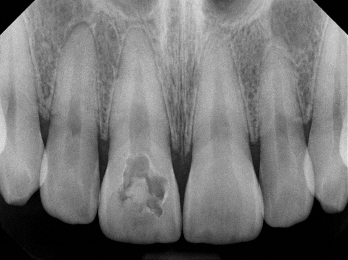
Which dietary factor is most strongly associated with colon carcinoma?
Which of the following is a true statement regarding Barrett's esophagus?
Which of the following is NOT a feature of Crohn's disease?
Crohn's disease is associated with which of the following infectious agents?
Pseudopolyps are commonly associated with which of the following conditions?
What is the pattern of inheritance for Gardner syndrome?
What is the most common primary malignant neoplasm of the small bowel?
Explanation: The question refers to **Gastrointestinal Stromal Tumors (GIST)**, which are the most common mesenchymal neoplasms of the gastrointestinal tract. **Why CD 117 is the Correct Answer:** The hallmark of GIST is the expression of **CD 117**, a proto-oncogene product known as **c-KIT**. Approximately 95% of GISTs are positive for CD 117. This marker represents a receptor tyrosine kinase that, when mutated, leads to constitutive activation and tumor cell proliferation. These tumors are thought to originate from the **Interstitial Cells of Cajal (ICC)**, the "pacemakers" of the gut, which also normally express CD 117 [1]. **Analysis of Incorrect Options:** * **CD 34:** While CD 34 is expressed in about 60–70% of GISTs, it is less specific and less sensitive than CD 117. It is also found in various other vascular and soft tissue tumors. * **CD 56:** This is a marker for neural/neuroendocrine differentiation. It is used to identify tumors like Small Cell Carcinoma or Neuroblastoma, not GIST. * **CD 24:** This is a cell adhesion molecule associated with various epithelial cancers (like breast or colorectal adenocarcinoma) but is not a diagnostic marker for GIST. **High-Yield Clinical Pearls for NEET-PG:** * **DOG1 (Discovered On GIST 1):** This is a highly sensitive and specific marker, often positive even in CD 117-negative GISTs. * **Genetics:** Most GISTs have mutations in the **KIT gene**; a subset has mutations in the **PDGFRA gene** [1]. * **Treatment:** The discovery of CD 117/c-KIT led to the use of **Imatinib (Gleevec)**, a tyrosine kinase inhibitor, which revolutionized the treatment of GIST. * **Location:** The **Stomach** is the most common site (60%), followed by the small intestine [1]. **References:** [1] Kumar V, Abbas AK, et al.. Robbins and Cotran Pathologic Basis of Disease. 9th ed. The Gastrointestinal Tract, pp. 782-783.
Explanation: **Explanation:** **Hirschsprung Disease (Congenital Aganglionic Megacolon)** is a developmental disorder characterized by the failure of neural crest cells to migrate from the cecum to the rectum during embryonic development [1]. **Why Option B is Correct:** The hallmark of the disease is the **absence of intrinsic enteric ganglion cells** (both the Meissner/submucosal plexus and the Auerbach/myenteric plexus) in the distal segment of the colon [1], [2]. This aganglionosis leads to a functional obstruction because the affected segment fails to undergo coordinated peristaltic contractions, resulting in proximal dilation (megacolon) [1]. **Why Incorrect Options are Wrong:** * **Option A:** Muscle atrophy is not the primary pathology. In fact, the proximal (normal) segment often shows compensatory **hypertrophy** as it attempts to push stool past the distal obstructed segment. * **Option C:** The **extrinsic** nerve supply (parasympathetic fibers) is actually **increased** and hypertrophied in the aganglionic segment, but these fibers cannot coordinate peristalsis without the intrinsic plexuses. **High-Yield Clinical Pearls for NEET-PG:** * **Gold Standard Diagnosis:** Rectal Suction Biopsy (shows absence of ganglion cells and increased **Acetylcholinesterase** staining) [2]. * **Most Common Site:** Always involves the **rectum** (starts distally and extends proximally). * **Genetic Association:** Strongly linked to mutations in the **RET proto-oncogene** [2]. * **Clinical Presentation:** Delayed passage of meconium (>48 hours) in a neonate and "blast sign" (explosive release of gas/stool) on digital rectal exam. * **Associated Condition:** Down Syndrome (Trisomy 21) is seen in approximately 10% of cases [1]. **References:** [1] Cross SS. Underwood's Pathology: A Clinical Approach. 6th ed. (Basic Pathology) introduces the student to key general principles of pathology, both as a medical science and as a clinical activity with a vital role in patient care. Part 2 (Disease Mechanisms) provides fundamental knowledge about the cellular and molecular processes involved in diseases, providing the rationale for their treatment. Part 3 (Systematic Pathology) deals in detail with specific diseases, with emphasis on the clinically important aspects., pp. 94-95. [2] Kumar V, Abbas AK, et al.. Robbins and Cotran Pathologic Basis of Disease. 9th ed. The Gastrointestinal Tract, p. 759.
Explanation: ### Explanation In **Ulcerative Colitis (UC)**, the development of colorectal cancer (CRC) follows a distinct molecular pathway known as the **Inflammation-Dysplasia-Carcinoma sequence**. Unlike sporadic CRC, which typically arises from pre-existing adenomatous polyps, UC-associated cancer arises from areas of **flat, non-polypoid dysplasia** within the chronically inflamed mucosa. #### Why "Dysplastic sites" is correct: Chronic inflammation leads to repeated cycles of mucosal injury and regeneration, causing oxidative stress and DNA damage. This results in widespread (field effect) genetic mutations (e.g., early *TP53* mutations). These changes manifest histologically as **dysplasia**, which is the direct precursor to invasive adenocarcinoma in inflammatory bowel disease (IBD). #### Why other options are incorrect: * **A. Pseudopolyps:** These are non-neoplastic islands of regenerating mucosa surrounded by areas of ulceration. They are inflammatory in nature and have **no malignant potential**. * **C. Familial polyposis (FAP):** This is a genetic syndrome caused by a germline mutation in the *APC* gene [1]. While it leads to CRC, it is a distinct clinical entity unrelated to the inflammatory pathogenesis of UC. * **D. Multiple adenomatous polyps:** This describes the precursor for sporadic CRC or polyposis syndromes [2]. In UC, cancer often arises from **flat mucosa** rather than discrete, stalked polyps [2]. #### High-Yield Clinical Pearls for NEET-PG: * **Risk Factors for CRC in UC:** Duration of disease (>8–10 years), extent of involvement (Pancolitis > Left-sided), and co-existence of Primary Sclerosing Cholangitis (PSC). * **Molecular Difference:** In UC, *TP53* mutations occur **early**, and *APC* mutations occur **late** (the opposite of the sporadic "Vogelstein" adenoma-carcinoma sequence). * **Surveillance:** Regular colonoscopic biopsies are mandatory to detect "Dysplasia-Associated Lesion or Mass" (DALM). **References:** [1] Kumar V, Abbas AK, et al.. Robbins and Cotran Pathologic Basis of Disease. 9th ed. The Gastrointestinal Tract, pp. 821-822. [2] Cross SS. Underwood's Pathology: A Clinical Approach. 6th ed. Common Clinical Problems From Alimentary System Disease, pp. 372-373.
Explanation: **Explanation:** The development of colorectal carcinoma (CRC) is multifactorial, but dietary habits play a pivotal role in its pathogenesis. [1] **Why Low Fiber Intake is Correct:** A diet low in vegetable fiber and high in refined carbohydrates/fat is the most significant dietary risk factor for CRC. [1] The underlying medical concept involves two mechanisms: 1. **Increased Transit Time:** Low fiber leads to decreased stool bulk and slower transit time, allowing prolonged contact between the colonic mucosa and potential fecal carcinogens. [1] 2. **Altered Microbiota:** High fiber intake promotes the growth of beneficial bacteria that ferment fiber into **short-chain fatty acids (like butyrate)**, which have protective, anti-proliferative effects. Conversely, a low-fiber, high-fat diet increases the synthesis of bile acids, which can be converted into carcinogens by intestinal bacteria. **Analysis of Incorrect Options:** * **High fiber intake:** This is a **protective factor**, not a risk factor. It increases stool bulk and dilutes carcinogens. * **Consumption of smoked fish:** This is primarily associated with **Gastric Carcinoma** due to the presence of benzopyrenes and nitrosamines, rather than colon cancer. **NEET-PG High-Yield Pearls:** * **Most common site:** Historically the rectum/sigmoid, but there is an increasing trend toward "proximal shift" (Right-sided/Cecal). * **Precursor Lesion:** Most CRCs arise from the **Adenoma-Carcinoma sequence** (APC gene mutation → KRAS → TP53). * **Protective Agents:** Aspirin and other NSAIDs (by inhibiting COX-2, which is overexpressed in colon cancers and promotes epithelial proliferation). * **Gold Standard Screening:** Colonoscopy (starting at age 45 for average-risk individuals). **References:** [1] Kumar V, Abbas AK, et al.. Robbins and Cotran Pathologic Basis of Disease. 9th ed. The Gastrointestinal Tract, pp. 817-819.
Explanation: **Explanation:** **Barrett’s Esophagus (BE)** is a classic example of **metaplasia**, a reversible change where one adult cell type is replaced by another to withstand chronic stress [1]. In BE, the normal non-keratinized stratified squamous epithelium of the distal esophagus is replaced by **non-ciliated columnar epithelium with goblet cells** (intestinal metaplasia) due to chronic gastroesophageal reflux disease (GERD) [1]. **Analysis of Options:** * **Option D (Correct):** The core pathology is the metaplastic transformation of the esophageal mucosa in response to acid injury. * **Option A:** Most patients are actually **asymptomatic** or present with standard GERD symptoms (heartburn, regurgitation) [2]. The severity of symptoms does not always correlate with the presence or extent of Barrett’s. * **Option B:** BE is caused by chronic acid reflux; therefore, an **abnormal pH profile** (increased acid exposure) is characteristic, not a normal one. * **Option C:** BE is a precursor to **Adenocarcinoma**, not squamous cell carcinoma [1]. Chronic irritation from alcohol and smoking typically leads to squamous cell carcinoma, whereas acid reflux leads to adenocarcinoma [3]. **High-Yield Facts for NEET-PG:** * **Pathognomonic Feature:** Presence of **Goblet cells** on biopsy is essential for diagnosis [1]. * **Endoscopic Appearance:** Characterized by "salmon-pink" velvety tongues of mucosa extending upwards from the gastroesophageal junction. * **Risk Factor:** It increases the risk of esophageal adenocarcinoma by 30–40 fold [1]. * **Surveillance:** Periodic endoscopy with biopsies (Seattle Protocol) is required to monitor for dysplasia [2]. **References:** [1] Cross SS. Underwood's Pathology: A Clinical Approach. 6th ed. Common Clinical Problems From Alimentary System Disease, pp. 348-349. [2] Kumar V, Abbas AK, et al.. Robbins and Cotran Pathologic Basis of Disease. 9th ed. The Gastrointestinal Tract, pp. 764-765. [3] Kumar V, Abbas AK, et al.. Robbins and Cotran Pathologic Basis of Disease. 9th ed. The Gastrointestinal Tract, pp. 765-766.
Explanation: **Explanation:** The correct answer is **A. Lymphoid hyperplasia**. While lymphoid aggregates are commonly seen in the submucosa and subserosa of the bowel wall in Crohn's disease [1], "Lymphoid hyperplasia" is specifically the hallmark histological feature of **Yersinia enterocolitica** infection or **Aphthous ulcers** in early stages. In the context of Inflammatory Bowel Disease (IBD), lymphoid hyperplasia is not a defining diagnostic feature, whereas the other options are classic characteristics of Crohn’s. **Analysis of Options:** * **B. Skip Lesions:** This is a hallmark of Crohn’s disease. Unlike Ulcerative Colitis (UC), which involves the rectum and spreads continuously, Crohn’s presents as sharply demarcated diseased segments separated by normal-appearing "skip" areas [2]. * **C. Transmural Involvement:** Crohn’s affects all layers of the bowel wall (mucosa to serosa) [1]. This leads to complications like fistulas, sinus tracts, and "creeping fat." In contrast, UC is limited to the mucosa and submucosa [3]. * **D. Crypt Abscess:** While more characteristic and prominent in Ulcerative Colitis [3], crypt abscesses (neutrophils within the crypt lumen) can also occur in the active phase of Crohn’s disease. Therefore, it *is* a feature, though less specific. **NEET-PG High-Yield Pearls:** * **Granulomas:** Non-caseating granulomas are the most specific histological finding for Crohn’s (seen in ~35% of cases) [2]. * **Gross Appearance:** Look for "Cobblestone mucosa," "String sign of Kantor" on X-ray, and "Creeping fat" [1]. * **Serology:** Crohn’s is associated with **ASCA** (Anti-Saccharomyces cerevisiae antibodies), while UC is associated with **p-ANCA**. * **Site:** Most common site is the **terminal ileum** [1]. **References:** [1] Cross SS. Underwood's Pathology: A Clinical Approach. 6th ed. Common Clinical Problems From Alimentary System Disease, pp. 365-367. [2] Kumar V, Abbas AK, et al.. Robbins and Cotran Pathologic Basis of Disease. 9th ed. The Gastrointestinal Tract, pp. 806-807. [3] Kumar V, Abbas AK, et al.. Robbins and Cotran Pathologic Basis of Disease. 9th ed. The Gastrointestinal Tract, p. 809.
Explanation: ### Explanation **Correct Option: A. Clostridium difficile** In the context of Inflammatory Bowel Disease (IBD), particularly Crohn’s disease and Ulcerative Colitis, patients are at a significantly increased risk for **Clostridium difficile infection (CDI)**. This association is attributed to altered gut microbiota (dysbiosis), frequent antibiotic use, and immunosuppressive therapy [1]. CDI is a major cause of disease "flares" and is associated with increased morbidity and hospitalization in Crohn's patients. Current clinical guidelines recommend testing for *C. difficile* in all IBD patients presenting with a sudden worsening of symptoms. **Analysis of Incorrect Options:** * **B. Mycobacterium paratuberculosis:** While the "Map hypothesis" has long suggested a link between *M. avium paratuberculosis* (MAP) and Crohn’s disease due to its role in Johne’s disease (a similar granulomatous enteritis in cattle), it remains a **controversial** and unproven association. It is not considered a definitive causative or associated agent in standard medical examinations. * **C. Cytomegalovirus (CMV):** CMV is more commonly associated with **Ulcerative Colitis**, particularly in steroid-refractory cases, where it can cause deep ulcerations and toxic megacolon. * **D. Mycoplasma:** There is no established clinical or pathological association between Mycoplasma species and the pathogenesis or exacerbation of Crohn’s disease. **High-Yield Pearls for NEET-PG:** * **Transmural Involvement:** Crohn’s involves all layers of the bowel wall (leads to fistulas/strictures), whereas UC is limited to mucosa/submucosa [1]. * **Granulomas:** Non-caseating granulomas are a hallmark of Crohn’s (seen in ~35% of cases) but are absent in UC [1]. * **Skip Lesions:** Crohn's is characterized by discontinuous "skip lesions" and a "cobblestone" appearance. * **Serology:** Crohn’s is associated with **ASCA** (Anti-Saccharomyces cerevisiae antibodies), while UC is associated with **p-ANCA**. **References:** [1] Kumar V, Abbas AK, et al.. Robbins and Cotran Pathologic Basis of Disease. 9th ed. The Gastrointestinal Tract, pp. 805-807.
Explanation: **Explanation:** **1. Why Ulcerative Colitis (UC) is the correct answer:** Pseudopolyps (inflammatory polyps) are a hallmark endoscopic and pathological finding in **Ulcerative Colitis**. They are not true neoplastic growths; rather, they represent islands of **regenerating residual mucosa** surrounded by areas of extensive ulceration and mucosal denudation [1]. Because UC involves continuous superficial inflammation, the remaining healthy mucosa bulges upward, appearing like polyps against the "flat" background of ulcerated tissue. **2. Why the other options are incorrect:** * **Crohn’s Disease:** While inflammatory polyps can occasionally occur, Crohn’s is more characteristically associated with a **"cobblestone appearance"** due to deep fissuring ulcers and intervening normal mucosa [4]. * **Colon Cancer:** This typically presents as true neoplastic polyps (adenomas) or malignant masses, not inflammatory pseudopolyps [3]. * **Diverticulosis:** This involves herniation of the mucosa through the muscular layers of the colon wall (outpouchings), which is structurally the opposite of polypoid projections. **3. NEET-PG High-Yield Pearls:** * **Lead Pipe Appearance:** Seen in chronic UC on barium enema due to loss of haustrations. * **Backwash Ileitis:** Involvement of the terminal ileum in UC (despite it being primarily a colonic disease) [2]. * **Microscopic hallmark of UC:** Crypt abscesses (neutrophils in the crypt lumen) [1]. * **Malignancy Risk:** Long-standing UC with extensive pseudopolyps and inflammation significantly increases the risk of Colorectal Carcinoma [3]. * **Extraintestinal manifestation:** Primary Sclerosing Cholangitis (PSC) is most strongly associated with UC. **References:** [1] Kumar V, Abbas AK, et al.. Robbins and Cotran Pathologic Basis of Disease. 9th ed. The Gastrointestinal Tract, p. 809. [2] Cross SS. Underwood's Pathology: A Clinical Approach. 6th ed. Common Clinical Problems From Alimentary System Disease, pp. 367-368. [3] Kumar V, Abbas AK, et al.. Robbins and Cotran Pathologic Basis of Disease. 9th ed. The Gastrointestinal Tract, pp. 809-810. [4] Kumar V, Abbas AK, et al.. Robbins and Cotran Pathologic Basis of Disease. 9th ed. The Gastrointestinal Tract, pp. 806-807.
Explanation: **Explanation:** **Gardner syndrome** is a phenotypic variant of **Familial Adenomatous Polyposis (FAP)**. It is inherited in an **Autosomal Dominant** pattern, caused by a germline mutation in the **APC (Adenomatous Polyposis Coli) gene** [1] located on chromosome **5q21**. 1. **Why Autosomal Dominant is correct:** In Gardner syndrome, a single copy of the mutated APC gene inherited from one parent is sufficient to predispose an individual to developing hundreds to thousands of adenomatous colonic polyps [1]. The "two-hit hypothesis" applies here: the patient is born with one mutated allele (first hit), and the subsequent somatic loss of the second wild-type allele (second hit) leads to adenoma formation. 2. **Why other options are incorrect:** * **Autosomal recessive:** While *MUTYH-associated polyposis (MAP)* is an autosomal recessive polyposis syndrome, Gardner syndrome follows the classic dominant inheritance of FAP. * **X-linked:** There are no major hereditary colorectal cancer syndromes that are linked to the X or Y chromosomes. **Clinical Pearls for NEET-PG:** * **The Triad:** Gardner syndrome is characterized by the clinical triad of **Colonic Polyposis**, **Extra-colonic soft tissue tumors** (specifically Desmoid tumors and sebaceous cysts), and **Skeletal anomalies** (Osteomas, typically of the mandible or skull). * **Dental Abnormalities:** Impacted teeth and supernumerary teeth are high-yield diagnostic clues. * **CHRPE:** Congenital Hypertrophy of Retinal Pigment Epithelium is a specific ocular finding that can be a screening marker. * **Malignant Potential:** Like FAP, the risk of progression to Colorectal Carcinoma is nearly **100%** by age 40 if a prophylactic colectomy is not performed [1]. **References:** [1] Kumar V, Abbas AK, et al.. Robbins and Cotran Pathologic Basis of Disease. 9th ed. The Gastrointestinal Tract, pp. 821-822.
Explanation: ### Explanation **Correct Option: A. Adenocarcinoma** In the small intestine, **adenocarcinoma** is historically and statistically the most common primary malignant neoplasm, accounting for approximately 30–40% of cases [2]. These tumors most frequently occur in the **duodenum** (specifically the periampullary region). They often arise from pre-existing adenomas, following the same adenoma-carcinoma sequence seen in the colon [4], and are frequently associated with conditions like Familial Adenomatous Polyposis (FAP) [1] and Celiac disease. **Analysis of Incorrect Options:** * **B. Carcinoid (Neuroendocrine Tumors):** While some recent registries suggest the incidence of NETs is rising and may surpass adenocarcinoma in certain populations, adenocarcinoma remains the traditional "textbook" answer for the most common primary malignancy. Carcinoids are most commonly found in the **ileum**, though more than 40% of all GI carcinoids occur in the small intestine overall [2]. * **C. Gastrointestinal Stromal Tumor (GIST):** These are mesenchymal tumors arising from the **Interstitial Cells of Cajal**. While they occur in the small bowel, they are less common than epithelial malignancies; those in the small intestine tend to be more aggressive than gastric GISTs [3]. * **D. Lymphoma:** The small bowel is the most common site for extranodal lymphoma (usually B-cell type), but it ranks behind adenocarcinoma in overall frequency. It is strongly associated with **MALT** and **Celiac disease** (EATL). **NEET-PG High-Yield Pearls:** * **Most common site for Small Bowel Adenocarcinoma:** Duodenum. * **Most common site for Small Bowel Carcinoid:** Ileum. * **Most common site for Small Bowel Lymphoma:** Ileum (due to higher concentration of lymphoid tissue/Peyer's patches). * **Peutz-Jeghers Syndrome:** Increases the risk of small bowel adenocarcinoma, though the polyps themselves are hamartomatous. * **Clinical Presentation:** Often presents late with occult bleeding or intestinal obstruction (napkin-ring appearance). **References:** [1] Kumar V, Abbas AK, et al.. Robbins and Cotran Pathologic Basis of Disease. 9th ed. The Gastrointestinal Tract, p. 817. [2] Kumar V, Abbas AK, et al.. Robbins and Cotran Pathologic Basis of Disease. 9th ed. The Gastrointestinal Tract, pp. 780-781. [3] Kumar V, Abbas AK, et al.. Robbins and Cotran Pathologic Basis of Disease. 9th ed. The Gastrointestinal Tract, pp. 783-784. [4] Cross SS. Underwood's Pathology: A Clinical Approach. 6th ed. Common Clinical Problems From Alimentary System Disease, pp. 371-372.
Oral Cavity and Esophageal Pathology
Practice Questions
Gastritis and Peptic Ulcer Disease
Practice Questions
Inflammatory Bowel Disease
Practice Questions
Malabsorption Syndromes
Practice Questions
Vascular Disorders of Intestine
Practice Questions
Diverticular Disease
Practice Questions
Intestinal Obstruction
Practice Questions
Gastrointestinal Infections
Practice Questions
Polyps and Neoplasms
Practice Questions
Appendiceal Pathology
Practice Questions
Get full access to all questions, explanations, and performance tracking.
Start For Free