A patient presents with diarrhea, malabsorption, abdominal pain, and arthralgia. Intestinal biopsy reveals PAS-positive organisms within macrophages. What is the most likely diagnosis?
A 50-year-old male presents with a history of Gastroesophageal reflux disease. What is the most common pathological finding associated with this condition?
What is the most common type of gastric polyp?
Which marker is used for the early diagnosis of Hirschsprung disease?
What is the most important prognostic indicator in esophageal carcinoma?
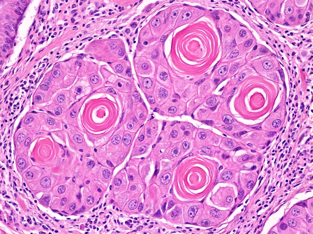
A 60-year-old patient with a prolonged history of tobacco chewing presents with symptoms of weight loss and dysphagia. An esophageal biopsy from a projectile mass is shown below. What is your likely diagnosis?

Meckel Diverticulum is a remnant of which embryonic structure?
What is characteristic of a typhoid ulcer?
Which of the following anaemias is a risk factor for the development of gastric carcinoma?
Type B gastritis is characterized by predominance in which region of the stomach?
Explanation: **Explanation:** The clinical presentation of malabsorption, diarrhea, and abdominal pain, combined with systemic features like **arthralgia**, is classic for **Whipple’s disease** [1]. The definitive diagnostic finding is the presence of **PAS-positive, diastase-resistant bacilli** (Tropheryma whipplei) within the lysosomes of **macrophages** in the lamina propria of the small intestine [1]. These macrophages accumulate and cause lymphatic obstruction, leading to fat malabsorption (steatorrhea) [1]. **Analysis of Options:** * **Celiac Sprue:** Characterized by villous atrophy, crypt hyperplasia, and increased intraepithelial lymphocytes [3]. It does not show PAS-positive macrophages and typically presents with sensitivity to gluten [2], [3]. * **Zollinger-Ellison Syndrome:** Caused by a gastrinoma leading to excessive gastric acid. It presents with multiple peptic ulcers and diarrhea, but biopsy would show gastric mucosal hyperplasia, not PAS-positive organisms. * **Crohn’s Disease:** A transmural inflammatory bowel disease characterized by non-caseating granulomas, "skip lesions," and "cobblestone" appearance. It does not feature PAS-positive macrophages. **High-Yield Clinical Pearls for NEET-PG:** * **Causative Agent:** *Tropheryma whipplei* (a Gram-positive actinomycete) [1]. * **Triad/Tetrad:** Malabsorption, Weight loss, Arthralgia, and Lymphadenopathy [1]. * **CNS Involvement:** Can cause dementia or **oculomasticatory myorhythmia** (pathognomonic). * **Biopsy Tip:** The PAS stain highlights the glycoprotein cell wall of the partially digested bacteria within macrophages. * **Treatment:** Long-term antibiotics (usually Ceftriaxone followed by Trimethoprim-sulfamethoxazole). **References:** [1] Kumar V, Abbas AK, et al.. Robbins and Cotran Pathologic Basis of Disease. 9th ed. The Gastrointestinal Tract, pp. 798-799. [2] Cross SS. Underwood's Pathology: A Clinical Approach. 6th ed. Common Clinical Problems From Alimentary System Disease, pp. 361-362. [3] Cross SS. Underwood's Pathology: A Clinical Approach. 6th ed. Common Clinical Problems From Alimentary System Disease, pp. 360-361.
Explanation: **Explanation:** The correct answer is **Columnar metaplasia**, specifically known as **Barrett’s Esophagus**. **Why it is correct:** Gastroesophageal Reflux Disease (GERD) involves the chronic backflow of gastric acid and bile into the lower esophagus. The normal lining of the esophagus is **stratified squamous epithelium**, which is designed to withstand friction but not acid [3]. In response to chronic acid injury, the tissue undergoes an adaptive change called **metaplasia** [1]. It transforms into **simple columnar epithelium with goblet cells** (intestinal metaplasia), which is more resistant to acidic environments [1]. **Analysis of Incorrect Options:** * **A. Squamous metaplasia:** This is incorrect because the esophagus is already lined by squamous epithelium. Squamous metaplasia typically occurs in the lungs (due to smoking) or the cervix. * **C. Dysplasia:** While Barrett’s esophagus can progress to dysplasia (disordered growth), it is a subsequent step and not the primary or most common pathological finding of the initial reflux-induced change [2]. * **D. Malignancy:** Long-standing GERD increases the risk of **Adenocarcinoma**, but this is a late-stage complication, not the most common pathological finding [4]. **High-Yield Clinical Pearls for NEET-PG:** * **Definition:** Barrett’s Esophagus is defined endoscopically by salmon-pink tongues of mucosa and histologically by **intestinal metaplasia (Goblet cells)** [1]. * **Risk:** It is a pre-malignant condition; patients require regular surveillance biopsies to check for high-grade dysplasia [2]. * **Cancer Association:** GERD/Barrett’s leads to **Adenocarcinoma** (typically in the lower 1/3rd), whereas smoking/alcohol leads to **Squamous Cell Carcinoma** (typically in the middle 1/3rd) [4]. **References:** [1] Cross SS. Underwood's Pathology: A Clinical Approach. 6th ed. Common Clinical Problems From Alimentary System Disease, pp. 348-349. [2] Kumar V, Abbas AK, et al.. Robbins and Cotran Pathologic Basis of Disease. 9th ed. The Gastrointestinal Tract, pp. 764-765. [3] Kumar V, Abbas AK, et al.. Robbins and Cotran Pathologic Basis of Disease. 9th ed. The Gastrointestinal Tract, pp. 762-763. [4] Kumar V, Abbas AK, et al.. Robbins and Cotran Pathologic Basis of Disease. 9th ed. The Gastrointestinal Tract, pp. 765-766.
Explanation: **Explanation:** **1. Why Hyperplastic Polyp is Correct:** Hyperplastic polyps (along with inflammatory polyps) represent approximately **75% to 90%** of all gastric polyps, making them the most common type encountered in clinical practice [1]. They typically arise in the background of chronic gastritis (often associated with *H. pylori* infection), which triggers reactive mucosal proliferation and tissue repair. Histologically, they are characterized by elongated, distorted, and "corkscrew" shaped foveolar glands [2]. **2. Analysis of Incorrect Options:** * **B. Hamartomatous polyp:** These are rare in the stomach and are usually associated with specific syndromes like Peutz-Jeghers syndrome or Juvenile Polyposis syndrome [1]. * **C. Malignant polyp:** While gastric polyps (especially adenomatous types) can undergo malignant transformation, the majority of gastric polyps are benign/non-neoplastic [1]. * **D. Familial polyposis:** This refers to Familial Adenomatous Polyposis (FAP). While patients with FAP often develop numerous Fundic Gland Polyps (the most common "syndromic" polyp), they do not represent the most common type in the general population. **3. NEET-PG High-Yield Pearls:** * **Fundic Gland Polyps:** These are the most common polyps in patients taking **Proton Pump Inhibitors (PPIs)** due to increased gastrin levels. * **Malignant Potential:** Hyperplastic polyps have a very low risk of malignancy (usually <1%), whereas **Gastric Adenomas** (the true neoplastic polyps) have a high risk (up to 30%) and are often associated with intestinal metaplasia [3]. * **Size Matters:** Any gastric polyp larger than **1.5 cm** should be excised and examined histologically to rule out dysplasia or occult malignancy [3]. **References:** [1] Kumar V, Abbas AK, et al.. Robbins and Cotran Pathologic Basis of Disease. 9th ed. The Gastrointestinal Tract, pp. 776-777. [2] Kumar V, Abbas AK, et al.. Robbins and Cotran Pathologic Basis of Disease. 9th ed. The Gastrointestinal Tract, pp. 775-776. [3] Cross SS. Underwood's Pathology: A Clinical Approach. 6th ed. Common Clinical Problems From Alimentary System Disease, pp. 354-355.
Explanation: **Explanation:** **Hirschsprung Disease (Congenital Megacolon)** is characterized by the absence of ganglion cells in the submucosal (Meissner) and myenteric (Auerbach) plexuses due to a failure of neural crest cell migration [1]. **Why Acetylcholinesterase (AChE) is the correct answer:** In the absence of inhibitory ganglion cells, there is a compensatory **hypertrophy and proliferation of extrinsic cholinergic nerve fibers** in the affected segment. These hypertrophied fibers produce high levels of the enzyme **Acetylcholinesterase**. Histochemical staining for AChE on a rectal suction biopsy reveals an intense positive reaction (dark staining) in the lamina propria and muscularis mucosae [2]. This serves as a highly sensitive and specific marker for early diagnosis, especially when traditional H&E staining is inconclusive in neonates [2]. **Analysis of Incorrect Options:** * **B. Adrenaline:** While the autonomic nervous system is involved, adrenergic markers are not used for the histopathological diagnosis of Hirschsprung disease. * **C. VIP (Vasoactive Intestinal Peptide):** VIP is a neurotransmitter found in the gastrointestinal tract. While VIP-containing nerve fibers are decreased in Hirschsprung disease, it is not a standard or reliable diagnostic marker compared to AChE. **High-Yield Clinical Pearls for NEET-PG:** * **Gold Standard Diagnosis:** Rectal Suction Biopsy (must include the submucosa). * **Key Histological Finding:** Absence of ganglion cells + presence of hypertrophied nerve bundles [2]. * **Calretinin Immunohistochemistry:** A newer, increasingly popular marker. In Hirschsprung disease, Calretinin staining is **absent** (negative), whereas it is positive in a normal bowel. * **Associated Mutation:** *RET* proto-oncogene (most common) [2]. * **Clinical Presentation:** Failure to pass meconium within 48 hours, abdominal distension, and "blast sign" on digital rectal examination [1]. **References:** [1] Cross SS. Underwood's Pathology: A Clinical Approach. 6th ed. (Basic Pathology) introduces the student to key general principles of pathology, both as a medical science and as a clinical activity with a vital role in patient care. Part 2 (Disease Mechanisms) provides fundamental knowledge about the cellular and molecular processes involved in diseases, providing the rationale for their treatment. Part 3 (Systematic Pathology) deals in detail with specific diseases, with emphasis on the clinically important aspects., pp. 94-95. [2] Kumar V, Abbas AK, et al.. Robbins and Cotran Pathologic Basis of Disease. 9th ed. The Gastrointestinal Tract, p. 759.
Explanation: **Explanation:** The prognosis of esophageal carcinoma (both Squamous Cell Carcinoma and Adenocarcinoma) is primarily determined by the **TNM staging system**. Among the options provided, the **depth of invasion (T stage)** is the most critical prognostic factor [1], [2]. 1. **Why Depth of Invasion is Correct:** The esophagus lacks a serosal layer (except for the intra-abdominal segment), which facilitates the early transmural spread of tumors. As the tumor invades deeper into the esophageal wall (from mucosa to submucosa and then muscularis propria), the risk of lymph node metastasis increases significantly due to the rich submucosal lymphatic network [3]. Therefore, the depth of penetration is the strongest predictor of survival and surgical resectability [1]. 2. **Why Other Options are Incorrect:** * **Length of involvement:** While a larger tumor size may suggest advanced disease, it is not as reliable a predictor of survival as the depth of wall penetration. * **Histological grading:** The degree of differentiation (well, moderately, or poorly differentiated) provides some information but is secondary to the anatomical stage (TNM) in determining clinical outcome [2]. * **Immunohistochemistry (IHC):** IHC is used for diagnosis and identifying molecular targets (like HER2 in GE junction tumors) but is not a primary prognostic indicator. **High-Yield Clinical Pearls for NEET-PG:** * **Most common site:** Middle third (Squamous Cell CA); Lower third (Adenocarcinoma). * **Most common histological type (Worldwide):** Squamous Cell Carcinoma. * **Most common histological type (USA/West):** Adenocarcinoma (associated with Barrett’s esophagus). * **Lymphatic Spread:** The esophagus has a unique longitudinal lymphatic drainage, often leading to "skip metastasis." * **Overall Prognosis:** Generally poor, as most patients present with advanced-stage disease (dysphagia occurs only when >2/3rd of the lumen is obstructed) [1], [3]. **References:** [1] Cross SS. Underwood's Pathology: A Clinical Approach. 6th ed. Common Clinical Problems From Alimentary System Disease, pp. 349-350. [2] Cross SS. Underwood's Pathology: A Clinical Approach. 6th ed. (Basic Pathology) introduces the student to key general principles of pathology, both as a medical science and as a clinical activity with a vital role in patient care. Part 2 (Disease Mechanisms) provides fundamental knowledge about the cellular and molecular processes involved in diseases, providing the rationale for their treatment. Part 3 (Systematic Pathology) deals in detail with specific diseases, with emphasis on the clinically important aspects., pp. 236-237. [3] Kumar V, Abbas AK, et al.. Robbins and Cotran Pathologic Basis of Disease. 9th ed. The Gastrointestinal Tract, pp. 765-766.
Explanation: ***Squamous cell carcinoma*** - **Tobacco chewing** is a major risk factor for **esophageal squamous cell carcinoma**, especially in the upper and middle esophagus. - Histologically characterized by **keratin pearls**, **intercellular bridges**, and **dyskeratosis** in the epithelial cells. *Adenocarcinoma* - Primarily associated with **gastroesophageal reflux disease (GERD)** and **Barrett's esophagus**, not tobacco chewing. - Typically occurs in the **lower esophagus** and shows **glandular architecture** with mucin production. *Carcinoid tumor* - Arises from **neuroendocrine cells** and is extremely rare in the esophagus compared to stomach or small bowel. - Presents with **carcinoid syndrome** (flushing, diarrhea) and shows **uniform cells** with **salt-and-pepper chromatin** pattern. *Small cell cancer* - Very rare in the esophagus and more commonly seen in the **lungs** of heavy smokers. - Shows **small blue cells** with scant cytoplasm and high **mitotic rate**, different from squamous morphology.
Explanation: **Explanation:** **1. Why the Correct Answer is Right:** Meckel diverticulum is the most common congenital anomaly of the gastrointestinal tract. It occurs due to the **failure of the vitellointestinal duct** (also known as the **omphalomesenteric duct**) to involute completely [1], [2]. During early embryonic life (around the 5th to 8th week), this duct connects the primitive midgut lumen to the yolk sac. Normally, it obliterates and disappears; however, its persistence at the ileal end results in a "true" diverticulum (containing all layers of the bowel wall) [1], [2]. **2. Why Incorrect Options are Wrong:** * **A. Vitelloumbilical duct:** This is a misnomer. While the duct connects the gut to the umbilicus, the standard anatomical term is vitellointestinal or omphalomesenteric duct. * **C & D. Vitelloportal / Vitellodiaphragmatic duct:** These are fabricated terms and do not represent recognized embryonic structures in human development. **3. Clinical Pearls for NEET-PG (Rule of 2s):** * **Prevalence:** Occurs in **2%** of the population. * **Location:** Usually located within **2 feet** (60 cm) of the ileocecal valve [2]. * **Length:** Approximately **2 inches** long. * **Age:** Often becomes symptomatic before age **2**. * **Ectopic Tissue:** Most commonly contains **Gastric mucosa** (leading to painless bleeding/peptic ulceration) or **Pancreatic tissue** [1], [2]. * **Diagnosis:** The investigation of choice for a bleeding Meckel diverticulum is the **Technetium-99m pertechnetate scan** (Meckel scan), which identifies ectopic gastric mucosa. **References:** [1] Kumar V, Abbas AK, et al.. Robbins and Cotran Pathologic Basis of Disease. 9th ed. The Gastrointestinal Tract, pp. 759-760. [2] Cross SS. Underwood's Pathology: A Clinical Approach. 6th ed. Common Clinical Problems From Alimentary System Disease, pp. 359-360.
Explanation: **Explanation:** Typhoid fever, caused by *Salmonella typhi*, primarily affects the lymphoid tissue of the small intestine, specifically the **Peyer’s patches** in the terminal ileum [1]. Understanding the morphology of typhoid ulcers is high-yield for NEET-PG. 1. **Ulceration of Peyer's Patches:** The pathogenesis involves the infiltration of Peyer’s patches by macrophages (typhoid cells), leading to hyperplasia, necrosis, and subsequent sloughing of the overlying mucosa [1]. This results in the formation of classic typhoid ulcers. 2. **Longitudinal Orientation:** Because Peyer’s patches are arranged along the long axis of the bowel, the resulting ulcers are **longitudinal (oval)** in shape. This is a critical distinguishing feature from **Tubercular ulcers**, which are typically transverse (circumferential) because they follow the path of the lymphatics. 3. **Risk of Perforation:** These ulcers are deep and involve the lymphoid tissue. During the third week of the disease, the necrotic tissue sloughs off, which can lead to complications such as intestinal hemorrhage or **perforation**, usually in the terminal ileum. **Why "All of the above" is correct:** Since typhoid ulcers are derived from Peyer's patches (A), are oriented longitudinally (B), and carry a significant risk of perforation (C), all statements are pathologically accurate. **High-Yield Clinical Pearls for NEET-PG:** * **Microscopy:** Look for **"Typhoid cells"** (activated macrophages containing erythrocytes and bacilli—erythrophagocytosis) and **Mallory nodules** in the liver/spleen. * **Widal Test:** Becomes positive in the 2nd week. * **Blood Culture:** Most sensitive in the 1st week (Mnemonic: **BASU** – Blood, Agglutination/Widal, Stool, Urine for weeks 1, 2, 3, and 4 respectively). **References:** [1] Cross SS. Underwood's Pathology: A Clinical Approach. 6th ed. Common Clinical Problems From Alimentary System Disease, pp. 362-363.
Explanation: **Explanation:** **Pernicious Anaemia (Option A)** is the correct answer because it is an autoimmune condition characterized by the destruction of gastric parietal cells [1, 2]. This leads to **Type A Chronic Atrophic Gastritis**, primarily affecting the fundus and body of the stomach [2]. The chronic inflammation and subsequent loss of specialized glandular tissue result in **intestinal metaplasia**, a well-recognized precancerous lesion. Patients with pernicious anaemia have a 3-fold to 6-fold increased risk of developing **Gastric Adenocarcinoma** (specifically the intestinal type) and gastric carcinoid tumors [2]. **Why other options are incorrect:** * **Megaloblastic Anaemia (Option B):** While pernicious anaemia is a *cause* of megaloblastic anaemia [1, 3], not all megaloblastic anaemias (e.g., those caused by dietary Folate deficiency) lead to gastric cancer. The risk is specific to the underlying gastric atrophy found in pernicious anaemia, not the macrocytosis itself. * **Aplastic Anaemia (Option C):** This is a bone marrow failure syndrome. It does not involve the gastric mucosa and has no association with gastric malignancy. * **Haemolytic Anaemia (Option D):** This involves the premature destruction of RBCs. While it can lead to complications like gallstones (pigment stones), it is not a risk factor for gastric carcinoma. **High-Yield Clinical Pearls for NEET-PG:** * **Precancerous conditions for Gastric Cancer:** Chronic atrophic gastritis (H. pylori or Autoimmune) [2], Adenomatous gastric polyps (risk >2cm) [5], Post-gastrectomy stumps (after 15-20 years), and Menetrier’s disease. * **Autoantibodies:** Pernicious anaemia is associated with antibodies against **Parietal cells** (H+/K+ ATPase) and **Intrinsic Factor** [1, 4]. * **Site:** Autoimmune gastritis (Pernicious anaemia) affects the **Body/Fundus**, whereas H. pylori gastritis typically starts in the **Antrum** [2]. **References:** [1] Cross SS. Underwood's Pathology: A Clinical Approach. 6th ed. Common Clinical Problems From Blood And Bone Marrow Disease, pp. 592-593. [2] Kumar V, Abbas AK, et al.. Robbins and Cotran Pathologic Basis of Disease. 9th ed. The Gastrointestinal Tract, pp. 771-772. [3] Kumar V, Abbas AK, et al.. Robbins and Cotran Pathologic Basis of Disease. 9th ed. The Gastrointestinal Tract, pp. 772-773. [4] Kumar V, Abbas AK, et al.. Robbins and Cotran Pathologic Basis of Disease. 9th ed. Red Blood Cell and Bleeding Disorders, pp. 655-656. [5] Kumar V, Abbas AK, et al.. Robbins and Cotran Pathologic Basis of Disease. 9th ed. The Gastrointestinal Tract, pp. 777-778.
Explanation: **Explanation:** Chronic gastritis is broadly classified into two main types: **Type A (Autoimmune)** and **Type B (Bacterial/H. pylori)** [1]. **Why Antral Predominant is Correct:** Type B gastritis is caused by *Helicobacter pylori* infection. In the initial stages and the most common presentation, the bacteria colonize the **antrum** of the stomach [1]. This is because the antrum has a higher pH (less acidic) compared to the acid-secreting body and fundus, providing a more favorable environment for *H. pylori* to thrive. This antral-predominant pattern is typically associated with high acid production and an increased risk of duodenal ulcers. **Analysis of Incorrect Options:** * **Body and Fundus Predominant (Options A & B):** These regions are the primary sites for **Type A Gastritis**. Type A is an autoimmune condition where antibodies target parietal cells and intrinsic factor, which are located in the oxyntic mucosa of the body and fundus [1]. * **All (Option D):** While long-standing *H. pylori* infection can eventually progress to involve the entire stomach (Pangastritis), the hallmark and characteristic starting point for Type B is the antrum [1]. **High-Yield Clinical Pearls for NEET-PG:** * **Type A (Autoimmune):** Associated with **A**ntibodies, **A**chlorhydria, **A**nemia (Pernicious), and **A**adenocarcinoma risk. It involves the **A**natomical body/fundus [1]. * **Type B (Bacterial):** Most common type worldwide. Associated with *H. pylori*. * **Histology:** Look for "lymphoid follicles" in the lamina propria [2], which is highly suggestive of *H. pylori* infection. * **Complications:** Type B increases the risk of Peptic Ulcer Disease, Gastric Adenocarcinoma, and MALToma [1]. **References:** [1] Kumar V, Abbas AK, et al.. Robbins and Cotran Pathologic Basis of Disease. 9th ed. The Gastrointestinal Tract, pp. 770-772. [2] Cross SS. Underwood's Pathology: A Clinical Approach. 6th ed. Common Clinical Problems From Alimentary System Disease, pp. 356-357.
Oral Cavity and Esophageal Pathology
Practice Questions
Gastritis and Peptic Ulcer Disease
Practice Questions
Inflammatory Bowel Disease
Practice Questions
Malabsorption Syndromes
Practice Questions
Vascular Disorders of Intestine
Practice Questions
Diverticular Disease
Practice Questions
Intestinal Obstruction
Practice Questions
Gastrointestinal Infections
Practice Questions
Polyps and Neoplasms
Practice Questions
Appendiceal Pathology
Practice Questions
Get full access to all questions, explanations, and performance tracking.
Start For Free