Toxic megacolon is a complication of which of the following conditions?
Which of the following statements is false regarding familial polyposis colon cancer syndrome?
Which of the following conditions shows the most specific change in a biopsy?
Mixed tumors of the salivary glands are most commonly found in which location?
Which of the following is NOT typically seen in Ulcerative colitis?
Biopsy of the lower esophagus in a patient with chronic reflux demonstrates epithelial metaplasia. Which of the following cell types was most likely observed in the involved areas?
Which of the following polyps is not premalignant?
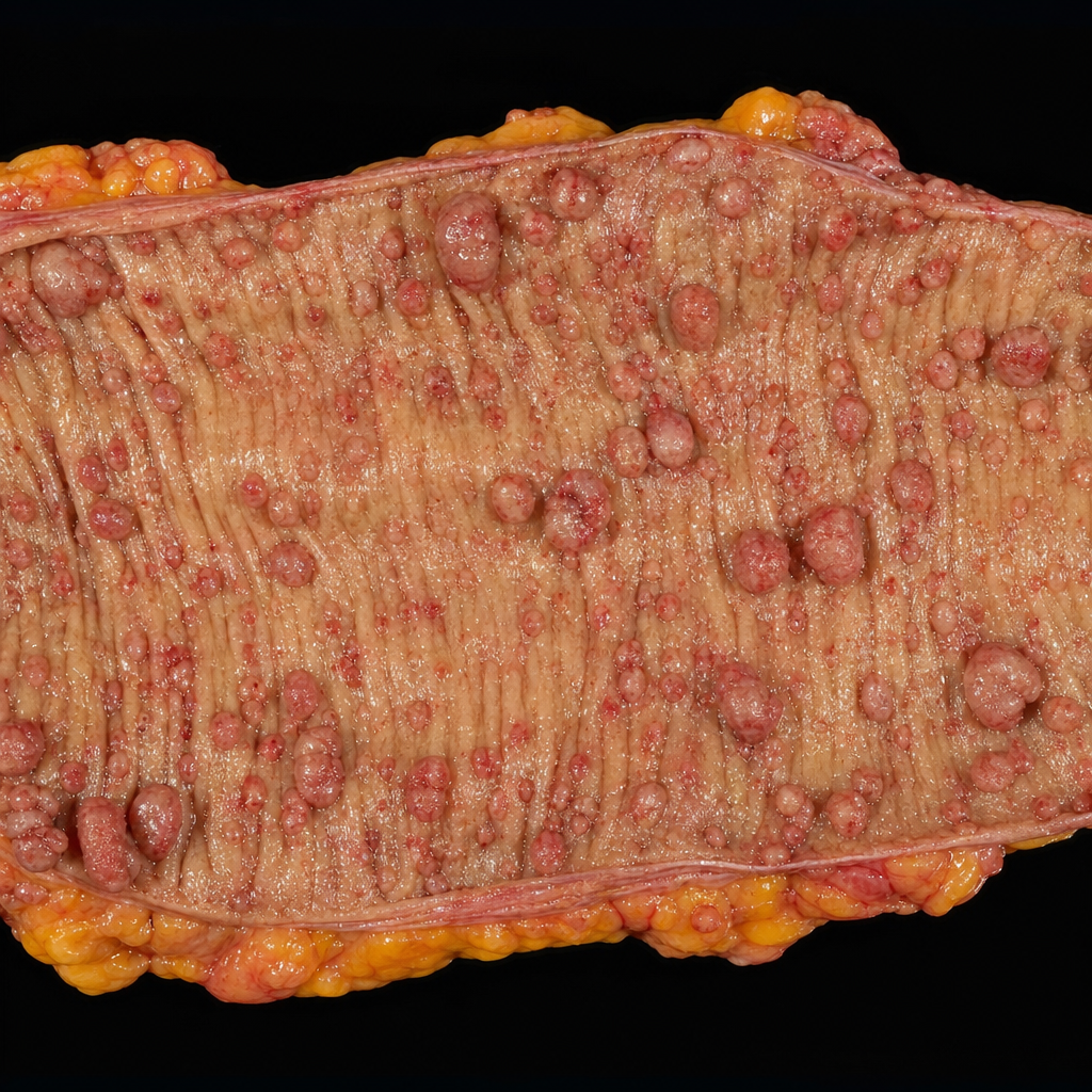
This is a specimen of which condition?

What is the most common tumor of the small intestine?
Which of the following is NOT true regarding the intestinal type of gastric carcinoma?
Explanation: **Explanation:** **Toxic Megacolon** is a life-threatening complication characterized by total or segmental non-obstructive colonic dilatation (usually >6 cm) associated with systemic toxicity. **Why Ulcerative Colitis (UC) is the correct answer:** Toxic megacolon is most classically associated with **Ulcerative Colitis**. In UC, the inflammatory process, which is usually mucosal, can sometimes penetrate deep into the muscularis propria [3]. This leads to the release of inflammatory mediators (like Nitric Oxide) that inhibit smooth muscle tone and damage the myenteric plexus. The resulting neuromuscular paralysis causes the colon to dilate rapidly, risking perforation [3]. **Why the other options are incorrect:** * **Crohn’s Disease:** While it can cause toxic megacolon, it is significantly **less common** than in UC because the transmural fibrosis characteristic of Crohn’s often prevents the bowel wall from over-distending [2]. * **Aganglionic Megacolon (Hirschsprung Disease):** This is a congenital functional obstruction due to the absence of ganglion cells in the distal colon. While the colon dilates (megacolon), it is a chronic mechanical issue, not an acute inflammatory "toxic" state [1]. * **Ischemic Colitis:** This usually presents with acute abdominal pain and hematochezia in elderly patients. While severe ischemia can lead to gangrene, it rarely presents as the classic toxic megacolon syndrome seen in IBD. **High-Yield Clinical Pearls for NEET-PG:** * **Diagnostic Criteria:** Radiographic evidence of colonic dilatation **>6 cm** + signs of systemic toxicity (fever, tachycardia, leukocytosis). * **Trigger:** Often precipitated by **antimotility agents** (loperamide), opioids, or colonoscopy during an acute flare. * **Management:** Initial management is medical (NPO, IV fluids, steroids); however, **emergency total proctocolectomy** is indicated if there is no improvement within 24–72 hours or if perforation occurs. * **Most common site of dilatation:** The **transverse colon** (due to gas accumulation in the supine position). **References:** [1] Kumar V, Abbas AK, et al.. Robbins and Cotran Pathologic Basis of Disease. 9th ed. The Gastrointestinal Tract, p. 809. [2] Kumar V, Abbas AK, et al.. Robbins and Cotran Pathologic Basis of Disease. 9th ed. The Gastrointestinal Tract, pp. 806-807. [3] Cross SS. Underwood's Pathology: A Clinical Approach. 6th ed. Common Clinical Problems From Alimentary System Disease, pp. 367-368.
Explanation: **Explanation:** **1. Why Option A is the correct (false) statement:** Familial Adenomatous Polyposis (FAP) is an **Autosomal Dominant** condition, not recessive. It is caused by a germline mutation in the **APC (Adenomatous Polyposis Coli) gene** located on chromosome **5q21** [1]. According to the "Knudson Two-Hit Hypothesis," an individual inherits one defective copy, and a somatic mutation in the second allele leads to the development of hundreds to thousands of adenomatous polyps [1]. **2. Analysis of other options:** * **Option B (Associated with fibromas and osteomas):** This describes **Gardner Syndrome**, a clinical variant of FAP. It includes intestinal polyps plus extra-colonic manifestations like osteomas (usually of the mandible/skull), epidermal cysts, and desmoid tumors (fibromas). * **Option C (Associated with brain tumors):** This describes **Turcot Syndrome**, another variant of FAP. It is characterized by the association of colonic polyposis with CNS tumors (classically **Medulloblastomas** in FAP-associated Turcot, or Glioblastomas in Lynch syndrome). * **Option D (100% chance of developing colon carcinoma):** This is a hallmark of FAP. If left untreated (without prophylactic colectomy), the progression from adenoma to adenocarcinoma is inevitable, usually by the age of 40-50 [1]. **High-Yield Clinical Pearls for NEET-PG:** * **Minimum Requirement:** Diagnosis requires at least **100 polyps** [1]. * **Screening:** Start colonoscopy/sigmoidoscopy at age **10–12 years**. * **CHRPE:** Congenital Hypertrophy of Retinal Pigment Epithelium is a highly specific early extra-colonic sign of FAP. * **Molecular Pathway:** FAP follows the **APC-Wnt/β-catenin pathway** (the classic Adenoma-Carcinoma sequence). **References:** [1] Kumar V, Abbas AK, et al.. Robbins and Cotran Pathologic Basis of Disease. 9th ed. The Gastrointestinal Tract, pp. 821-822.
Explanation: **Explanation:** The correct answer is **Abetalipoproteinemia** because it presents with a pathognomonic (highly specific) histological finding: **vacuolated enterocytes filled with lipid droplets** (clear cytoplasm) on a small bowel biopsy, especially after a fatty meal. **Why Abetalipoproteinemia is the most specific:** This autosomal recessive disorder involves a mutation in the **Microsomal Triglyceride Transfer Protein (MTP)**. MTP is essential for loading lipids onto Apolipoprotein B (ApoB-48 in the gut and ApoB-100 in the liver). Without functional MTP, enterocytes can absorb dietary fats but cannot assemble or export them as chylomicrons. Consequently, triglycerides accumulate within the enterocyte cytoplasm, creating a distinctive "foamy" appearance while the **villous architecture remains normal**. **Why other options are less specific:** * **Tropical Sprue:** Shows non-specific changes like villous atrophy, crypt hyperplasia, and increased intraepithelial lymphocytes. These findings overlap significantly with Celiac disease. * **Giardiasis:** Diagnosis depends on seeing the pear-shaped trophozoites; however, the mucosal architecture is often completely normal or shows mild, non-specific inflammation. * **Malnutrition:** Typically results in non-specific villous blunting and thinning of the mucosa, which can be seen in various malabsorptive states. **High-Yield Clinical Pearls for NEET-PG:** * **Blood Smear:** Look for **Acanthocytes** (spur cells) due to altered red cell membrane lipids [1]. * **Clinical Triad:** Fat malabsorption (steatorrhea), Retinitis pigmentosa, and Ataxia (due to Vitamin E deficiency) [1]. * **Lab Profile:** Extremely low levels of VLDL, LDL, and total cholesterol (ApoB is undetectable). * **Treatment:** High-dose fat-soluble vitamins (especially Vitamin E) and restriction of long-chain fatty acids. **References:** [1] Kumar V, Abbas AK, et al.. Robbins and Cotran Pathologic Basis of Disease. 9th ed. The Gastrointestinal Tract, pp. 791-792.
Explanation: **Explanation:** **Mixed tumors**, also known as **Pleomorphic Adenomas**, are the most common benign neoplasms of the salivary glands [1]. They are termed "mixed" because they consist of both epithelial and mesenchymal components (myxoid, chondroid, or osteoid tissue) derived from a single germ layer. * **Why Option C is Correct:** The **Parotid gland** is the most frequent site for salivary gland tumors in general, and specifically for Pleomorphic Adenomas [1]. Approximately **60-80%** of all pleomorphic adenomas occur in the parotid gland, typically presenting as a slow-growing, painless, mobile mass at the angle of the jaw [1]. * **Why Options A, B, and D are Incorrect:** * **Option A:** While they can occur in the submandibular gland, it is much less common (approx. 10%) [1]. * **Option B:** Pleomorphic adenomas are **benign**. However, if left untreated for years, they can undergo malignant transformation into *Carcinoma ex pleomorphic adenoma* [1]. * **Option D:** Salivary calculi (Sialolithiasis) are most commonly associated with the **Submandibular gland** (Wharton’s duct) due to its alkaline, calcium-rich secretions and upward drainage path; they are not a characteristic feature of mixed tumors. **High-Yield Clinical Pearls for NEET-PG:** * **Most common salivary tumor:** Pleomorphic Adenoma (Parotid) [1]. * **Most common malignant salivary tumor:** Mucoepidermoid Carcinoma [3]. * **Warthin’s Tumor (Adenolymphoma):** Second most common benign tumor; strongly associated with **smoking** and almost exclusive to the parotid [2]. * **Histology:** Look for "islands of epithelium in a chondromyxoid stroma." * **Surgical Note:** They have a high recurrence rate if enucleated due to "pseudopod" extensions; hence, superficial parotidectomy is the preferred treatment [1]. **References:** [1] Kumar V, Abbas AK, et al.. Robbins and Cotran Pathologic Basis of Disease. 9th ed. Head and Neck, pp. 751-753. [2] Kumar V, Abbas AK, et al.. Robbins and Cotran Pathologic Basis of Disease. 9th ed. Head and Neck, p. 753. [3] Kumar V, Abbas AK, et al.. Robbins and Cotran Pathologic Basis of Disease. 9th ed. Head and Neck, pp. 753-755.
Explanation: **Explanation:** The correct answer is **B. Skip lesions**. **Ulcerative Colitis (UC)** is characterized by **continuous, diffuse mucosal inflammation** that starts in the rectum and extends proximally without any interruptions [2]. **Skip lesions** (areas of diseased tissue separated by normal-appearing mucosa) are a hallmark feature of **Crohn’s Disease**, not Ulcerative Colitis [4]. **Analysis of Options:** * **A. Pseudopolyps:** These are common in UC [1]. They are islands of regenerating, bulging residual mucosa surrounded by areas of extensive ulceration and mucosal atrophy. * **C. Rectum involvement:** The rectum is involved in nearly **100% of cases** of UC [2]. The disease typically begins as proctitis and spreads in a retrograde, continuous fashion. * **D. Malabsorption:** While more common in Crohn’s disease (due to small bowel involvement), chronic, extensive UC can lead to malabsorption and nutritional deficiencies due to severe mucosal damage and rapid transit time. **NEET-PG High-Yield Pearls:** * **Depth of Involvement:** UC is limited to the **mucosa and submucosa** [2], whereas Crohn’s is **transmural** [4]. * **Microscopic Hallmark:** **Crypt abscesses** (neutrophils within the crypt lumen) are characteristic of UC [1]. * **Lead Pipe Appearance:** On barium enema, loss of haustrations due to chronic inflammation leads to a "lead pipe" appearance. * **Complications:** UC carries a higher risk of **Toxic Megacolon** and **Colorectal Carcinoma** compared to Crohn’s. * **Smoking Paradox:** Smoking is a risk factor for Crohn’s but appears to be **protective** against Ulcerative Colitis [3]. **References:** [1] Kumar V, Abbas AK, et al.. Robbins and Cotran Pathologic Basis of Disease. 9th ed. The Gastrointestinal Tract, p. 809. [2] Cross SS. Underwood's Pathology: A Clinical Approach. 6th ed. Common Clinical Problems From Alimentary System Disease, pp. 367-368. [3] Kumar V, Abbas AK, et al.. Robbins and Cotran Pathologic Basis of Disease. 9th ed. The Gastrointestinal Tract, pp. 807-809. [4] Cross SS. Underwood's Pathology: A Clinical Approach. 6th ed. Common Clinical Problems From Alimentary System Disease, pp. 366-367.
Explanation: **Explanation:** The clinical scenario describes **Barrett’s Esophagus**, a classic example of **metaplasia** resulting from chronic gastroesophageal reflux disease (GERD). [1] **1. Why the correct answer is right:** Metaplasia is a reversible change in which one adult cell type is replaced by another cell type better suited to withstand a specific stress. [1] In the lower esophagus, the normal **non-keratinized stratified squamous epithelium** is sensitive to gastric acid. To protect itself, the tissue undergoes intestinal metaplasia, transforming into **nonciliated columnar epithelium** containing **goblet cells**. [1] These columnar cells are more resistant to the acidic environment because they secrete protective mucus. **2. Why the incorrect options are wrong:** * **Ciliated columnar epithelium:** This is characteristic of the respiratory tract (e.g., trachea, bronchi). It is not a feature of intestinal metaplasia in the GI tract. [2] * **Keratinizing squamous epithelium:** This is found in the skin (epidermis). While the esophagus is normally squamous, it is *non-keratinized*. Keratinization is not the adaptive response to acid reflux. [1] **3. High-Yield Clinical Pearls for NEET-PG:** * **Definition:** Barrett’s esophagus is defined histologically by the presence of **intestinal metaplasia** (specifically the presence of **Goblet cells**) in the esophageal mucosa. [1] * **Risk of Malignancy:** Barrett’s esophagus is a significant pre-malignant condition that increases the risk of **Esophageal Adenocarcinoma** [1] (Note: Squamous cell carcinoma is associated with smoking/alcohol, not GERD). * **Endoscopic Appearance:** It appears as "velvety red" or "salmon-pink" tongues of mucosa extending upward from the gastroesophageal junction. * **Key Marker:** CDX2 is a transcription factor often expressed in intestinal metaplasia. **References:** [1] Cross SS. Underwood's Pathology: A Clinical Approach. 6th ed. Common Clinical Problems From Alimentary System Disease, pp. 348-349. [2] Cross SS. Underwood's Pathology: A Clinical Approach. 6th ed. (Basic Pathology) introduces the student to key general principles of pathology, both as a medical science and as a clinical activity with a vital role in patient care. Part 2 (Disease Mechanisms) provides fundamental knowledge about the cellular and molecular processes involved in diseases, providing the rationale for their treatment. Part 3 (Systematic Pathology) deals in detail with specific diseases, with emphasis on the clinically important aspects., pp. 91-92.
Explanation: **Explanation:** The question asks which of the listed conditions is **not** a premalignant **polyp**. While Ulcerative Colitis (UC) significantly increases the risk of colorectal carcinoma, the malignancy in UC arises from **flat, dysplastic mucosa** (dysplasia-associated lesion or mass - DALM) rather than a discrete premalignant polyp [1]. The "polyps" seen in UC are typically **pseudopolyps** (inflammatory polyps), which are islands of regenerating mucosa amidst areas of ulceration and have **no malignant potential** [1]. **Analysis of Options:** * **Juvenile Polyposis Syndrome (Option A):** Although individual juvenile polyps are hamartomatous, the *syndrome* carries a 30-50% risk of adenocarcinoma due to the accumulation of mutations (SMAD4/BMPR1A) and co-existing adenomatous changes [1]. * **Peutz-Jeghers Syndrome (Option B):** These are hamartomatous polyps [1]. While the polyps themselves are benign, the syndrome is associated with a markedly increased risk of various visceral cancers (colorectal, pancreatic, breast, and ovarian). * **Familial Polyposis Coli (Option D):** This is a classic premalignant condition. It involves thousands of **adenomatous polyps**, with a 100% risk of progression to colorectal cancer if left untreated [1]. **NEET-PG High-Yield Pearls:** * **Pseudopolyps:** Characteristic of Ulcerative Colitis; they are non-neoplastic [1]. * **Hamartomatous Polyps:** Found in Juvenile Polyposis and Peutz-Jeghers; they have low malignant potential individually but high risk within a syndrome [1]. * **Adenomatous Polyps:** Always considered premalignant (Tubular < Tubulovillous < Villous) [1]. * **UC Malignancy:** Risk increases with the duration of disease (>8-10 years) and extent (Pancolitis). Unlike sporadic CRC, UC-associated cancer is often multifocal and arises from flat dysplasia [1]. **References:** [1] Kumar V, Abbas AK, et al.. Robbins and Cotran Pathologic Basis of Disease. 9th ed. The Gastrointestinal Tract, pp. 807-822.
Explanation: ***Familial adenomatous polyposis (FAP)*** - Characterized by a **carpet-like appearance** of hundreds to thousands of small **adenomatous polyps** densely covering the colonic mucosa - The **uniform distribution** and **sheer number** of small polyps throughout the colon is pathognomonic for FAP *Ulcerative colitis* - Shows **diffuse mucosal inflammation** with superficial ulcerations and **pseudopolyps** formed by regenerating mucosa - Lacks the characteristic **carpet of true adenomatous polyps** seen in FAP; inflammation is limited to **mucosa and submucosa** *Crohn's disease* - Exhibits **skip lesions** with normal bowel between affected segments and **cobblestone appearance** due to deep ulcers - Shows **transmural inflammation** with thickened bowel wall, not the superficial carpet of polyps characteristic of FAP *Peutz-Jeghers syndrome* - Features **few large hamartomatous polyps** (typically 5-20) rather than hundreds of small adenomatous polyps - Associated with **mucocutaneous pigmentation** around lips and mouth, and polyps have a different histological appearance than adenomatous polyps
Explanation: The small intestine is a unique segment of the GI tract where primary neoplasms are relatively rare compared to the stomach and colon. **Explanation of the Correct Answer:** **A. Leiomyoma:** While many textbooks focus on malignant tumors, **Leiomyoma** is historically and statistically considered the **most common benign tumor** of the small intestine. Since benign tumors of the small intestine are more frequent than malignant ones, Leiomyoma is often cited as the overall most common primary tumor. These are intramural, slow-growing smooth muscle tumors, most frequently found in the jejunum. **Explanation of Incorrect Options:** * **B. Lymphoma:** This is the most common malignancy in children (specifically Burkitt lymphoma) or in patients with Celiac disease (EATL) [1]. However, it is not the most common tumor overall. * **C. Adenocarcinoma:** This is the **most common primary malignancy** of the small intestine (typically occurring in the duodenum). While clinically significant, its incidence is lower than that of benign mesenchymal tumors. * **D. Hemangioma:** These are vascular tumors that can cause GI bleeding, but they are significantly less common than leiomyomas. **High-Yield Clinical Pearls for NEET-PG:** 1. **Most common site for Small Bowel Adenocarcinoma:** Duodenum (near the Ampulla of Vater). 2. **Most common site for Carcinoid Tumor:** Ileum (specifically the distal ileum/appendix) [1]. 3. **GIST vs. Leiomyoma:** Most tumors previously labeled as "Leiomyomas" are now classified as **Gastrointestinal Stromal Tumors (GIST)**, which are **c-KIT (CD117) positive** [2]. 4. **Peutz-Jeghers Syndrome:** Associated with multiple hamartomatous polyps in the small intestine and increased risk of malignancy. **References:** [1] Kumar V, Abbas AK, et al.. Robbins and Cotran Pathologic Basis of Disease. 9th ed. The Gastrointestinal Tract, pp. 780-781. [2] Kumar V, Abbas AK, et al.. Robbins and Cotran Pathologic Basis of Disease. 9th ed. The Gastrointestinal Tract, pp. 783-784.
Explanation: **Explanation:** Gastric carcinoma is classified by the **Lauren classification** into two main histological types: **Intestinal** and **Diffuse** [1]. Understanding the distinction between these two is high-yield for NEET-PG. **Why Option C is the correct answer (False statement):** While both types of gastric cancer can metastasize, the **Diffuse type** (characterized by signet-ring cells and linitis plastica) is notorious for early, extensive submucosal infiltration and widespread lymphatic spread [1]. In contrast, the **Intestinal type** tends to grow as a cohesive mass (polypoid or ulcerative) and spreads primarily via the bloodstream (hematogenous) to the liver [2]. **Analysis of Incorrect Options (True statements):** * **Option A:** The intestinal type is characterized by cohesive cells forming **well-defined glands**, mimicking colonic adenocarcinoma [2]. * **Option B:** It most commonly arises in the **distal stomach** (antrum and lesser curvature) and is strongly associated with environmental factors like *H. pylori* and intestinal metaplasia [1], [2]. * **Option D:** This type typically affects the **elderly population** (mean age ~55 years) and shows a strong male predominance (2:1) [1]. **High-Yield Clinical Pearls for NEET-PG:** * **Intestinal Type:** Associated with *H. pylori*, chronic gastritis, and dietary nitrates. Incidence is decreasing globally [1]. * **Diffuse Type:** Associated with **CDH1 gene mutations** (E-cadherin loss) [2]. It has a younger age of onset, equal male-to-female ratio, and a worse prognosis [1]. * **Virchow’s Node:** Left supraclavicular lymphadenopathy, a classic sign of metastatic gastric cancer. * **Sister Mary Joseph Nodule:** Periumbilical metastasis, more common in intestinal types. **References:** [1] Kumar V, Abbas AK, et al.. Robbins and Cotran Pathologic Basis of Disease. 9th ed. The Gastrointestinal Tract, pp. 779-780. [2] Kumar V, Abbas AK, et al.. Robbins and Cotran Pathologic Basis of Disease. 9th ed. The Gastrointestinal Tract, p. 779.
Oral Cavity and Esophageal Pathology
Practice Questions
Gastritis and Peptic Ulcer Disease
Practice Questions
Inflammatory Bowel Disease
Practice Questions
Malabsorption Syndromes
Practice Questions
Vascular Disorders of Intestine
Practice Questions
Diverticular Disease
Practice Questions
Intestinal Obstruction
Practice Questions
Gastrointestinal Infections
Practice Questions
Polyps and Neoplasms
Practice Questions
Appendiceal Pathology
Practice Questions
Get full access to all questions, explanations, and performance tracking.
Start For Free