Which intestinal polyp has the potential to grow into cancer?
Anal cancer is associated with which of the following viruses?
A patient presents with symmetrical, persistent, and painless swelling of the lacrimal and salivary glands. The patient reports no or minimal xerostomia and shows a positive response to steroid therapy. What is the most likely diagnosis?
Hourglass deformity is seen in which of the following conditions?
What is the most common site of ulcerative colitis?
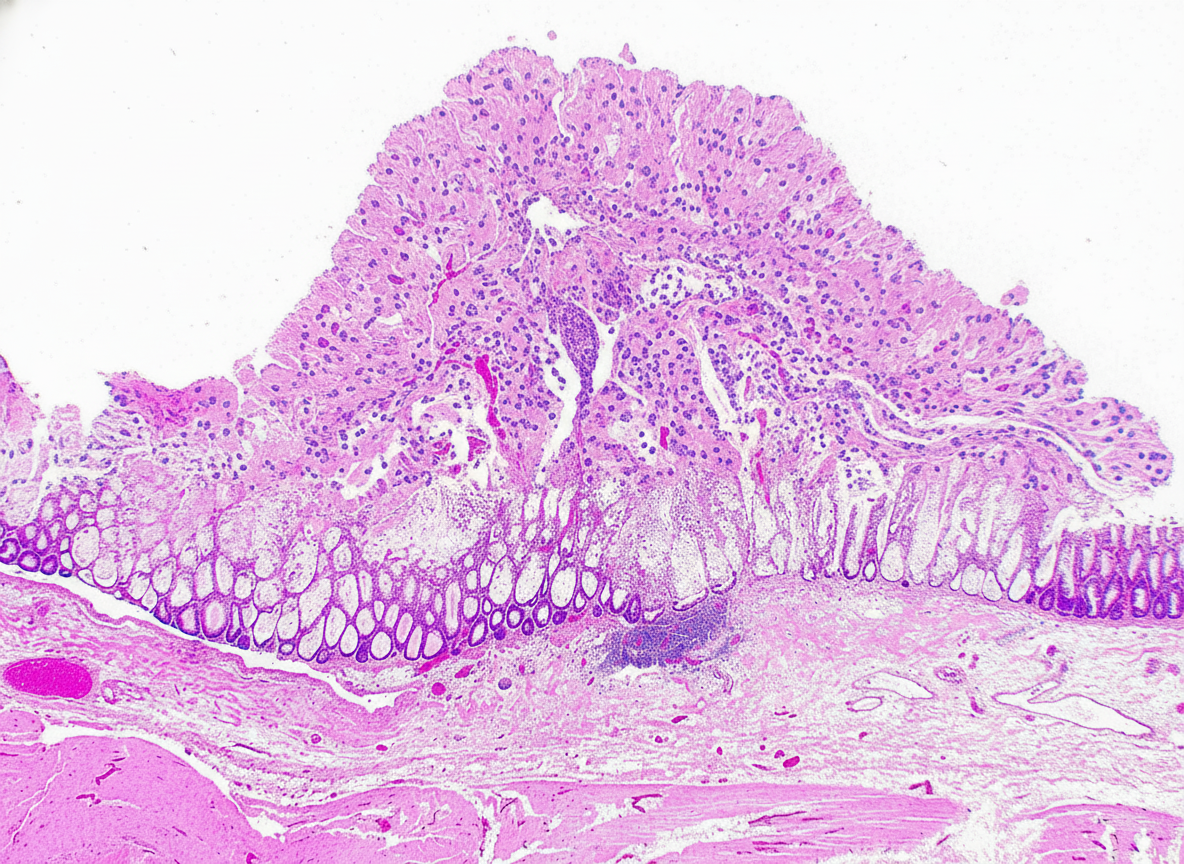
Which of the following is NOT true regarding Barrett's esophagus?
A patient has an increased number of columnar cells in the lower esophagus. Which of the following histological changes is present?
An elderly male with a history of long-term antibiotic intake presents with profuse watery diarrhea and abdominal cramps. The colonoscopic biopsy findings are shown in the image. What is the most likely diagnosis?

A 54-year-old man presents with dysphagia, heartburn, belching, and epigastric pain. A barium swallow shows a sliding hiatal hernia and a stricture situated higher than usual in the mid-esophagus. Endoscopic findings suggest Barrett's esophagus with marked esophagitis and linear ulcerations. Biopsy confirms columnar epithelium at the affected area and normal squamous epithelium above. What statement is TRUE regarding this condition?
Which condition involves the entire thickness of the bowel wall with skin lesions?
Explanation: **Explanation:** The potential for an intestinal polyp to undergo malignant transformation depends on its histological nature. **1. Why Adenomatous Polyp is Correct:** Adenomatous polyps (Adenomas) are **true neoplasms** and are considered **premalignant** [2]. They arise from epithelial proliferation and dysplasia. The progression to adenocarcinoma occurs via the **"Adenoma-Carcinoma Sequence,"** involving mutations in the *APC* gene, *KRAS*, and *TP53* [2]. The risk of malignancy increases with: * **Size:** >2 cm (highest risk) [1]. * **Histology:** Villous architecture (highest risk) > Tubulovillous > Tubular [3]. * **Degree of Dysplasia:** High-grade dysplasia [1]. **2. Why Other Options are Incorrect:** * **Hyperplastic Polyps:** These are non-neoplastic proliferations of mature goblet and absorptive cells. They typically lack malignant potential (though "Serrated Adenomas" are a distinct, premalignant subtype often confused with them). * **Juvenile Polyps:** These are **inflammatory/hamartomatous** lesions common in children. Solitary juvenile polyps have no malignant potential. (Note: Juvenile Polyposis *Syndrome* increases cancer risk due to the sheer number of polyps, but the individual polyp itself is benign). * **Hamartomatous Polyps:** These consist of disorganized native tissue (e.g., Peutz-Jeghers polyps). They are non-neoplastic and do not transform into cancer, although the associated syndromes carry an increased risk of extra-intestinal malignancies. **High-Yield Clinical Pearls for NEET-PG:** * **Most common type of polyp:** Hyperplastic polyp. * **Most common neoplastic polyp:** Tubular adenoma [3]. * **Highest risk of cancer:** Villous adenoma ("Villous is Villainous") [3]. * **Familial Adenomatous Polyposis (FAP):** 100% risk of progression to colorectal cancer; caused by germline *APC* mutation [2]. **References:** [1] Kumar V, Abbas AK, et al.. Robbins and Cotran Pathologic Basis of Disease. 9th ed. The Gastrointestinal Tract, pp. 815-817. [2] Kumar V, Abbas AK, et al.. Robbins and Cotran Pathologic Basis of Disease. 9th ed. The Gastrointestinal Tract, pp. 821-822. [3] Cross SS. Underwood's Pathology: A Clinical Approach. 6th ed. Common Clinical Problems From Alimentary System Disease, pp. 371-372.
Explanation: **Explanation:** **1. Why Human Papilloma Virus (HPV) is correct:** Anal cancer, specifically **Squamous Cell Carcinoma (SCC)** of the anal canal, is strongly associated with persistent infection by high-risk strains of **Human Papilloma Virus (HPV)**, most notably **types 16 and 18** [1], [2]. The pathogenesis involves the viral oncoproteins **E6 and E7**, which inhibit the host’s tumor suppressor proteins **p53 and Rb**, respectively [3], [4]. This leads to uncontrolled cell cycle progression and the development of Anal Intraepithelial Neoplasia (AIN), the precursor lesion to invasive cancer [4]. Risk factors include receptive anal intercourse and immunosuppression (e.g., HIV/AIDS) [1]. **2. Why the other options are incorrect:** * **Epstein-Barr Virus (EBV):** Associated with Burkitt lymphoma, Nasopharyngeal carcinoma, and Hodgkin lymphoma, but not typically with anal SCC [2]. * **HTLV-1:** This retrovirus is the causative agent of Adult T-cell Leukemia/Lymphoma (ATLL) and Tropical Spastic Paraparesis [2]. * **Polyoma virus:** Specifically, the JC virus causes Progressive Multifocal Leukoencephalopathy (PML), and the BK virus causes nephropathy in transplant patients. Merkel cell polyomavirus is linked to skin cancer, but not anal cancer. **3. Clinical Pearls for NEET-PG:** * **The "Zone of Transition":** Most anal cancers arise at the **cloacogenic zone** (transition between columnar rectal mucosa and squamous anal mucosa), similar to the transformation zone of the cervix. * **HIV Link:** HIV-positive individuals have a significantly higher incidence of HPV-related anal SCC [1]. * **Screening:** Anal Pap smears are used for screening high-risk populations. * **Other HPV-related cancers:** Cervical, Vulvar, Vaginal, Penile, and Oropharyngeal (tonsillar) SCC [1], [2]. **References:** [1] Kumar V, Abbas AK, et al.. Robbins and Cotran Pathologic Basis of Disease. 9th ed. Diseases of the Immune System, pp. 261-262. [2] Cross SS. Underwood's Pathology: A Clinical Approach. 6th ed. (Basic Pathology) introduces the student to key general principles of pathology, both as a medical science and as a clinical activity with a vital role in patient care. Part 2 (Disease Mechanisms) provides fundamental knowledge about the cellular and molecular processes involved in diseases, providing the rationale for their treatment. Part 3 (Systematic Pathology) deals in detail with specific diseases, with emphasis on the clinically important aspects., pp. 219-220. [3] Kumar V, Abbas AK, et al.. Robbins and Cotran Pathologic Basis of Disease. 9th ed. Neoplasia, pp. 334-335. [4] Kumar V, Abbas AK, et al.. Robbins and Cotran Pathologic Basis of Disease. 9th ed. The Female Genital Tract, pp. 1007-1008.
Explanation: **Explanation:** The clinical presentation of symmetrical, painless enlargement of the lacrimal and salivary glands is known as **Mikulicz syndrome**. When this occurs as an isolated entity (often associated with IgG4-related disease), it is termed **Mikulicz disease**. **1. Why Mikulicz Disease is Correct:** Mikulicz disease is characterized by the enlargement of the lacrimal, parotid, and submandibular glands. Unlike Sjögren syndrome, patients with Mikulicz disease typically have **minimal or no xerostomia** (dry mouth) [1] [2] or xerophthalmia (dry eyes). A hallmark feature that distinguishes it from Sjögren syndrome is its **dramatic response to steroid therapy**. Pathologically, it is now considered a manifestation of **IgG4-related disease (IgG4-RD)**, showing storiform fibrosis and elevated serum IgG4 levels. **2. Why Other Options are Incorrect:** * **Primary Sjögren Syndrome (Sicca Syndrome):** While it involves gland enlargement [3], the defining feature is severe functional impairment leading to **sicca symptoms** (dry eyes and mouth) [1] [2]. It is an autoimmune destruction of glands, often associated with Anti-Ro (SS-A) and Anti-La (SS-B) antibodies [4], and does not respond as significantly to steroids as Mikulicz disease. * **Secondary Sjögren Syndrome:** This occurs in the presence of another autoimmune disease (most commonly Rheumatoid Arthritis). The clinical focus is on the underlying connective tissue disease and the resulting sicca symptoms. **Clinical Pearls for NEET-PG:** * **Mikulicz Syndrome vs. Disease:** *Syndrome* is a clinical triad (enlargement of lacrimal/salivary glands) seen in various conditions like Sarcoidosis, Leukemia, or Lymphoma. *Disease* is the specific IgG4-related entity. * **Histology:** Look for "Epimyoepithelial islands" in Sjögren’s; look for "IgG4+ plasma cells and storiform fibrosis" in Mikulicz disease. * **Key Differentiator:** If the question mentions **"Steroid Responsive"** and **"No Xerostomia,"** always lean towards Mikulicz disease/IgG4-RD. **References:** [1] Cross SS. Underwood's Pathology: A Clinical Approach. 6th ed. Common Clinical Problems From Alimentary System Disease, pp. 345-346. [2] Kumar V, Abbas AK, et al.. Robbins and Cotran Pathologic Basis of Disease. 9th ed. Head and Neck, pp. 749-750. [3] Kumar V, Abbas AK, et al.. Robbins and Cotran Pathologic Basis of Disease. 9th ed. Diseases of the Immune System, pp. 235-236. [4] Kumar V, Abbas AK, et al.. Robbins and Cotran Pathologic Basis of Disease. 9th ed. Diseases of the Immune System, pp. 234-235.
Explanation: **Explanation:** **1. Why Peptic Ulcer Disease (PUD) is correct:** The "Hourglass stomach" deformity is a classic complication of chronic gastric ulcers, typically located on the **lesser curvature** [1]. It occurs due to chronic inflammation and subsequent **fibrosis (cicatrization)**. As the ulcer heals, the fibrous tissue contracts, pulling the greater curvature toward the lesser curvature. This creates a central constriction that divides the stomach into two distinct pouches, resembling an hourglass. **2. Analysis of Incorrect Options:** * **Carcinoma of the stomach:** Typically presents as a "Leather bottle stomach" (**Linitis Plastica**) in the diffuse type (Signet ring cell carcinoma). This involves a rigid, thickened, and non-distensible stomach wall due to submucosal infiltration, rather than a localized central constriction. * **Duodenal atresia:** Characterized by the **"Double bubble sign"** on X-ray. It is a congenital obstruction of the duodenum resulting in a dilated stomach and a dilated proximal duodenum. * **Congenital Hypertrophic Pyloric Stenosis (CHPS):** Presents with a "String sign" or "Beak sign" on contrast studies and an "Olive-shaped mass" on palpation. It involves hypertrophy of the pyloric sphincter, leading to gastric outlet obstruction, not a mid-stomach constriction. **3. NEET-PG High-Yield Pearls:** * **Leather bottle stomach:** Linitis Plastica (Gastric Cancer). * **Cup and spill/Cascade stomach:** A functional or structural variation where the fundus folds back on the body. * **Watermelon stomach:** Gastric Antral Vascular Ectasia (GAVE). * **Most common site for Peptic Ulcer:** First part of the duodenum (Duodenal Ulcer) and Lesser curvature/Antrum (Gastric Ulcer) [1]. **References:** [1] Kumar V, Abbas AK, et al.. Robbins and Cotran Pathologic Basis of Disease. 9th ed. The Gastrointestinal Tract, pp. 773-774.
Explanation: **Explanation:** **Ulcerative Colitis (UC)** is a chronic inflammatory bowel disease characterized by diffuse, mucosal inflammation limited to the colon. **Why Rectum is the correct answer:** The hallmark of Ulcerative Colitis is that it **always involves the rectum** (proctitis) and extends proximally in a **continuous, symmetrical fashion** without skip lesions [1]. In approximately 95% of cases, the rectum is involved at the time of diagnosis, making it the most common and characteristic site of the disease. **Analysis of Incorrect Options:** * **B. Caecum:** While UC can involve the caecum (as part of pancolitis), it is rarely the primary or most common site. An exception is "backwash ileitis," where the terminal ileum is affected secondary to total colonic involvement [1]. * **C. Small Intestine:** UC is primarily a disease of the **large intestine**. Small bowel involvement is a distinguishing feature of Crohn’s Disease, not UC (except for the aforementioned backwash ileitis). * **D. Appendix:** While appendiceal inflammation can sometimes be seen in UC (appendiceal "skip" lesion), it is not the most common site [1]. Interestingly, prior appendectomy is considered a protective factor against developing UC. **High-Yield Clinical Pearls for NEET-PG:** * **Distribution:** Continuous involvement; starts in the rectum and moves proximally [1]. * **Depth:** Limited to the **mucosa and submucosa** (unlike Crohn’s, which is transmural) [1]. * **Microscopy:** Characterized by **Crypt abscesses** (neutrophils in crypt lumens) and crypt distortion. * **Gross Features:** Pseudopolyps (regenerating mucosa) and "Lead-pipe" appearance on barium enema due to loss of haustrations. * **Complications:** Toxic megacolon and a significantly higher risk of **Adenocarcinoma** compared to Crohn’s. **References:** [1] Cross SS. Underwood's Pathology: A Clinical Approach. 6th ed. Common Clinical Problems From Alimentary System Disease, pp. 367-368.
Explanation: **Explanation:** **1. Why Option A is the correct answer:** Barrett’s esophagus is a premalignant condition, but it is associated with an increased risk of **Adenocarcinoma**, not squamous cell carcinoma (SCC) [1]. In Barrett’s, the normal stratified squamous epithelium is replaced by columnar epithelium. Malignant transformation of this glandular (columnar) tissue leads to adenocarcinoma. Squamous cell carcinoma is typically associated with smoking, alcohol, and caustic injury, rather than chronic acid reflux [2]. **2. Analysis of incorrect options:** * **Option B (Metaplasia):** This is a true statement. Barrett’s is a classic example of **adaptive metaplasia**, where one adult cell type (squamous) is replaced by another (columnar) to better withstand the stress of chronic gastric acid exposure [3]. * **Option C (Columnar epithelium):** This is true. Specifically, the diagnosis requires the presence of **specialized intestinal metaplasia**, characterized by the presence of **Goblet cells** within the columnar mucosa [3]. * **Option D (Lower esophagus):** This is true. Barrett’s occurs as a complication of Chronic Gastroesophageal Reflux Disease (GERD); therefore, the changes are most commonly found in the distal (lower) third of the esophagus [1]. **Clinical Pearls for NEET-PG:** * **Endoscopic Appearance:** Appears as "salmon-pink" velvety tongues of mucosa extending upwards from the gastroesophageal junction. * **Gold Standard Diagnosis:** Endoscopy followed by biopsy showing **Goblet cells** (essential for diagnosis in the US, though some UK guidelines differ) [3]. * **Molecular Marker:** Increased expression of **p53** and **p16** are early markers of progression toward dysplasia. * **Screening:** Patients with long-standing GERD require surveillance to detect high-grade dysplasia or early adenocarcinoma [3]. **References:** [1] Kumar V, Abbas AK, et al.. Robbins and Cotran Pathologic Basis of Disease. 9th ed. The Gastrointestinal Tract, pp. 765-766. [2] Kumar V, Abbas AK, et al.. Robbins and Cotran Pathologic Basis of Disease. 9th ed. The Gastrointestinal Tract, pp. 766-767. [3] Cross SS. Underwood's Pathology: A Clinical Approach. 6th ed. Common Clinical Problems From Alimentary System Disease, pp. 348-349.
Explanation: **Explanation:** The correct answer is **Metaplasia**. **Why Metaplasia is correct:** Metaplasia is a reversible change in which one adult cell type (epithelial or mesenchymal) is replaced by another adult cell type [3]. In the lower esophagus, chronic gastroesophageal reflux disease (GERD) causes the normal **stratified squamous epithelium** to be replaced by **simple columnar epithelium** (often with goblet cells) [1]. This specific transformation is known as **Barrett’s Esophagus**. The body undergoes this change because columnar cells are more resistant to the corrosive effects of gastric acid and pepsin [1]. **Why the other options are incorrect:** * **Dysplasia:** This refers to disordered growth and maturation of an epithelium, characterized by a loss of architectural uniformity and cellular pleomorphism [4]. While Barrett’s esophagus can progress to dysplasia, the initial change from squamous to columnar cells is metaplasia. * **Anaplasia:** This is a hallmark of malignancy characterized by a total lack of differentiation. It represents "backward formation" to a primitive cell type and is not a compensatory response to irritation. * **Normal histology:** The normal lining of the esophagus is non-keratinized stratified squamous epithelium. The presence of columnar cells in this location is pathological. **NEET-PG High-Yield Pearls:** * **Barrett’s Esophagus** is the single most important risk factor for **Esophageal Adenocarcinoma** [1]. * The most common type of metaplasia is **Squamous Metaplasia** (e.g., in the respiratory tract of smokers), but Barrett’s is a classic example of **Columnar Metaplasia** [3]. * Metaplasia is reversible if the stimulus (acid reflux) is removed, but if the stimulus persists, it can progress to dysplasia and eventually neoplasia [2]. **References:** [1] Cross SS. Underwood's Pathology: A Clinical Approach. 6th ed. Common Clinical Problems From Alimentary System Disease, pp. 348-349. [2] Kumar V, Abbas AK, et al.. Robbins and Cotran Pathologic Basis of Disease. 9th ed. Cellular Responses to Stress and Toxic Insults: Adaptation, Injury, and Death, p. 49. [3] Cross SS. Underwood's Pathology: A Clinical Approach. 6th ed. (Basic Pathology) introduces the student to key general principles of pathology, both as a medical science and as a clinical activity with a vital role in patient care. Part 2 (Disease Mechanisms) provides fundamental knowledge about the cellular and molecular processes involved in diseases, providing the rationale for their treatment. Part 3 (Systematic Pathology) deals in detail with specific diseases, with emphasis on the clinically important aspects., pp. 91-92. [4] Kumar V, Abbas AK, et al.. Robbins and Cotran Pathologic Basis of Disease. 9th ed. The Gastrointestinal Tract, pp. 764-765.
Explanation: ***Pseudomembranous Colitis*** - History of **long-term antibiotic use** leading to **C. difficile** overgrowth causing profuse watery diarrhea is pathognomonic for pseudomembranous colitis. - Colonoscopic biopsy shows characteristic **volcano/summit lesions** with **pseudomembranes** composed of fibrin, mucus, and inflammatory debris overlying damaged epithelium. *Crohn's disease* - Characterized by **transmural inflammation** with **skip lesions** and **granulomas**, not pseudomembranes. - Typically presents with **bloody diarrhea**, **weight loss**, and **perianal disease** rather than watery diarrhea following antibiotic use. *Ulcerative colitis* - Shows **continuous mucosal inflammation** limited to mucosa and submucosa with **crypt abscesses**. - Presents with **bloody diarrhea** and **tenesmus**, not the watery diarrhea pattern seen with antibiotic-associated colitis. *Amoebic colitis* - Caused by **Entamoeba histolytica** showing **flask-shaped ulcers** and **trophozoites** in tissue. - Associated with **travel history** to endemic areas and **bloody mucoid stools**, not antibiotic exposure history.
Explanation: **Explanation:** **1. Why the correct answer is right:** Barrett’s esophagus (BE) is a metaplastic transformation where the normal stratified squamous epithelium of the distal esophagus is replaced by columnar epithelium (often with goblet cells) due to chronic gastroesophageal reflux disease (GERD) [2]. The primary goal of medical management is to **control acid reflux and prevent esophagitis**. By reducing acid exposure through Proton Pump Inhibitors (PPIs) or surgical interventions like Nissen fundoplication, clinicians aim to alleviate symptoms, promote healing of ulcerations, and potentially slow the progression of the metaplasia-dysplasia-carcinoma sequence [1]. **2. Why the incorrect options are wrong:** * **Option A:** Adenocarcinoma is actually **significantly more common** in Barrett’s esophagus [4]. BE is the most important precursor lesion for esophageal adenocarcinoma, increasing the risk by 30–40 times compared to the general population [2]. * **Option B:** Most patients (approx. 80-90%) with Barrett’s esophagus **do have associated GERD** [3]. Chronic reflux is the primary driver of the metaplastic changes. * **Option C:** Ectopic gastric lining (or metaplastic columnar epithelium) does **not** protect against aspiration. In fact, the underlying GERD associated with BE increases the risk of nocturnal regurgitation and recurrent aspiration pneumonitis [3]. **3. High-Yield Clinical Pearls for NEET-PG:** * **Definition:** Intestinal metaplasia (presence of **Goblet cells**) is the histological hallmark of Barrett’s esophagus [2]. * **Endoscopy:** Appears as "salmon-pink" velvety tongues of mucosa extending upward from the gastroesophageal junction. * **Screening/Surveillance:** Patients with BE require periodic endoscopic surveillance with biopsies (Seattle Protocol) to monitor for high-grade dysplasia [1]. * **Stricture Location:** While peptic strictures usually occur at the GE junction, in Barrett’s, they can occur higher up at the new squamocolumnar junction. **References:** [1] Kumar V, Abbas AK, et al.. Robbins and Cotran Pathologic Basis of Disease. 9th ed. The Gastrointestinal Tract, pp. 764-765. [2] Cross SS. Underwood's Pathology: A Clinical Approach. 6th ed. Common Clinical Problems From Alimentary System Disease, pp. 348-349. [3] Kumar V, Abbas AK, et al.. Robbins and Cotran Pathologic Basis of Disease. 9th ed. The Gastrointestinal Tract, pp. 762-763. [4] Kumar V, Abbas AK, et al.. Robbins and Cotran Pathologic Basis of Disease. 9th ed. The Gastrointestinal Tract, pp. 765-766.
Explanation: **Explanation:** The correct answer is **Crohn’s disease (CD)** because of its characteristic **transmural inflammation** and associated **extraintestinal manifestations** [2]. 1. **Why Crohn’s Disease is Correct:** * **Transmural Involvement:** Unlike other inflammatory conditions, CD involves all layers of the bowel wall (mucosa, submucosa, muscularis propria, and serosa) [2]. This leads to complications like deep fissures, fistulae, and "creeping fat." * **Skin Lesions:** CD is frequently associated with dermatological manifestations. The most common is **Erythema Nodosum** (tender red nodules on shins). Other lesions include **Pyoderma Gangrenosum** (more common in UC but occurs in CD) and "metastatic" Crohn’s (skin ulcerations distant from the GI tract) [1]. 2. **Why Other Options are Incorrect:** * **Ulcerative Colitis (UC):** Inflammation is strictly limited to the **mucosa and submucosa** (superficial) [2]. While UC also presents with skin lesions (like Pyoderma Gangrenosum), it does not involve the "entire thickness" of the wall. * **Irritable Bowel Syndrome (IBS):** This is a functional disorder. There is no structural damage, transmural inflammation, or associated systemic skin lesions. * **Option D:** Incorrect because the "entire thickness" (transmural) feature is unique to Crohn’s disease. **NEET-PG High-Yield Pearls:** * **Microscopy:** CD shows **Non-caseating granulomas** (pathognomonic in 35% of cases); UC shows **Crypt abscesses** [1]. * **Radiology:** CD shows the **"String sign of Kantor"** (due to strictures); UC shows the **"Lead pipe appearance"** (loss of haustra) [2]. * **Distribution:** CD is "skip lesions" from mouth to anus (mostly terminal ileum); UC is continuous involvement starting from the rectum [2]. **References:** [1] Kumar V, Abbas AK, et al.. Robbins and Cotran Pathologic Basis of Disease. 9th ed. The Gastrointestinal Tract, pp. 806-807. [2] Cross SS. Underwood's Pathology: A Clinical Approach. 6th ed. Common Clinical Problems From Alimentary System Disease, pp. 365-367.
Oral Cavity and Esophageal Pathology
Practice Questions
Gastritis and Peptic Ulcer Disease
Practice Questions
Inflammatory Bowel Disease
Practice Questions
Malabsorption Syndromes
Practice Questions
Vascular Disorders of Intestine
Practice Questions
Diverticular Disease
Practice Questions
Intestinal Obstruction
Practice Questions
Gastrointestinal Infections
Practice Questions
Polyps and Neoplasms
Practice Questions
Appendiceal Pathology
Practice Questions
Get full access to all questions, explanations, and performance tracking.
Start For Free