What is TRUE regarding Barrett's esophagus?
Multiple strictures in the intestine are found in which of the following conditions?
Absence of myenteric ganglion is seen in which condition?
Helicobacter pylori is typically found in which layer of the gastric mucosa?
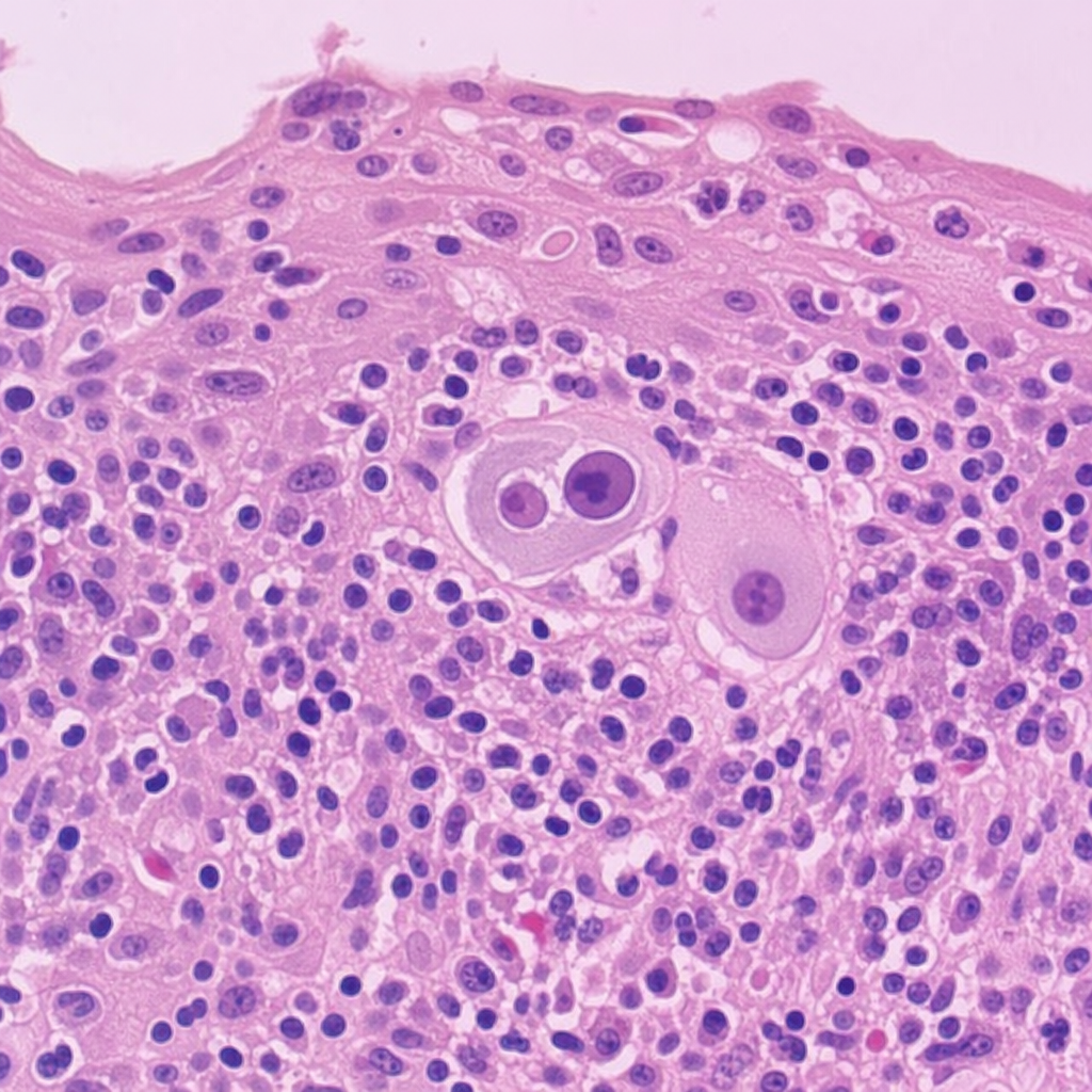
A 40-year-old immunocompromised patient presents with dysphagia. Upper GI endoscopy reveals multiple ulcers in the distal esophagus. Biopsy from the esophagus shows characteristic findings. What is the most likely diagnosis?
Which of the following is true regarding Crohn's disease?
Which of the following is NOT a characteristic of ulcerative colitis progressing to malignancy?
A biopsy of a small, rounded rectal polyp demonstrates glands and saw tooth crypts composed of a proliferation of goblet and columnar epithelial cells. No atypia is seen. This polyp is best classified as which of the following?
Which of the following statements concerning carcinoma of the esophagus is true?
Adenocarcinoma in the esophagus most commonly occurs in association with which of the following conditions?
Explanation: Barrett’s Esophagus (BE) is a complication of chronic Gastroesophageal Reflux Disease (GERD) characterized by the replacement of the normal stratified squamous epithelium of the distal esophagus with **metaplastic columnar epithelium** (containing goblet cells) [1]. **Why Option B is Correct:** Barrett’s esophagus is a well-established **premalignant condition**. The metaplastic columnar cells can undergo a sequence of genetic mutations leading to low-grade dysplasia, high-grade dysplasia, and eventually **Esophageal Adenocarcinoma** [2], [3]. Patients with BE have a 30 to 40-fold increased risk of developing adenocarcinoma compared to the general population [1]. **Why Other Options are Incorrect:** * **Option A:** BE is more common in **males** (M:F ratio of approx. 3:1) and typically affects Caucasian males between 40–60 years of age. * **Option C:** While conservative management (PPIs, lifestyle changes) treats the underlying GERD symptoms, it **does not reliably reverse** the metaplastic changes once they have occurred [2]. Definitive management for dysplasia involves endoscopic surveillance or ablation [1]. * **Option D:** The hallmark of BE is **Columnar metaplasia** (specifically intestinal metaplasia with goblet cells) [1]. Squamous epithelium is the *normal* lining; its replacement by columnar cells is the pathology. **High-Yield NEET-PG Pearls:** * **Endoscopic Appearance:** Characterized by "salmon-pink," velvety tongues of mucosa extending upwards from the GE junction. * **Microscopy:** Presence of **Goblet cells** is essential for the diagnosis of intestinal metaplasia [1]. * **Tumor Marker:** Overexpression of **p53** and **p16** is often associated with progression to malignancy. * **Screening:** Periodic endoscopic biopsy is mandatory to monitor for dysplasia [1], [2]. **References:** [1] Cross SS. Underwood's Pathology: A Clinical Approach. 6th ed. Common Clinical Problems From Alimentary System Disease, pp. 348-349. [2] Kumar V, Abbas AK, et al.. Robbins and Cotran Pathologic Basis of Disease. 9th ed. The Gastrointestinal Tract, pp. 764-765. [3] Kumar V, Abbas AK, et al.. Robbins and Cotran Pathologic Basis of Disease. 9th ed. The Gastrointestinal Tract, pp. 765-766.
Explanation: **Explanation:** **Radiation enteritis** is the correct answer because it is a chronic condition characterized by progressive obliterative endarteritis of the small vessels. This leads to chronic ischemia, transmural fibrosis, and collagen deposition within the intestinal wall. These fibrotic changes are often segmental and multifocal, resulting in the formation of **multiple, long, and tubular strictures**. **Analysis of Incorrect Options:** * **Duodenal Ulcer:** Typically presents as a solitary ulcer in the first part of the duodenum. While chronic healing can lead to scarring and gastric outlet obstruction (stenosis), it does not cause multiple intestinal strictures. * **Ulcerative Colitis:** This is a mucosal disease that primarily involves the rectum and colon continuously [1]. Because it lacks transmural involvement, strictures are rare. If a stricture is found in UC, it is highly suspicious for underlying adenocarcinoma. * **Gastric Erosion:** These are superficial mucosal defects that do not breach the muscularis mucosae. They heal without scarring and do not lead to stricture formation. **NEET-PG High-Yield Pearls:** * **Stricture Morphology:** Crohn’s disease (transmural inflammation) is the most common cause of "string sign" and multiple strictures in the ileum [1]. * **Tuberculous Enteritis:** Characterized by **transverse (circumferential) ulcers** which lead to short, "napkin-ring" strictures [3]. * **Typhoid Ulcers:** These are **longitudinal** (along the long axis of the bowel) and involve Peyer's patches; they typically do not cause strictures because they do not involve the entire circumference. * **Ischemic Enteritis:** Can also cause strictures, usually at "watershed" areas like the splenic flexure [2][4]. **References:** [1] Cross SS. Underwood's Pathology: A Clinical Approach. 6th ed. Common Clinical Problems From Alimentary System Disease, pp. 365-367. [2] Kumar V, Abbas AK, et al.. Robbins and Cotran Pathologic Basis of Disease. 9th ed. The Gastrointestinal Tract, pp. 786-787. [3] Cross SS. Underwood's Pathology: A Clinical Approach. 6th ed. Common Clinical Problems From Alimentary System Disease, pp. 363-364. [4] Cross SS. Underwood's Pathology: A Clinical Approach. 6th ed. Common Clinical Problems From Alimentary System Disease, pp. 369-370.
Explanation: **Explanation:** **Hirschsprung’s Disease (Congenital Megacolon)** is the correct answer because it is characterized by the **congenital absence of ganglion cells** in both the **Myenteric (Auerbach’s)** and **Submucosal (Meissner’s)** plexuses [1]. This occurs due to a failure in the craniocaudal migration of neural crest cells during embryonic development (weeks 5–12) [1]. The absence of these inhibitory neurons leads to a permanent state of contraction in the affected segment (usually the rectum and sigmoid), resulting in functional obstruction and proximal dilation of the healthy colon. **Analysis of Incorrect Options:** * **Crohn’s Disease & Ulcerative Colitis:** These are Inflammatory Bowel Diseases (IBD). While they involve transmural or mucosal inflammation respectively, the enteric nervous system remains intact. In fact, in some chronic cases of IBD, there may be reactive hypertrophy of nerve fibers, but not an absence of ganglia. * **Intussusception:** This is a mechanical condition where one segment of the intestine telescopes into another. It is typically caused by a "lead point" (like a polyp or Meckel’s diverticulum) and is not related to a primary neuronal deficit. **NEET-PG High-Yield Pearls:** * **Gold Standard Diagnosis:** Rectal suction biopsy (must include the submucosa to evaluate for Meissner’s plexus) [2]. * **Histopathology:** Absence of ganglion cells + **Hypertrophy of acetylcholinesterase-positive nerve fibers** [2]. * **Clinical Presentation:** Failure to pass meconium within the first 48 hours of birth and "blast sign" (explosive release of stool) on digital rectal exam. * **Genetics:** Strongly associated with mutations in the **RET proto-oncogene** and seen in 10% of children with **Down Syndrome** [1], [2]. **References:** [1] Cross SS. Underwood's Pathology: A Clinical Approach. 6th ed. (Basic Pathology) introduces the student to key general principles of pathology, both as a medical science and as a clinical activity with a vital role in patient care. Part 2 (Disease Mechanisms) provides fundamental knowledge about the cellular and molecular processes involved in diseases, providing the rationale for their treatment. Part 3 (Systematic Pathology) deals in detail with specific diseases, with emphasis on the clinically important aspects., pp. 94-95. [2] Kumar V, Abbas AK, et al.. Robbins and Cotran Pathologic Basis of Disease. 9th ed. The Gastrointestinal Tract, p. 759.
Explanation: **Explanation:** *Helicobacter pylori* is a gram-negative, spiral-shaped bacterium that specifically colonizes the stomach. Its primary niche is the **gastric mucosa**, where it resides within the overlying **mucus layer** and adheres to the apical surface of the surface mucous cells [1]. **Why Option B is Correct:** The gastric mucosa consists of the surface epithelium, the lamina propria, and the muscularis mucosae. *H. pylori* survives the acidic environment of the stomach by secreting **urease**, which creates a neutralizing ammonia cloud. It primarily dwells within the viscous mucus layer and the superficial pits of the mucosa [1]. While it attaches to the epithelium, it is anatomically categorized as a mucosal pathogen. **Why Other Options are Incorrect:** * **Option A (Epithelium):** While *H. pylori* adheres to epithelial cells via adhesins (like BabA), it does not typically invade the intracellular space of the epithelium. It is considered an extracellular organism. * **Option C (Muscularis mucosa):** This is the deep boundary of the mucosa. *H. pylori* remains superficial and does not penetrate this deep layer. * **Option D (Adventitia):** This is the outermost connective tissue layer of the stomach. *H. pylori* is a luminal/surface pathogen and never reaches the serosa or adventitia. **High-Yield Clinical Pearls for NEET-PG:** * **Site of Colonization:** Most commonly found in the **Antrum** [1]. * **Stains:** Silver stains (Warthin-Starry, Steiner) and Giemsa are the gold standards for visualization [1]. * **Virulence Factors:** **CagA** (associated with cancer) and **VacA** (cytotoxin). * **Associations:** Strongest risk factor for Peptic Ulcer Disease (PUD), Gastric Adenocarcinoma, and MALToma. * **Diagnosis:** Urea Breath Test (Non-invasive) is the test of choice for confirming eradication. **References:** [1] Kumar V, Abbas AK, et al.. Robbins and Cotran Pathologic Basis of Disease. 9th ed. The Gastrointestinal Tract, pp. 770-771.
Explanation: ### Explanation **1. Why Cytomegalovirus (CMV) Esophagitis is Correct:** In immunocompromised patients (e.g., HIV/AIDS, transplant recipients), CMV is a common cause of infectious esophagitis [1]. The classic endoscopic presentation of CMV esophagitis is **large, shallow, linear ulcers**, typically located in the distal esophagus. Histologically, CMV infects **endothelial and stromal cells** (not squamous cells), showing characteristic **large intranuclear "owl’s eye" inclusion bodies** and cytoplasmic inclusions. **2. Why the Other Options are Incorrect:** * **Candida esophagitis:** This is the most common cause of infectious esophagitis in immunocompromised patients [1]. However, it typically presents as **white, adherent, curd-like plaques** (pseudomembranes) rather than distinct ulcers [2], [3]. * **Herpes esophagitis (HSV-1):** While it also causes ulcers in immunocompromised hosts [1], HSV typically presents as **small, deep, "punched-out" ulcers** (volcano ulcers). Histologically, it affects squamous epithelial cells, showing Cowdry Type A inclusions, multinucleation, and nuclear molding (3Ms). * **Eosinophilic esophagitis:** This is an allergic/immune-mediated condition typically seen in atopic individuals [2]. Endoscopy reveals **stacked rings (trachealization)**, linear furrows, or white papules (microabscesses), not discrete ulcers. **3. High-Yield Clinical Pearls for NEET-PG:** * **Biopsy Site Matters:** For **CMV**, biopsy the **ulcer base** (where granulation tissue/endothelium is located). For **HSV**, biopsy the **ulcer edge** (where infected squamous cells are found). * **CMV Inclusion:** Large (cytomegaly) cell with a prominent basophilic intranuclear inclusion surrounded by a clear halo (**Owl’s eye appearance**). * **Drug of Choice:** Ganciclovir is the first-line treatment for CMV esophagitis, whereas Acyclovir is used for HSV. **References:** [1] Cross SS. Underwood's Pathology: A Clinical Approach. 6th ed. Common Clinical Problems From Alimentary System Disease, pp. 347-348. [2] Kumar V, Abbas AK, et al.. Robbins and Cotran Pathologic Basis of Disease. 9th ed. The Gastrointestinal Tract, pp. 761-762. [3] Kumar V, Abbas AK, et al.. Robbins and Cotran Pathologic Basis of Disease. 9th ed. Infectious Diseases, pp. 394-395.
Explanation: This question appears to be a **"Negative" or "Except" type question** common in NEET-PG, where the options B, C, and D are classic features of Crohn’s Disease, making **Scleroderma (Option A)** the outlier and the "correct" answer for being false. ### **Explanation** 1. **Why Scleroderma is the Correct Answer (False regarding Crohn's):** Scleroderma (Systemic Sclerosis) is a connective tissue disorder characterized by fibrosis. While it can affect the GI tract (causing esophageal dysmotility or small bowel bacterial overgrowth), it is **not** a pathological feature or a recognized complication of Crohn’s Disease. Crohn’s is an Inflammatory Bowel Disease (IBD), not a systemic fibrotic collagen vascular disease. 2. **Why Other Options are Wrong (True regarding Crohn's):** * **Transmural Involvement:** Unlike Ulcerative Colitis (mucosal/submucosal), Crohn’s involves **all layers** of the bowel wall [1], [2]. This leads to complications like fistulas, perforations, and strictures [2]. * **Cobblestone Appearance:** This is a classic macroscopic hallmark. It results from deep longitudinal and transverse ulcers intersecting with islands of edematous, normal-looking mucosa [2]. * **Skin Involvement:** Crohn’s has several extra-intestinal manifestations. The most common skin findings are **Erythema Nodosum** and **Pyoderma Gangrenosum**. Additionally, "Metastatic Crohn’s" can cause skin ulcers histologically similar to the bowel lesions [1]. ### **High-Yield Clinical Pearls for NEET-PG** * **Skip Lesions:** Discontinuous involvement of the GI tract (Mouth to Anus) [2]. * **Microscopy:** Non-caseating granulomas (pathognomonic, seen in 40-60% of cases) [1], [2]. * **String Sign of Kantor:** Radiologic finding due to terminal ileal strictures [2]. * **Creeping Fat:** Mesenteric fat wraps around the serosal surface of the bowel. * **Antibody:** ASCA (Anti-Saccharomyces cerevisiae antibodies) is positive in Crohn’s, whereas p-ANCA is associated with Ulcerative Colitis. **References:** [1] Kumar V, Abbas AK, et al.. Robbins and Cotran Pathologic Basis of Disease. 9th ed. The Gastrointestinal Tract, pp. 806-807. [2] Cross SS. Underwood's Pathology: A Clinical Approach. 6th ed. Common Clinical Problems From Alimentary System Disease, pp. 365-367.
Explanation: **Explanation:** In Ulcerative Colitis (UC), the risk of developing **Colorectal Carcinoma (CRC)** is a significant long-term complication. Understanding the distinction between inflammatory changes and neoplastic progression is crucial for NEET-PG. [1] **Why Option D is the Correct Answer (The "NOT" characteristic):** **Pseudopolyps** (inflammatory polyps) are a hallmark of the **active inflammatory phase** of UC. [1] They represent islands of regenerating mucosa surrounded by areas of ulceration and denudation. [1] Crucially, pseudopolyps are **non-neoplastic** and do not progress to malignancy. [2] In contrast, malignancy in UC typically arises from **flat, non-polypoid dysplastic lesions** (DALM - Dysplasia-Associated Lesion or Mass), making them harder to detect endoscopically than sporadic colon cancer. **Analysis of Incorrect Options:** * **Option A:** The risk of CRC is directly proportional to the **duration** of the disease (usually increasing significantly after 8–10 years) and the **extent** of involvement (pancolitis has a higher risk than left-sided colitis). * **Option B:** Once malignancy develops in the background of UC, the prognosis is generally **worse** than sporadic CRC, often because these tumors are more aggressive, multifocal, and higher grade. * **Option C:** This is a factual statement; the long-term prognosis of a UC patient is heavily dictated by the duration of the disease due to the cumulative risk of neoplastic transformation. **High-Yield Clinical Pearls for NEET-PG:** * **Surveillance:** Annual or biennial colonoscopy with biopsies is recommended after 8 years of disease. * **Molecular Pathway:** Unlike sporadic CRC (APC → KRAS → p53), UC-associated malignancy often shows **p53 mutations early** and APC mutations late. * **Backwash Ileitis:** Involvement of the terminal ileum in UC; it does not increase the risk of malignancy but indicates pancolitis. * **Protective Factor:** Smoking is paradoxically protective in UC (but a risk factor for Crohn’s). [1] **References:** [1] Kumar V, Abbas AK, et al.. Robbins and Cotran Pathologic Basis of Disease. 9th ed. The Gastrointestinal Tract, pp. 807-809. [2] Kumar V, Abbas AK, et al.. Robbins and Cotran Pathologic Basis of Disease. 9th ed. The Gastrointestinal Tract, p. 821.
Explanation: ### Explanation **Correct Answer: A. Hyperplastic polyp** The description provided is classic for a **Hyperplastic polyp**, the most common non-neoplastic polyp of the colon and rectum. [1] * **Underlying Concept:** These polyps result from decreased epithelial cell turnover and delayed shedding, leading to a "piling up" of cells. [1] * **Histopathology:** The hallmark feature is a **"sawtooth" or serrated appearance** of the surface epithelium and crypt lumens. [1] This is caused by the crowding of mature goblet and columnar cells. Crucially, these cells show **no cytologic atypia** (normal nuclei, no stratification), distinguishing them from neoplastic lesions. [1] They are typically small (<5mm) and found most frequently in the rectosigmoid region. **Why the other options are incorrect:** * **B. Peutz-Jeghers polyp:** These are **hamartomatous** polyps characterized by a "Christmas tree" branching pattern of smooth muscle (arborization) covered by normal-appearing mucosa. They are not defined by a simple sawtooth glandular pattern. * **C & D. Tubular/Tubulovillous adenoma:** These are **neoplastic (premalignant)** polyps. By definition, they must exhibit **dysplasia** (cytologic atypia), such as nuclear elongation, hyperchromasia, and loss of polarity. [2] The question explicitly states "no atypia is seen," ruling these out. **NEET-PG High-Yield Pearls:** * **Location:** Hyperplastic polyps are most common in the **left colon (rectosigmoid)**. * **Malignant Potential:** Traditional hyperplastic polyps have **no malignant potential**. However, they must be distinguished from *Serrated Adenomas*, which also have a sawtooth pattern but *do* harbor malignant potential via the "serrated pathway" (BRAF mutations/Microsatellite Instability). [1][2] * **Rule of Thumb:** Sawtooth + No Atypia = Hyperplastic Polyp; Sawtooth + Atypia/Dysplasia = Serrated Adenoma. [2] **References:** [1] Kumar V, Abbas AK, et al.. Robbins and Cotran Pathologic Basis of Disease. 9th ed. The Gastrointestinal Tract, pp. 811-813. [2] Kumar V, Abbas AK, et al.. Robbins and Cotran Pathologic Basis of Disease. 9th ed. The Gastrointestinal Tract, pp. 821-822.
Explanation: **Explanation:** The correct answer is **D: It occurs more commonly in patients with corrosive esophagitis.** **Why Option D is Correct:** Corrosive esophagitis, typically resulting from the accidental or intentional ingestion of strong acids or alkalis (lye), leads to severe mucosal injury and subsequent stricture formation [1]. Chronic inflammation and constant epithelial regeneration in these scarred areas significantly increase the risk of developing **Squamous Cell Carcinoma (SCC)**. The risk is estimated to be 1000–3000 times higher than the general population, often manifesting 20–40 years after the initial insult. **Analysis of Incorrect Options:** * **Option A:** While alcohol is a major risk factor for Squamous Cell Carcinoma, it is considered a **predisposing or risk factor**, not a "precipitating factor" (which implies an immediate trigger). However, in the context of NEET-PG, if multiple options are plausible, the strongest established pathological association (like corrosive injury) is preferred. * **Option B:** The most common histological type at the **cardioesophageal junction** (distal esophagus) is **Adenocarcinoma**, arising from Barrett’s esophagus [2]. Squamous carcinoma typically involves the upper and middle thirds [2]. * **Option C:** This statement is actually **true** (males are affected more than females). However, in many standard medical examinations, if a question asks for "the" true statement and includes a specific high-risk pathological association (Option D), it is prioritized over general demographic trends. *Note: In some versions of this classic question, Option C is phrased as "higher incidence in females," making it definitively false.* **High-Yield NEET-PG Pearls:** * **Most common type worldwide:** Squamous Cell Carcinoma. * **Most common type in the West/increasing incidence:** Adenocarcinoma. * **Plummer-Vinson Syndrome:** Triad of iron deficiency anemia, glossitis, and esophageal webs; strongly associated with SCC. * **Tylosis (Palmoplantar keratoderma):** An autosomal dominant condition with a near 100% lifetime risk of esophageal SCC. * **Barrett’s Esophagus:** Intestinal metaplasia (presence of Goblet cells) is the precursor for Adenocarcinoma [2]. **References:** [1] Cross SS. Underwood's Pathology: A Clinical Approach. 6th ed. Common Clinical Problems From Alimentary System Disease, pp. 347-348. [2] Kumar V, Abbas AK, et al.. Robbins and Cotran Pathologic Basis of Disease. 9th ed. The Gastrointestinal Tract, pp. 764-767.
Explanation: **Explanation:** **Correct Option: C. Barrett's esophagus** Adenocarcinoma of the esophagus is strongly associated with chronic gastroesophageal reflux disease (GERD). The underlying mechanism involves **Barrett’s esophagus**, a metaplastic process where the normal stratified squamous epithelium of the lower esophagus is replaced by **columnar epithelium with goblet cells** (intestinal metaplasia) due to acid injury [1]. This metaplastic tissue is unstable and can progress through a sequence of low-grade dysplasia to high-grade dysplasia [2], and eventually to **invasive adenocarcinoma** [1], [2]. It typically occurs in the **distal (lower) third** of the esophagus [3]. **Incorrect Options:** * **A & B (Midline/Upper esophagus):** These locations are the classic sites for **Squamous Cell Carcinoma (SCC)**, which is associated with smoking, alcohol, and caustic injury. Adenocarcinoma is almost exclusively a disease of the distal third [3]. * **D (Zenker's diverticulum):** This is a false diverticulum occurring at Killian’s triangle. While it can cause dysphagia and regurgitation, it is not a precursor to adenocarcinoma. **High-Yield Clinical Pearls for NEET-PG:** * **Most common esophageal cancer worldwide:** Squamous Cell Carcinoma. * **Most common esophageal cancer in Western countries (and rising in India):** Adenocarcinoma. * **Molecular Marker:** Overexpression of **p53** and **HER2/neu** is often seen in esophageal adenocarcinoma. * **Histology of Barrett’s:** Presence of **Goblet cells** is the diagnostic hallmark [1]. * **Risk Factors:** Obesity and GERD are the primary drivers for Adenocarcinoma, whereas smoking and alcohol are for SCC. **References:** [1] Cross SS. Underwood's Pathology: A Clinical Approach. 6th ed. Common Clinical Problems From Alimentary System Disease, pp. 348-349. [2] Kumar V, Abbas AK, et al.. Robbins and Cotran Pathologic Basis of Disease. 9th ed. The Gastrointestinal Tract, pp. 764-765. [3] Kumar V, Abbas AK, et al.. Robbins and Cotran Pathologic Basis of Disease. 9th ed. The Gastrointestinal Tract, pp. 765-766.
Oral Cavity and Esophageal Pathology
Practice Questions
Gastritis and Peptic Ulcer Disease
Practice Questions
Inflammatory Bowel Disease
Practice Questions
Malabsorption Syndromes
Practice Questions
Vascular Disorders of Intestine
Practice Questions
Diverticular Disease
Practice Questions
Intestinal Obstruction
Practice Questions
Gastrointestinal Infections
Practice Questions
Polyps and Neoplasms
Practice Questions
Appendiceal Pathology
Practice Questions
Get full access to all questions, explanations, and performance tracking.
Start For Free