All of the following are benign mesenchymal tumors of the stomach EXCEPT?
Which of the following gastric polyps does not undergo malignant transformation?
Tylosis is associated with which of the following conditions?
All of the following statements are true for telangiectasia of the colon EXCEPT:
Macrophages containing PAS-positive granules and rod-shaped bacilli are found in which condition affecting the small intestinal mucosa?
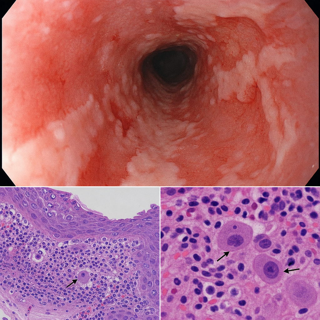
What is the most common stomach tumor that causes bleeding?
Early gastric cancer generally indicates?
Gastric carcinoma is associated with all EXCEPT:
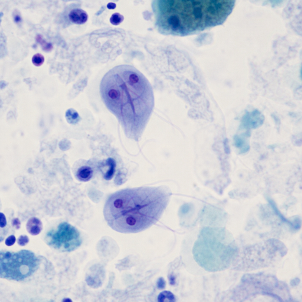
Which condition is a known cause of colonic carcinoma?
Which of the following is the most aggressive type of rectal carcinoma?
Explanation: **Explanation:** The core of this question lies in distinguishing between **tissue of origin** (epithelial vs. mesenchymal) [3]. **1. Why Adenoma is the correct answer:** An **Adenoma** is a benign tumor of **epithelial origin** (specifically from glandular epithelium) [3]. In the stomach, gastric adenomas are precursors to adenocarcinoma and are characterized by dysplastic epithelium [1]. Because the question asks for "mesenchymal" tumors, the Adenoma is the outlier. **2. Analysis of Incorrect Options (Mesenchymal Tumors):** Mesenchymal tumors arise from the connective tissues of the GI wall (smooth muscle, fat, nerves, or fibrous tissue) [3]. * **Leiomyoma (Option A):** A benign tumor arising from the **smooth muscle** (muscularis propria or muscularis mucosae) [1]. It is a classic mesenchymal tumor. * **Fibroma (Option C):** A benign tumor arising from **fibrous connective tissue** [3]. * **Lipoma (Option D):** A benign tumor arising from **adipose tissue**, typically found in the submucosal layer of the stomach [2]. **Clinical Pearls for NEET-PG:** * **Gastrointestinal Stromal Tumor (GIST):** This is the **most common** mesenchymal tumor of the abdomen (stomach is the most common site) [1]. It originates from the **Interstitial Cells of Cajal** and is characterized by the **c-KIT (CD117)** mutation. * **Rule of Thumb:** Any tumor ending in "-oma" preceded by a connective tissue prefix (Leiomyo-, Lipo-, Fibro-, Angio-, Neuro-) is mesenchymal. If it involves "Adeno-" or "Papillo-", it is epithelial [3]. * **Gastric Adenomas** are most commonly associated with **Familial Adenomatous Polyposis (FAP)** and carry a significant risk of malignant transformation if they are >2cm in size [1]. **References:** [1] Cross SS. Underwood's Pathology: A Clinical Approach. 6th ed. Common Clinical Problems From Alimentary System Disease, pp. 354-355. [2] Kumar V, Abbas AK, et al.. Robbins and Cotran Pathologic Basis of Disease. 9th ed. Bones, Joints, and Soft Tissue Tumors, p. 1222. [3] Cross SS. Underwood's Pathology: A Clinical Approach. 6th ed. (Basic Pathology) introduces the student to key general principles of pathology, both as a medical science and as a clinical activity with a vital role in patient care. Part 2 (Disease Mechanisms) provides fundamental knowledge about the cellular and molecular processes involved in diseases, providing the rationale for their treatment. Part 3 (Systematic Pathology) deals in detail with specific diseases, with emphasis on the clinically important aspects., pp. 208-209.
Explanation: ### Explanation **Correct Answer: D. Hyperplastic polyps** **1. Why Hyperplastic Polyps are the Correct Choice:** Hyperplastic polyps are the most common type of gastric polyp (representing ~75-85% of cases). They are **non-neoplastic** lesions that arise as an exaggerated regenerative response to chronic inflammation, such as *H. pylori* gastritis. Histologically, they consist of irregular, dilated, and elongated foveolar glands. Because they are inflammatory and reactive in nature rather than dysplastic, they have **no intrinsic malignant potential**. However, it is important to note that the surrounding inflamed mucosa may still be at risk for adenocarcinoma. **2. Analysis of Incorrect Options:** * **A & B (Tubular and Villous Adenomas):** These are true **neoplastic** polyps [1]. They contain dysplastic epithelium by definition [1]. The risk of malignancy is high (up to 30%), especially if the lesion is larger than 2 cm [1]. Villous adenomas generally carry a higher risk of harboring invasive carcinoma than tubular ones. * **C (Multiple Polyposis):** Syndromes like Familial Adenomatous Polyposis (FAP) involve the development of numerous adenomatous polyps. These carry a near 100% risk of malignant transformation if left untreated, as they follow the classic adenoma-carcinoma sequence. **3. NEET-PG High-Yield Pearls:** * **Fundic Gland Polyps:** These are common in patients taking **Proton Pump Inhibitors (PPIs)**. While usually sporadic and benign, they can be associated with FAP. * **Size Matters:** In gastric adenomas, a diameter **>2 cm** is the strongest predictor of focal malignancy [1]. * **Most Common Site:** Gastric adenomas are most frequently found in the **antrum** [1]. * **Management:** If a hyperplastic polyp is found, the clinician must biopsy the surrounding mucosa to check for *H. pylori* or atrophic gastritis. **References:** [1] Cross SS. Underwood's Pathology: A Clinical Approach. 6th ed. Common Clinical Problems From Alimentary System Disease, pp. 354-355.
Explanation: **Explanation:** **Tylosis** (also known as *Howel-Evans syndrome*) is a rare autosomal dominant genetic disorder characterized by focal hyperkeratosis of the palms and soles (palmoplantar keratoderma). **Why Oesophageal Carcinoma is Correct:** The association between Tylosis and **Squamous Cell Carcinoma (SCC) of the oesophagus** is one of the strongest genetic predispositions in gastrointestinal pathology. It is caused by a mutation in the **RHBDF2 gene** on chromosome 17q25. Individuals with this syndrome have a lifetime risk of developing oesophageal cancer approaching **90%** by age 70. This makes regular endoscopic surveillance mandatory for these patients. **Why Other Options are Incorrect:** * **B, C, and D:** While Uterine, Gastric, and Prostatic carcinomas have various genetic and environmental risk factors (e.g., *BRCA* mutations for prostate/uterine, *H. pylori* or *CDH1* for gastric), they have no established clinical or genetic link with palmoplantar keratoderma or the RHBDF2 mutation. **High-Yield Clinical Pearls for NEET-PG:** * **Type of Cancer:** Tylosis is specifically associated with **Squamous Cell Carcinoma**, not Adenocarcinoma. * **Triad:** Look for the triad of palmoplantar hyperkeratosis, oral leukoplakia, and oesophageal SCC in clinical vignettes. * **Other Oesophageal SCC Risk Factors:** Plummer-Vinson Syndrome (Iron deficiency anemia + glossitis + esophageal webs), Achalasia cardia, and corrosive injury (strictures). * **Genetic Locus:** Chromosome **17q25** (Tylosis Oesophageal Cancer locus).
Explanation: **Explanation:** **Telangiectasia of the colon**, also known as **Angiodysplasia**, is a common cause of lower gastrointestinal bleeding in the elderly. It consists of malformed, dilated submucosal and mucosal blood vessels [1]. **Why Option D is the Correct Answer (The False Statement):** Angiodysplasia is characteristically located in the **right colon**, specifically the **cecum and ascending colon** (approx. 75-80% of cases) [1]. It is rarely found in the rectum. Therefore, the statement that 50% involve the rectum is factually incorrect. **Analysis of Other Options:** * **Options A & B:** While angiodysplasia is most common in patients **over the age of 60** (due to degenerative changes in vessel walls over time), it can occasionally be seen in younger individuals (less than 40 years), often associated with systemic conditions or congenital predispositions [1]. * **Option C:** The **cecum** is indeed the most common site. This is attributed to Laplace’s Law; the cecum has the largest diameter of the colon, resulting in the highest wall tension, which leads to chronic intermittent venous obstruction and subsequent vessel dilation [1]. **Clinical Pearls for NEET-PG:** * **Association:** Frequently associated with **Aortic Stenosis** (Heyde’s Syndrome) and Chronic Kidney Disease. * **Presentation:** Usually presents as painless, chronic, or episodic hematochezia or iron deficiency anemia [1]. * **Diagnosis:** Gold standard is **Colonoscopy** (appears as bright red, stellate/fern-like vascular lesions). Angiography is used if colonoscopy is inconclusive. * **Pathogenesis:** Attributed to chronic, intermittent occlusion of submucosal veins as they pierce the muscularis propria [1]. **References:** [1] Kumar V, Abbas AK, et al.. Robbins and Cotran Pathologic Basis of Disease. 9th ed. The Gastrointestinal Tract, pp. 787-789.
Explanation: **Explanation:** **Whipple’s Disease (Correct Answer):** Whipple’s disease is a systemic infectious process caused by the gram-positive actinomycete **_Tropheryma whipplei_**. The hallmark histological finding is the infiltration of the small intestinal lamina propria by **foamy macrophages** [1]. These macrophages contain large cytoplasmic granules that are **PAS-positive** and diastase-resistant [1]. These granules represent partially digested bacterial cell walls. Electron microscopy reveals the characteristic **rod-shaped bacilli** (the causative organism) within these macrophages [1]. **Why the other options are incorrect:** * **Agammaglobulinemia:** This immunodeficiency typically shows a lack of plasma cells in the lamina propria and may be associated with *Giardia lamblia* infections, but not PAS-positive macrophage infiltration. * **Tropical Sprue:** Characterized by total or subtotal villous atrophy and increased intraepithelial lymphocytes, similar to Celiac disease, but lacks specific PAS-positive inclusions. * **Celiac Sprue:** Histology shows villous atrophy, crypt hyperplasia, and increased intraepithelial lymphocytes (IELs). It is an immune-mediated response to gluten, not a bacterial infiltration of macrophages. **High-Yield Clinical Pearls for NEET-PG:** * **Clinical Tetrad:** Malabsorption (diarrhea/weight loss), migratory large-joint arthritis, hyperpigmentation, and lymphadenopathy [1]. * **CNS Involvement:** Can present with "Oculomasticatory myorhythmia" (pathognomonic). * **Mnemonic:** **W**hipple’s = **W**eight loss, **W**ide (foamy) macrophages, **W**orking (PAS) stain, and **W**heel-shaped (rod) bacilli. * **Treatment:** Long-term antibiotics (usually Ceftriaxone followed by Trimethoprim-sulfamethoxazole). **References:** [1] Kumar V, Abbas AK, et al.. Robbins and Cotran Pathologic Basis of Disease. 9th ed. The Gastrointestinal Tract, pp. 798-799.
Explanation: **Explanation:** The correct answer is **Leiomyosarcoma**. **Why Leiomyosarcoma is correct:** While **Adenocarcinoma** is the most common gastric malignancy overall [1], **Leiomyosarcoma** (a mesenchymal tumor arising from the smooth muscle of the stomach wall) is classically associated with significant gastrointestinal bleeding. These tumors tend to grow intramurally and often undergo **central necrosis and mucosal ulceration**. This leads to the formation of a characteristic "umbilicated" or "crater-like" ulcer over the tumor mass, which frequently erodes into large intramural vessels, causing brisk hematemesis or melena [2]. **Analysis of Incorrect Options:** * **Adenocarcinoma (A):** This is the most common gastric cancer (90-95%) [1]. While it can cause chronic occult blood loss leading to iron deficiency anemia [2], it is less likely than a leiomyosarcoma to present primarily with massive, acute bleeding as its hallmark feature. * **Squamous Cell Carcinoma (B):** This is extremely rare in the stomach (usually occurring at the gastroesophageal junction or as an extension from the esophagus). It is not the most common cause of tumor-related bleeding. * **Fibrosarcoma (D):** This is an exceptionally rare mesenchymal tumor of the stomach and is not a standard clinical consideration for gastric bleeding. **High-Yield NEET-PG Pearls:** * **GIST vs. Leiomyosarcoma:** In modern pathology, most tumors previously labeled as leiomyosarcomas are now classified as **Gastrointestinal Stromal Tumors (GIST)** [3]. GISTs are the most common mesenchymal tumors of the GI tract, are **c-KIT (CD117) positive**, and also frequently present with GI bleeding due to mucosal ulceration. * **Most common site for GIST:** Stomach (60%) [3]. * **Morphology:** Look for "spindle cells" on histology for both GIST and leiomyosarcoma. * **Clinical Presentation:** If a question asks for the most common *malignancy*, think Adenocarcinoma; if it asks for the tumor most likely to *bleed/ulcerate*, think Leiomyosarcoma/GIST. **References:** [1] Kumar V, Abbas AK, et al.. Robbins and Cotran Pathologic Basis of Disease. 9th ed. The Gastrointestinal Tract, p. 779. [2] Kumar V, Abbas AK, et al.. Robbins and Cotran Pathologic Basis of Disease. 9th ed. The Gastrointestinal Tract, pp. 780-781. [3] Kumar V, Abbas AK, et al.. Robbins and Cotran Pathologic Basis of Disease. 9th ed. The Gastrointestinal Tract, pp. 776-777.
Explanation: **Explanation:** **1. Why the Correct Answer is Right:** Early Gastric Cancer (EGC) is defined by its **depth of invasion**, not by its size or the presence of lymph node metastasis [1]. By definition, it is a gastric adenocarcinoma that is confined to the **mucosa and/or submucosa**, regardless of whether lymph nodes are involved (N0 or N+) [1]. This distinction is critical because EGC has a significantly better prognosis (5-year survival rate >90%) compared to advanced gastric cancer, which penetrates into the muscularis propria [1]. **2. Why the Other Options are Wrong:** * **Option A:** "Detected early" is a vague clinical description. EGC is a specific histopathological diagnosis based on the layer of the stomach wall involved [1]. * **Option B:** While EGC includes tumors confined to the mucosa (T1a), it *also* includes those that have invaded the submucosa (T1b) [1]. Limiting the definition only to the mucosa is incomplete. * **Option D:** Size is not a criterion for EGC [1]. A lesion can be several centimeters wide, but as long as it does not penetrate deeper than the submucosa, it is still classified as Early Gastric Cancer. **3. NEET-PG High-Yield Pearls:** * **Classification:** EGC is often classified using the **Japanese Endoscopic Classification** (Type I: Protruded, Type II: Superficial, Type III: Excavated). * **Lymph Node Involvement:** Approximately 10-20% of EGC cases may have regional lymph node metastasis, but they are still classified as "Early" [1]. * **Most Common Site:** The lesser curvature of the antrum and pylorus [3]. * **Prognosis:** The 5-year survival rate for EGC is 90-95% [1], [2], whereas for advanced gastric cancer, it drops below 20%. **References:** [1] Cross SS. Underwood's Pathology: A Clinical Approach. 6th ed. Common Clinical Problems From Alimentary System Disease, pp. 355-356. [2] Cross SS. Underwood's Pathology: A Clinical Approach. 6th ed. Common Clinical Problems From Alimentary System Disease, pp. 349-350. [3] Kumar V, Abbas AK, et al.. Robbins and Cotran Pathologic Basis of Disease. 9th ed. The Gastrointestinal Tract, p. 779.
Explanation: **Explanation:** Gastric carcinoma is a multifactorial malignancy involving a complex interplay of genetic mutations and environmental triggers. **Why "Circulating autoimmune antibodies" is the correct answer:** While **Autoimmune Gastritis** (associated with anti-parietal cell and anti-intrinsic factor antibodies) is a known precursor to gastric adenocarcinoma due to chronic mucosal atrophy and intestinal metaplasia [1], the **antibodies themselves are not a feature of the carcinoma.** They are markers of the underlying autoimmune destruction of the gastric mucosa [1]. Once the carcinoma develops, it is driven by genetic instability, not by the persistence or action of these antibodies. **Analysis of Incorrect Options:** * **Inactivation of p53:** This is the most common genetic alteration in sporadic gastric cancer (found in >70% of cases). Loss of this tumor suppressor gene allows for unregulated cell cycle progression and inhibited apoptosis. * **Overexpression of c-erbB2 (HER2/neu):** This receptor tyrosine kinase is overexpressed in approximately 15-20% of gastric cancers, particularly the intestinal type [2]. It is a significant clinical marker as it dictates eligibility for **Trastuzumab** therapy. * **Overexpression of c-met:** The *MET* proto-oncogene encodes the Hepatocyte Growth Factor (HGF) receptor. Its amplification or overexpression is linked to advanced stages, poor prognosis, and increased metastatic potential in gastric cancer. **High-Yield Clinical Pearls for NEET-PG:** * **Lauren Classification:** Divides gastric cancer into **Intestinal** (associated with H. pylori, metaplasia, and *c-erbB2*) and **Diffuse** (associated with *CDH1* mutations/loss of E-cadherin and Signet ring cells) [2]. * **Virchow’s Node:** Left supraclavicular lymphadenopathy (Troisier sign) is a classic sign of metastatic gastric cancer. * **Sister Mary Joseph Nodule:** Periumbilical metastasis. * **Krukenberg Tumor:** Bilateral ovarian metastasis characterized by signet ring cells. **References:** [1] Kumar V, Abbas AK, et al.. Robbins and Cotran Pathologic Basis of Disease. 9th ed. The Gastrointestinal Tract, pp. 772-773. [2] Kumar V, Abbas AK, et al.. Robbins and Cotran Pathologic Basis of Disease. 9th ed. The Gastrointestinal Tract, p. 779.
Explanation: **Explanation:** **Ulcerative Colitis (UC)** is a well-established risk factor for the development of **Colorectal Carcinoma (CRC)**. The risk is primarily driven by chronic mucosal inflammation, which leads to a "dysplasia-carcinoma sequence." Unlike sporadic CRC (which follows the APC-adenoma-carcinoma pathway), colitis-associated cancer often arises from flat, non-adenomatous dysplastic lesions and frequently involves early **p53 mutations** and late **APC mutations**. The risk increases significantly with the duration of the disease (usually after 8–10 years), the anatomical extent (pancolitis carries higher risk than proctitis) [1], [2], and the presence of co-existing Primary Sclerosing Cholangitis (PSC). **Analysis of Incorrect Options:** * **Crohn’s Disease:** While Crohn’s does increase the risk of malignancy, the risk is statistically lower than in UC. In the context of NEET-PG, if both are options, UC is the "more correct" and classic association for colonic carcinoma. * **Erythema Multiforme:** This is an acute, self-limiting hypersensitivity reaction (often triggered by HSV or drugs) affecting the skin and mucous membranes; it has no association with colonic malignancy. * **Bullous Pemphigoid:** This is an autoimmune subepidermal blistering disease of the skin. While some bullous disorders are paraneoplastic, Bullous Pemphigoid is not a causative factor for colonic carcinoma. **High-Yield Clinical Pearls for NEET-PG:** * **Surveillance:** Patients with UC require screening colonoscopies with biopsies starting 8 years after diagnosis. * **Backwash Ileitis:** In UC, involvement of the terminal ileum increases the risk of CRC [2]. * **Protective Factors:** Regular use of 5-ASA (Mesalamine) and Folate supplementation are thought to reduce the risk of CRC in UC patients. **References:** [1] Kumar V, Abbas AK, et al.. Robbins and Cotran Pathologic Basis of Disease. 9th ed. The Gastrointestinal Tract, pp. 807-809. [2] Cross SS. Underwood's Pathology: A Clinical Approach. 6th ed. Common Clinical Problems From Alimentary System Disease, pp. 367-368.
Explanation: **Explanation:** The aggressiveness of colorectal carcinomas is often determined by their histological subtype and the production of extracellular mucin. **Why Secondary Mucoid Carcinoma is Correct:** Mucoid (mucinous) carcinoma is defined by the presence of extracellular mucin pools comprising more than 50% of the tumor volume. **Secondary mucoid carcinoma** refers to a conventional adenocarcinoma that has undergone mucinous transformation. These tumors are highly aggressive because the abundant extracellular mucin acts as a "hydrostatic pressure" mechanism, facilitating the dissection of tissue planes and promoting deep mural invasion. This leads to a higher stage at presentation, increased rates of lymph node metastasis, and a poorer prognosis compared to standard adenocarcinoma. **Analysis of Incorrect Options:** * **A. Adenocarcinoma:** This is the most common type of rectal cancer [2]. While malignant, the "not otherwise specified" (NOS) type generally has a better prognosis than its mucinous or signet-ring counterparts. * **C. Signet ring carcinoma:** While signet ring cell carcinoma (intracellular mucin) is extremely aggressive and often carries a worse prognosis than mucinous carcinoma, in the specific context of rectal cancer classifications used in standard pathology textbooks (like Boyd’s), secondary mucoid transformation is highlighted for its rapid spread and poor response to therapy. * **D. Squamous cell carcinoma:** Rare in the rectum (more common in the anal canal). While aggressive, it does not match the invasive potential of mucoid variants in the rectal ampulla. **High-Yield Pearls for NEET-PG:** * **Mucin Types:** *Intracellular* mucin characterizes Signet ring cells; *Extracellular* mucin characterizes Mucoid carcinoma. * **Genetic Association:** Mucinous carcinomas are frequently associated with **Microsatellite Instability (MSI)** and the BRAF mutation [1]. * **Staging:** The most important prognostic factor for rectal carcinoma remains the **TNM stage** (depth of invasion and nodal status), rather than histology alone [1]. **References:** [1] Kumar V, Abbas AK, et al.. Robbins and Cotran Pathologic Basis of Disease. 9th ed. The Gastrointestinal Tract, pp. 815-822. [2] Cross SS. Underwood's Pathology: A Clinical Approach. 6th ed. Common Clinical Problems From Alimentary System Disease, pp. 373-374.
Oral Cavity and Esophageal Pathology
Practice Questions
Gastritis and Peptic Ulcer Disease
Practice Questions
Inflammatory Bowel Disease
Practice Questions
Malabsorption Syndromes
Practice Questions
Vascular Disorders of Intestine
Practice Questions
Diverticular Disease
Practice Questions
Intestinal Obstruction
Practice Questions
Gastrointestinal Infections
Practice Questions
Polyps and Neoplasms
Practice Questions
Appendiceal Pathology
Practice Questions
Get full access to all questions, explanations, and performance tracking.
Start For Free