All of the following are TRUE regarding myoglobinuria, EXCEPT:
A 25-year old patient who had a Road traffic accident was initially conscious but later became unconscious and subsequently died. On postmortem examination, multiple petechial hemorrhages are seen in the corpus callosum, what is the probable diagnosis?
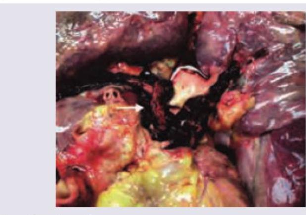
A 22-year-old biker had a road traffic accident with bilateral tibia fracture. After 7 days, his condition suddenly worsens and leads to death. Pathological specimen is provided. What could be the likely cause of death?

What is the Virchow method of organ removal in autopsy?
Explanation: **Explanation:** The key to answering this question lies in understanding the physiological handling of myoglobin versus hemoglobin. **1. Why Option D is the correct (False) statement:** Myoglobin is a small monomeric protein (17,000 Daltons) with a very low renal threshold. Once released from damaged muscle, it is rapidly cleared from the circulation by the kidneys. Consequently, even in severe rhabdomyolysis, the **serum remains clear (normal color)** because myoglobin does not accumulate in the plasma. In contrast, **hemoglobinuria** is associated with pink or red serum (hemoglobinemia) because hemoglobin binds to haptoglobin, creating a large complex that lingers in the circulation [1]. **2. Analysis of Incorrect Options (True Statements):** * **Option A:** Myoglobin is the primary oxygen-binding pigment found in all striated muscles, including both skeletal and cardiac muscle fibers. * **Option B:** For myoglobin to be visible to the naked eye (causing red-brown or "cola-colored" urine), approximately **200 grams** of muscle destruction is required. * **Option C:** The Benzidine test (and the orthotoluidine dipstick) detects the peroxidase activity of the heme group. Since myoglobin contains heme, it will yield a **positive** result, similar to hemoglobin. **Clinical Pearls for NEET-PG:** * **Differential Diagnosis:** To distinguish myoglobinuria from hemoglobinuria, observe the serum. **Clear serum + Red urine = Myoglobinuria**; **Red serum + Red urine = Hemoglobinuria.** * **Renal Impact:** Myoglobin is nephrotoxic; in acidic urine, it precipitates with Tamm-Horsfall protein to form "pigment casts," leading to Acute Tubular Necrosis (ATN) [1]. * **Biochemical Marker:** Serum Creatine Kinase (CK) levels are significantly elevated in cases of myoglobinuria. **References:** [1] Kumar V, Abbas AK, et al.. Robbins and Cotran Pathologic Basis of Disease. 9th ed. Red Blood Cell and Bleeding Disorders, pp. 639-640.
Explanation: ***Diffuse axonal injury*** - The characteristic finding of **multiple petechial hemorrhages** (microhemorrhages) specifically in deep structures like the **corpus callosum** and brainstem is the pathological hallmark of **Diffuse Axonal Injury** (DAI) [1]. - DAI results from severe acceleration/deceleration (shearing) forces during trauma, leading to widespread axonal disruption, which explains the progression from initial consciousness to coma [1]. *EDH* - Epidural Hematoma (EDH) involves an arterial bleed, typically from the **middle meningeal artery**, causing a hematoma external to the dura mater [1]. - Although EDH often presents with a clinical **lucid interval**, the post-mortem findings are limited to the hematoma collection external to the brain substance, not deep parenchymal petechiae [1]. *SDH* - Subdural hematoma (SDH) is caused by the tearing of **bridging veins** and collects between the dura and arachnoid mater. - While SDH can cause delayed deterioration due to haematoma expansion, the specific microscopic deep white matter hemorrhages described are the defining feature of **DAI**, not SDH. *Contusion* - Cerebral contusions present as focal areas of hemorrhagic necrosis, typically at the site of impact (coup) or opposite side (contrecoup), commonly affecting the frontal and temporal poles [1]. - Unlike DAI, contusions are macroscopic hemorrhagic lesions visible on gross examination, not the characteristic microscopic petechial hemorrhages in deep white matter structures like the corpus callosum [1]. **References:** [1] Kumar V, Abbas AK, et al.. Robbins and Cotran Pathologic Basis of Disease. 9th ed. The Central Nervous System, pp. 1262-1264.
Explanation: ***Fat embolism*** - **Fat embolism syndrome (FES)** is a common complication of **long bone fractures**, especially of the tibia and femur, which release fat globules into the circulation [1]. - The sudden worsening and death 7 days post-injury, following a bilateral tibia fracture, is highly suggestive of **fat embolism**, as symptoms typically manifest within 1-3 days but can be delayed [1]. *Septic shock* - Septic shock would typically present with signs of **infection** (fever, elevated WBC count) and **hemodynamic instability** (hypotension, organ dysfunction) which are not mentioned. - While possible in trauma, 7 days is a relatively short period for overwhelming sepsis to develop and cause sudden death without prior signs, especially in a young individual. *Pulmonary thromboembolism* - **Pulmonary thromboembolism (PTE)** usually arises from **deep vein thrombosis (DVT)**, which is a risk after trauma and immobilization. - While possible, the specific context of **bilateral tibia fracture** makes **fat embolism** a more classic and immediate consideration for sudden death in the acute post-trauma period. *Air embolism* - **Air embolism** typically occurs due to **iatrogenic causes** (e.g., central line insertion, surgery) or severe **chest trauma** leading to communication between airways and vessels. - It causes immediate and dramatic symptoms, usually leading to rapid death, which doesn't fit the 7-day delayed presentation described. **References:** [1] Cross SS. Underwood's Pathology: A Clinical Approach. 6th ed. (Basic Pathology) introduces the student to key general principles of pathology, both as a medical science and as a clinical activity with a vital role in patient care. Part 2 (Disease Mechanisms) provides fundamental knowledge about the cellular and molecular processes involved in diseases, providing the rationale for their treatment. Part 3 (Systematic Pathology) deals in detail with specific diseases, with emphasis on the clinically important aspects., pp. 146-147.
Explanation: ***Organs removed one by one in autopsy*** - The **Virchow method** (also called the **in situ technique**) involves removing and dissecting each organ **individually** from the body. - Each organ is examined separately in isolation, allowing detailed assessment of individual organ pathology without disturbing anatomical relationships during removal. - This method is particularly useful in **infectious diseases**, **poisoning cases**, and when specific organ systems require focused examination. - Named after Rudolf Virchow, the father of cellular pathology. *Organs removed all at once* - This describes the **Rokitansky method** (en masse technique), where organs are removed together maintaining their anatomical connections. - Useful for demonstrating inter-organ relationships and vascular connections, but distinct from the Virchow approach. *Examination without removal of organs* - This represents **in situ examination** (sometimes called the **Ghon technique**), where organs are examined within the body cavity. - While useful for preliminary assessment, it does not allow comprehensive dissection and histological sampling required in most autopsies. *Organs removed en masse with neck and tongue structures* - This describes the **Letulle method** (modified en masse technique), where thoracic, abdominal, and pelvic organs are removed together as a single block along with the tongue and neck structures. - Different from Virchow's individual organ approach and useful for maintaining cervical-thoracic continuity.
Death Investigation
Practice Questions
Postmortem Changes
Practice Questions
Forensic Traumatology
Practice Questions
Asphyxial Deaths
Practice Questions
Thermal Injuries
Practice Questions
Gunshot and Explosive Injuries
Practice Questions
Forensic Toxicology
Practice Questions
Identification of Unknown Remains
Practice Questions
Sexual Assault Investigation
Practice Questions
Medicolegal Autopsy
Practice Questions
Get full access to all questions, explanations, and performance tracking.
Start For Free