All of the following are small round blue cell tumours except
A 42-year-old man complains of head¬ache when he wakes up in the morning. It lasts into the afternoon and then subsides. Headache has been present for a few months. CT scan shows a frontal lobe mass with focal areas of calcification. You strongly suspect the pa¬tient has
Which of the following is the ovarian counterpart of testicular seminoma?
Craniopharyngioma arises from -
Hormonal secretions are tightly controlled by the time of day due to an inbuilt biological clock in human body. This rhythmic secretion is controlled by:
A 30-year-old woman presents with a history of amenorrhea and impaired vision of six months' duration. Physical examination shows normal findings except for pale optic discs and diminished visual acuity. The most likely diagnosis is -
Most radiosensitive tumor among the following is
Which of the following testicular tumors is not a germ cell neoplasm?
Melatonin is secreted from which cells of pineal gland?
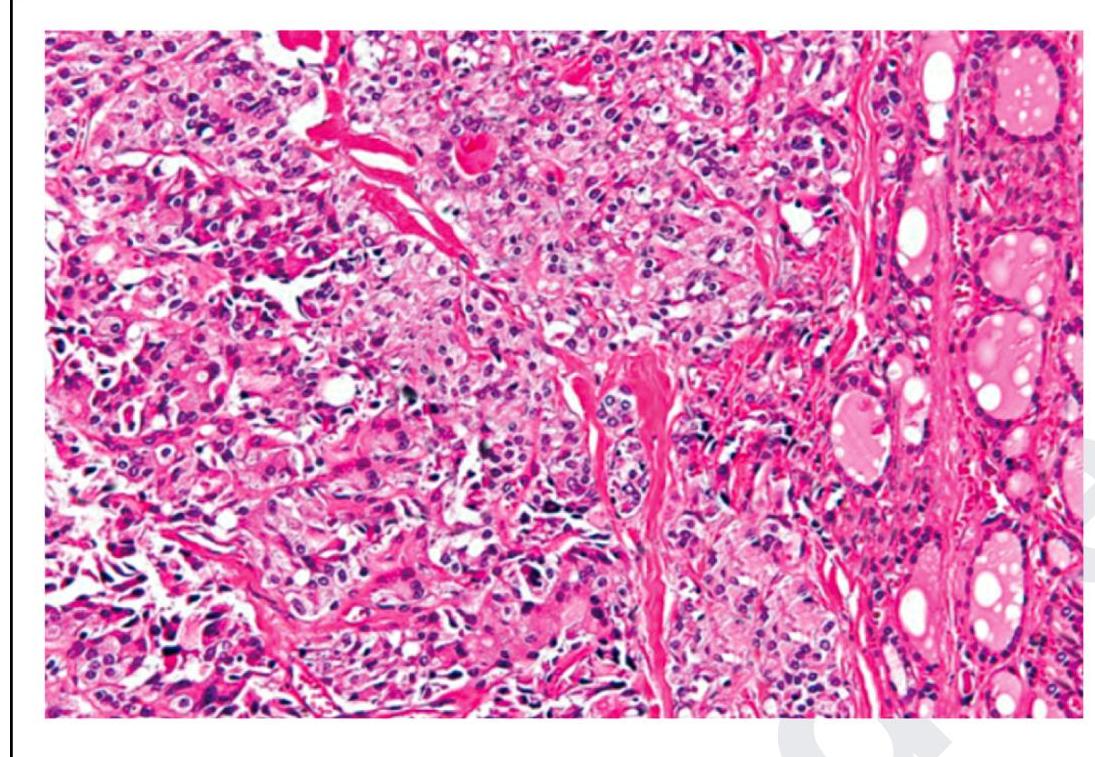
The following is a histopathological image of thyroid pathology. What is the diagnosis?

Explanation: ***Wilms tumour*** - **Wilms tumour** (nephroblastoma) is a **triphasic tumor** composed of blastemal, stromal, and epithelial elements [1], leading to a more varied histological appearance than mere small round blue cells. - While it can contain small, undifferentiated cells, its characteristic histology with **immature tubules** and mesenchymal elements differentiates it from classic small round blue cell tumors [1]. *Ewing's sarcoma* - **Ewing's sarcoma** is a classic example of a **small round blue cell tumor**, characterized by uniform, primitive cells with scant cytoplasm [2]. - It is classically associated with specific **chromosomal translocations**, particularly t(11;22). *Rhabdomyosarcoma* - **Rhabdomyosarcoma** is the most common soft tissue sarcoma in children and is also considered a **small round blue cell tumor** due to its primitive, undifferentiated cells [3]. - These tumors show evidence of **skeletal muscle differentiation**, which can be identified through immunohistochemistry (e.g., desmin, myogenin) [3]. *Retinoblastoma* - **Retinoblastoma** is a malignant tumor of the retina composed of primitive **neuroectodermal cells** that appear small, round, and blue [1]. - It classically forms characteristic **rosettes** (Flexner-Wintersteiner rosettes), which are a histological hallmark within its small round blue cell morphology [1]. **References:** [1] Cross SS. Underwood's Pathology: A Clinical Approach. 6th ed. (Basic Pathology) introduces the student to key general principles of pathology, both as a medical science and as a clinical activity with a vital role in patient care. Part 2 (Disease Mechanisms) provides fundamental knowledge about the cellular and molecular processes involved in diseases, providing the rationale for their treatment. Part 3 (Systematic Pathology) deals in detail with specific diseases, with emphasis on the clinically important aspects., pp. 211-212. [2] Cross SS. Underwood's Pathology: A Clinical Approach. 6th ed. Common Clinical Problems From Osteoarticular And Connective Tissue Disease, pp. 671-672. [3] Kumar V, Abbas AK, et al.. Robbins and Cotran Pathologic Basis of Disease. 9th ed. Bones, Joints, and Soft Tissue Tumors, pp. 1224-1225.
Explanation: ***An oligodendroglioma*** - This type of glioma is known for its **slow growth** and often presents with **calcifications** on imaging, particularly in the **frontal lobes**. [1] - **Morning headaches** that improve throughout the day are characteristic of increased intracranial pressure, which can be caused by a slow-growing mass like an oligodendroglioma. *An astrocytoma* - While astrocytomas are common brain tumors, **calcification** is less typical, especially in the more aggressive grades. - The presented symptoms do not exclusively point to an astrocytoma over other glial tumors. *A meningioma* - Meningiomas are typically **extra-axial tumors** that arise from the meninges and are often seen with **dural tails** and are more common in women. - Although meningiomas can calcify, the description of a "frontal lobe mass" favors an intra-axial lesion in this context. *A pinealoma* - Pinealomas originate in the **pineal gland**, located deep within the brain, and would typically present with symptoms related to compression of the tectum (e.g., **Parinaud syndrome**). - A frontal lobe mass with calcifications is not consistent with the typical location or presentation of a pinealoma.
Explanation: ***Dysgerminoma*** - **Dysgerminoma** is the most common malignant germ cell tumor of the ovary and is histologically identical to testicular **seminoma** [1]. - Both tumors arise from **primordial germ cells** and share similar morphology, including large, uniform cells with clear cytoplasm and prominent nucleoli, often arranged in nests and separated by fibrous septa with lymphocytic infiltration [1], [3]. *Dermoid* - **Dermoid cysts**, also known as mature cystic teratomas, are germ cell tumors composed of well-differentiated tissues from all three germ layers (ectoderm, mesoderm, endoderm) [2]. - They are typically benign and do not have a direct testicular counterpart that is histologically identical to seminoma. *Brenner tumor* - **Brenner tumors** are uncommon epithelial ovarian tumors characterized by nests of transitional epithelial cells resembling urothelium, separated by a fibrous stroma. - They are not germ cell tumors and do not have a testicular counterpart to seminoma. *Endodermal sinus tumor* - The **endodermal sinus tumor** (yolk sac tumor) is another type of malignant germ cell tumor of the ovary, but it is characterized by structures resembling the primitive yolk sac and the presence of **Schiller-Duval bodies**. - While it has a testicular counterpart, it is not histologically identical to seminoma; its testicular counterpart is also called a yolk sac tumor and is distinct from seminoma [3]. **References:** [1] Kumar V, Abbas AK, et al.. Robbins and Cotran Pathologic Basis of Disease. 9th ed. The Female Genital Tract, pp. 1034-1035. [2] Kumar V, Abbas AK, et al.. Robbins and Cotran Pathologic Basis of Disease. 9th ed. The Female Genital Tract, pp. 1035-1036. [3] Kumar V, Abbas AK, et al.. Robbins and Cotran Pathologic Basis of Disease. 9th ed. The Lower Urinary Tract and Male Genital System, pp. 979-980.
Explanation: Rathke's pouch - Craniopharyngiomas arise from remnants of Rathke's pouch, an embryonic structure that forms the anterior pituitary gland. - Rathke's pouch is an ectodermal outpouching from the roof of the primitive oral cavity (stomodeum) that migrates upward during embryonic development. - These squamous epithelial remnants can persist along the path of migration and give rise to craniopharyngiomas, typically in the suprasellar region. - The tumor's location near the pituitary stalk is due to the developmental pathway, but the embryological origin is specifically Rathke's pouch. Cerebellum - Tumors arising from the cerebellum are typically medulloblastomas or cerebellar astrocytomas, which are distinctly different from craniopharyngiomas [2]. - The cerebellum is a hindbrain derivative responsible for coordination and balance. - Cerebellar tumors present with ataxia and motor deficits, not the endocrine dysfunction characteristic of craniopharyngiomas. Pineal gland - Tumors of the pineal gland, such as pineoblastomas [1] or germinomas, originate from pinealocytes or germ cells, respectively. - These tumors are associated with Parinaud's syndrome (upgaze palsy) due to compression of the tectal plate. - Pineal region tumors are anatomically and embryologically distinct from craniopharyngiomas. Posterior pituitary - The posterior pituitary (neurohypophysis) develops from a downward extension of the hypothalamus, not from Rathke's pouch. - It is composed of axons from hypothalamic neurons and glial cells (pituicytes). - Tumors specifically from the posterior pituitary are extremely rare and differ histologically from craniopharyngiomas, which arise from epithelial remnants of Rathke's pouch.
Explanation: ***Suprachiasmatic nucleus*** - The **suprachiasmatic nucleus (SCN)**, located in the hypothalamus, is the primary pacemaker of the body's **circadian rhythms**, controlling the timing of hormonal secretions, sleep-wake cycles, and other daily oscillations. - It receives direct input from the **retina** about light-dark cycles, allowing it to synchronize the body's internal clock with the external environment. *Ventrolateral nucleus* - The **ventrolateral preoptic nucleus (VLPO)** is involved in **sleep regulation** and promoting non-REM sleep, but it does not act as the primary circadian pacemaker. - It receives input from the SCN and collaborates in regulating sleep, but its role is primarily inhibitory to wakefulness. *Supraoptic nucleus* - The **supraoptic nucleus** is primarily involved in the production and secretion of **vasopressin (ADH)** and **oxytocin**, which are neurohormones regulating fluid balance and social bonding, respectively. - It does not directly control the rhythmic aspect of general hormonal secretions or act as the central circadian clock. *Posterolateral nucleus* - This term is less commonly used in the context of circadian rhythm control; however, if referring to a thalamic nucleus, the **posterolateral nucleus** is generally associated with sensory processing, particularly somatosensory information. - It has no known role as a central pacemaker for hormonal secretions or circadian rhythms.
Explanation: ***Pituitary adenoma*** - Pituitary adenomas, particularly **macroadenomas**, can cause **bitemporal hemianopsia** due to compression of the **optic chiasm**, leading to impaired vision and pale optic discs [1]. - They also frequently secrete hormones, with **prolactinomas** being a common type, leading to **amenorrhea** in women due to inhibition of GnRH. *Benign intracranial hypertension* - This condition is primarily characterized by **increased intracranial pressure** leading to headache, pulsatile tinnitus, and **papilledema**, which presents as a swollen optic disc, not pale. - While it can cause visual field defects, **amenorrhea** is not a typical associated symptom. *Hypothalamic glioma* - Hypothalamic gliomas can cause visual field defects and endocrine dysfunction due to their location near the **optic chiasm** and **hypothalamus**. - However, they are more common in children and usually present with symptoms like **diabetes insipidus**, growth abnormalities, or precocious puberty, rather than isolated amenorrhea and optic disc pallor. *Craniopharyngioma* - Craniopharyngiomas are **suprasellar tumors** that can compress the optic chiasm, causing visual disturbances, and impact the pituitary stalk and hypothalamus, leading to endocrine dysfunction. - They often present with symptoms of **hydrocephalus**, **growth retardation**, or **diabetes insipidus**, and are more frequently diagnosed in childhood or adolescence, although they can occur in adults.
Explanation: ***Dysgerminoma*** - **Dysgerminomas** are highly **radiosensitive** tumors, meaning they respond very well to radiation therapy. - This characteristic is often exploited in their treatment, especially for widespread disease or as adjuvant therapy. *Osteogenic sarcoma* - **Osteogenic sarcomas** (osteosarcomas) are generally **radioresistant**, requiring high doses of radiation for local control, often with limited success. - Treatment primarily relies on **surgery** and **chemotherapy**. *Parotid carcinoma* - **Parotid carcinomas** exhibit variable radiosensitivity depending on their histology, but generally are not considered among the most radiosensitive tumors. - Postoperative **radiotherapy** is often used for high-risk features rather than as primary monotherapy. *Bronchogenic carcinoma* - **Bronchogenic carcinomas** (lung cancers) show variable radiosensitivity. **Small cell lung carcinoma** is more radiosensitive than **non-small cell lung carcinoma**, but neither is considered as radiosensitive as dysgerminoma. - Treatment often involves **multimodality therapy** including chemotherapy, surgery, and radiation, with radiation efficacy depending on tumor type and stage.
Explanation: ***Sertoli cell tumor*** - Sertoli cell tumors are classified as **sex-cord stromal tumors** [1][2], not germ cell neoplasms, and arise from **Sertoli cells** in the testis. - They are characterized by **hormonally active** properties and may lead to conditions like **gynecomastia** due to estrogen production. *Seminoma* - Seminomas are a type of **germ cell tumor** [2][3], derived from the germ cells in the testes, and typically present with **elevated AFP** and **hCG** levels. - Known for their **slow growth** and better prognosis compared to non-seminomatous germ cell tumors. *Yolk sac tumor* - Also a germ cell neoplasm [3], yolk sac tumors typically produce **alpha-fetoprotein (AFP)**, indicating their germinal origin. - Commonly occur in **younger males** and present as a **rapidly growing** tumor with a poor prognosis if not treated early. *Teratoma* - Teratomas are categorized as germ cell tumors [3] that can contain differentiated tissues and arise from **primitive germ cells**. - They are generally classified as either **mature** or **immature**, with the immature type being more aggressive and occurring primarily in **younger patients**. **References:** [1] Cross SS. Underwood's Pathology: A Clinical Approach. 6th ed. Common Clinical Problems From Diseases Of The Urinary And Male Genital Tracts, pp. 513-514. [2] Cross SS. Underwood's Pathology: A Clinical Approach. 6th ed. Common Clinical Problems From Diseases Of The Urinary And Male Genital Tracts, pp. 510-512. [3] Kumar V, Abbas AK, et al.. Robbins and Cotran Pathologic Basis of Disease. 9th ed. The Lower Urinary Tract and Male Genital System, pp. 979-980.
Explanation: ***Pinealocytes*** - **Pinealocytes** are the primary secretory cells within the **pineal gland** responsible for synthesizing and secreting **melatonin**. - These cells exhibit rhythmic activity, secreting more melatonin during periods of **darkness** to regulate the **sleep-wake cycle**. *Glial cells* - **Glial cells** (specifically astroglial cells and microglial cells) are present in the pineal gland but primarily provide **structural support** and **immune surveillance**, not melatonin synthesis. - Their main function is to maintain the **neural environment** and respond to injury or infection. *Zymogen cells* - **Zymogen cells** are typically found in glands like the pancreas or stomach, where they secrete **inactive enzyme precursors**. - They are not associated with the pineal gland or **melatonin production**. *Oncocytes* - **Oncocytes** are cells characterized by abundant **mitochondria** and are typically found in endocrine glands like the thyroid or parathyroid, often in **neoplastic conditions**. - They are not the **normal secretory cells** of the pineal gland.
Explanation: ***Medullary carcinoma of thyroid*** - This image shows sheets and nests of **polygonal to spindle-shaped cells**, which are characteristic of medullary thyroid carcinoma, especially when mixed with an **amyloid stroma** (seen as amorphous eosinophilic material) [2]. - The presence of **neuroendocrine features** and the production of **calcitonin** are hallmarks of these C-cell tumors [1], [2]. *Papillary carcinoma of thyroid* - Characterized by **papillary architecture**, **ground-glass (Orphan Annie eye) nuclei**, nuclear grooves, and intranuclear cytoplasmic inclusions. - These features are not prominently seen in the provided image. *Follicular carcinoma of thyroid* - Defined by an invasive growth pattern of **well-differentiated follicular cells** forming follicles, with either capsular or vascular invasion [2]. - The image does not show classic follicular architectural patterns or clear evidence of invasion in the absence of a capsule. *Anaplastic carcinoma of thyroid* - This is a highly aggressive and undifferentiated tumor with **marked pleomorphism**, bizarre giant cells, and high mitotic activity [2]. - While there is some pleomorphism, the overall pattern and cellular morphology in the image are more consistent with medullary carcinoma than the extreme anaplasia. **References:** [1] Kumar V, Abbas AK, et al.. Robbins and Cotran Pathologic Basis of Disease. 9th ed. The Endocrine System, pp. 1102-1103. [2] Cross SS. Underwood's Pathology: A Clinical Approach. 6th ed. Common Clinical Problems From Liver And Biliary System Disease, pp. 428-431.
Get full access to all questions, explanations, and performance tracking.
Start For Free