The function of which of the following is increased by an elevated parathyroid hormone concentration:
Hypophosphatemia is seen in:
The commonest cause of primary hyperparathyroidism is what?
All are true about primary hyperparathyroidism except which of the following?
Hypocalcemia in a child may be associated with
Primary Hyperparathyroidism is associated with -
Primary hyperparathyroidism is suggested by all of the following, except which of the following?
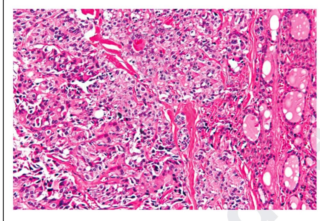
The following is a histopathological image of thyroid pathology. What is the diagnosis?

Chvostek sign could be seen after -
Most sensitive investigation for preoperative localization of abnormal parathyroid glands is
Explanation: ***Osteoclasts*** - **Parathyroid hormone (PTH)** primarily acts to increase serum calcium levels by stimulating **osteoclasts**, leading to bone resorption and release of calcium and phosphate into the bloodstream. - While PTH does not directly act on osteoclasts, it binds to receptors on osteoblasts, which then release factors that activate osteoclasts. *Action of osteoblasts only* - PTH indirectly affects **osteoblasts** by binding to their receptors, but this action primarily leads to **RANKL expression**, which then stimulates osteoclast activity, not a direct increase in osteoblastic bone formation. - Chronic elevation of PTH, as seen in primary hyperparathyroidism, can paradoxically lead to a net loss of bone mass due to increased osteoclastic activity. *Phosphate reabsorptive pathways in the renal tubules* - PTH actually **decreases reabsorption of phosphate** in the renal tubules, leading to phosphaturia. This helps to prevent calcium-phosphate precipitation by lowering serum phosphate levels while raising calcium. - This is a key mechanism by which PTH increases serum calcium—by both mobilizing it from bone and reducing its renal excretion, while simultaneously promoting renal phosphate excretion. *Hepatic formation of 25-hydroxycholecalciferol* - The **liver** is responsible for the hydroxylation of vitamin D3 (cholecalciferol) to **25-hydroxycholecalciferol (calcidiol)**, a process that is not directly regulated by PTH. - PTH primarily stimulates the **kidneys** to convert 25-hydroxycholecalciferol to its active form, **1,25-dihydroxyvitamin D (calcitriol)**, which then enhances intestinal calcium absorption.
Explanation: ***Hyperparathyroidism*** - In **primary hyperparathyroidism**, the excess **parathyroid hormone (PTH)** leads to increased phosphate excretion by the kidneys [1], [4]. - This results in **hypophosphatemia** as the body attempts to maintain **calcium-phosphate balance**, often at the expense of phosphate levels [1]. *Hyperthyroidism* - While hyperthyroidism can affect **bone metabolism**, it is typically associated with **normal or slightly elevated phosphate levels**, not hypophosphatemia [3]. - The main electrolyte disturbances are usually related to **calcium** (e.g., hypercalcemia) due to increased bone turnover [3]. *Hypoparathyroidism* - **Hypoparathyroidism** is characterized by **low or absent PTH**, leading to decreased renal phosphate excretion. - This results in **hyperphosphatemia**, along with **hypocalcemia** [2]. *Pseudohypoparathyroidism* - In **pseudohypoparathyroidism**, there is **PTH resistance** at target tissues, even with high or normal PTH levels [2]. - This leads to symptoms resembling hypoparathyroidism, including **hyperphosphatemia** and **hypocalcemia** [2].
Explanation: ***Solitary parathyroid adenoma*** - A **solitary parathyroid adenoma** is the cause of **80-85%** of primary hyperparathyroidism cases [2]. - This benign tumor leads to excessive parathyroid hormone (PTH) secretion, causing **hypercalcemia** [3]. *Parathyroid carcinoma* - **Parathyroid carcinoma** is a very rare cause of primary hyperparathyroidism, accounting for less than **1%** of cases. - While it also causes hypercalcemia, its clinical presentation is often more severe with significantly higher PTH and calcium levels, often with a palpable neck mass. *Chronic kidney disease* - **Chronic kidney disease** is a common cause of **secondary hyperparathyroidism**, not primary hyperparathyroidism [2]. - In secondary hyperparathyroidism, low calcium and vitamin D levels (due to impaired kidney function) stimulate the parathyroid glands to produce more PTH [1], [2]. *Parathyroid hyperplasia* - **Parathyroid hyperplasia**, involving enlargement of all four parathyroid glands, accounts for about **10-15%** of primary hyperparathyroidism cases. - It is the second most common cause after solitary adenomas, but still less frequent.
Explanation: ***Decreased calcium*** - Primary hyperparathyroidism is characterized by **excessive parathyroid hormone (PTH)** secretion, which leads to **hypercalcemia (increased calcium levels)**, not decreased calcium [1]. - PTH's main actions are to raise serum calcium by increasing **bone resorption**, **renal calcium reabsorption**, and **calcitriol synthesis** [2]. *Nephrolithiasis* - **Hypercalcemia** from primary hyperparathyroidism leads to increased calcium excretion in the urine, increasing the risk of **calcium oxalate stones** (nephrolithiasis) [3]. *Increased alkaline phosphatase* - PTH stimulates osteoclastic activity, leading to **increased bone turnover** and release of enzymes like **alkaline phosphatase**, particularly in cases with significant bone involvement. - Alkaline phosphatase levels may also be elevated due to **osteitis fibrosa cystica**, a severe form of bone disease associated with primary hyperparathyroidism. *Loss of lamina dura* - Chronic hyperparathyroidism can cause characteristic changes in bone, including the **resorption of the lamina dura** around the teeth, which is visible on dental radiographs. - This is a direct consequence of PTH-mediated **osteoclastic activity** in areas of high bone turnover.
Explanation: ***All of the options*** - **Hypocalcemia** can stem from various causes, and all the listed conditions (DiGeorge syndrome, magnesium deficiency, and hypoparathyroidism) are known to cause it. - A comprehensive understanding of potential etiologies is crucial for accurate diagnosis and treatment of hypocalcemia in children. *Digeorge syndrome* - **DiGeorge syndrome** is a genetic disorder associated with abnormal development of the **thymus** and **parathyroid glands**, leading to **hypoparathyroidism** and subsequent hypocalcemia. - This condition is characterized by a deletion on **chromosome 22q11.2**, resulting in various clinical manifestations including **cardiac defects** and **immune deficiencies**. *Magnesium deficiency* - **Magnesium deficiency (hypomagnesemia)** can impair the release of **parathyroid hormone (PTH)** and reduce target organ responsiveness to PTH, leading to **hypocalcemia**. - Adequate magnesium levels are essential for the proper functioning of the **parathyroid glands** and calcium homeostasis. *Hypoparathyroidism* - **Hypoparathyroidism** is a condition where the **parathyroid glands** produce insufficient amounts of **parathyroid hormone (PTH)**, which is crucial for regulating calcium levels. - Insufficient PTH leads to decreased reabsorption of calcium in the kidneys and reduced calcium release from bones, resulting in **hypocalcemia**.
Explanation: ***Increased serum PTH and Hypercalcemia*** - **Primary hyperparathyroidism** results from an autonomous overproduction of **parathyroid hormone (PTH)**, usually by an adenoma or hyperplasia [1]. - This excess PTH leads to increased calcium reabsorption from bones and kidneys, causing **hypercalcemia** [2][4]. *Decreased serum PTH and Hyporcalcemia* - This combination is characteristic of **hypoparathyroidism**, where insufficient PTH production leads to low calcium levels [3]. - Primary hyperparathyroidism, by definition, involves *increased* PTH [4]. *Decreased serum PTH and Hypercalcemia* - This might be seen in cases of **non-PTH-mediated hypercalcemia**, such as malignancy, where high calcium levels suppress normal PTH secretion [4]. - However, in primary hyperparathyroidism, PTH would be **elevated** [1]. *Increased serum PTH and Hyporcalcemia* - This scenario typically points to **secondary hyperparathyroidism**, where the parathyroid glands are overactive in response to chronic low calcium levels (e.g., due to **chronic kidney disease** or **Vitamin D deficiency**) [1][3]. - In primary hyperparathyroidism, the elevated PTH directly *causes* hypercalcemia [4].
Explanation: ***Low urinary calcium*** - In primary hyperparathyroidism, **urinary calcium levels are typically elevated** due to increased calcium reabsorption in the kidneys [2]. - **Low urinary calcium levels** would suggest a different condition, such as **hypoparathyroidism** or a renal issue affecting calcium excretion [5]. *Increased PTH* - Primary hyperparathyroidism is characterized by **elevated parathyroid hormone (PTH)** levels, as the parathyroid glands are overactive [1][3]. - High PTH contributes to increased serum calcium and bone resorption [2]. *Increased serum calcium* - A hallmark of primary hyperparathyroidism is **hypercalcemia**, resulting from increased bone resorption and renal tubular reabsorption of calcium [1][2]. - The condition often leads to symptoms such as **kidney stones** and **bone pain** due to elevated serum calcium levels [3][4]. *Increased C-AMP* - Elevated levels of **cyclic AMP (C-AMP)** in urine are observed in primary hyperparathyroidism due to the stimulatory effect of PTH on renal tubular reabsorption of calcium. - Increased C-AMP correlates with the action of PTH in promoting calcium release from the bones [2].
Explanation: ***Medullary carcinoma of thyroid*** - This image shows sheets and nests of **polygonal to spindle-shaped cells**, which are characteristic of medullary thyroid carcinoma, especially when mixed with an **amyloid stroma** (seen as amorphous eosinophilic material) [2]. - The presence of **neuroendocrine features** and the production of **calcitonin** are hallmarks of these C-cell tumors [1], [2]. *Papillary carcinoma of thyroid* - Characterized by **papillary architecture**, **ground-glass (Orphan Annie eye) nuclei**, nuclear grooves, and intranuclear cytoplasmic inclusions. - These features are not prominently seen in the provided image. *Follicular carcinoma of thyroid* - Defined by an invasive growth pattern of **well-differentiated follicular cells** forming follicles, with either capsular or vascular invasion [2]. - The image does not show classic follicular architectural patterns or clear evidence of invasion in the absence of a capsule. *Anaplastic carcinoma of thyroid* - This is a highly aggressive and undifferentiated tumor with **marked pleomorphism**, bizarre giant cells, and high mitotic activity [2]. - While there is some pleomorphism, the overall pattern and cellular morphology in the image are more consistent with medullary carcinoma than the extreme anaplasia. **References:** [1] Kumar V, Abbas AK, et al.. Robbins and Cotran Pathologic Basis of Disease. 9th ed. The Endocrine System, pp. 1102-1103. [2] Cross SS. Underwood's Pathology: A Clinical Approach. 6th ed. Common Clinical Problems From Liver And Biliary System Disease, pp. 428-431.
Explanation: ***Total Thyroidectomy*** - A total thyroidectomy involves the removal of the entire thyroid gland, which can inadvertently lead to the removal or damage of the **parathyroid glands** as well. - Damage to the parathyroid glands causes **hypoparathyroidism**, leading to **hypocalcemia**, which is characterized by neuromuscular excitability manifesting as a **Chvostek sign**. *Subtotal Thyroidectomy* - In a subtotal thyroidectomy, only a portion of the thyroid gland is removed, leaving some functional parathyroid tissue intact. - This procedure usually preserves enough parathyroid function to prevent severe **hypocalcemia** and the manifestation of a Chvostek sign. *Heller's Cardiomyotomy* - Heller's cardiomyotomy is a surgical procedure performed to treat **achalasia**, involving the cutting of muscle fibers in the esophagus. - This procedure does not involve the neck region or the parathyroid glands and therefore has no direct association with calcium regulation or the **Chvostek sign**. *Gastrojejunostomy* - A gastrojejunostomy is a surgical procedure that creates a bypass between the stomach and the jejunum, typically performed for conditions like gastric outlet obstruction or as part of bariatric surgery. - This operation is limited to the abdominal cavity and has no direct impact on calcium metabolism or the parathyroid glands that would elicit a **Chvostek sign**.
Explanation: ***(99mTc) labelled Sestamibi isotope scan*** - This scan uses a **radioactive tracer** that is preferentially taken up and retained by hyperfunctioning parathyroid tissue, making it highly sensitive for identifying **abnormal parathyroid glands**, especially parathyroid adenomas. - It is particularly useful for detecting **ectopic parathyroid glands** and in cases of persistent or recurrent hyperparathyroidism. *Neck ultrasound* - While useful for localizing parathyroid glands, its sensitivity can be limited by **operator dependence**, gland size, and location (e.g., retrosternal). - It is generally good for initial screening but not as sensitive as Sestamibi for identifying all abnormal glands, especially those located in challenging areas. *CT scan* - CT scans can visualize larger parathyroid adenomas, but their sensitivity is lower than Sestamibi scans for smaller lesions or those with **atypical locations**. - It is often used as a **second-line imaging modality** when Sestamibi is inconclusive or to complement findings. *MRI* - MRI can provide detailed anatomical information and identify parathyroid glands, but its sensitivity for detecting abnormal parathyroid tissue is generally **comparable to or slightly less** than CT and inferior to Sestamibi scanning. - It may be considered in cases of unclear findings from other modalities or when radiation exposure is a concern.
Get full access to all questions, explanations, and performance tracking.
Start For Free